Abstract
Purpose
The purpose of this pilot study was to evaluate the association between adenosine triphosphate-based chemotherapy response assays (ATP-CRAs) and subsets of tumor infiltrating lymphocytes (TILs) in gastric cancer.
Materials and Methods
In total, 15 gastric cancer tissue samples were obtained from gastrectomies performed between February 2007 and January 2011. Chemotherapy response assays were performed on tumor cells from these samples using 11 chemotherapeutic agents, including etoposide, doxorubicin, epirubicin, mitomycin, 5-fluorouracil (5-FU), oxaliplatin, irinotecan, docetaxel, paclitaxel, methotrexate, and cisplatin. TILs in the tissue samples were evaluated using antibodies specific for CD3, CD4, CD8, Foxp3, and Granzyme B.
Results
The highest cancer cell death rates were induced by etoposide (44.8%), 5-FU (43.1%), and mitomycin (39.9%). Samples from 10 patients who were treated with 5-FU were divided into 5-FU-sensitive and -insensitive groups according to median cell death rate. No difference was observed in survival between the two groups (P=0.216). Only two patients were treated with a chemotherapeutic agent determined by an ATP-CRA and there was no significant difference in overall survival compared with that of patients treated with their physician's choice of chemotherapeutic agent (P=0.105). However, a high number of CD3 TILs was a favorable prognostic factor (P=0.008). Pearson's correlation analyses showed no association between cancer cell death rates in response to chemotherapeutic agents and subsets of TILs.
Gastric cancer is the fifth leading cause of cancer deaths globally.1 The prognosis of gastric cancer is poor because even after curative resection, advanced cancer has a high risk of recurrence despite adjuvant chemotherapy.2345
To improve the response rate of chemotherapy, adenosine triphosphate-based chemotherapy response assays (ATP-CRAs) have been employed to individualize treatment.67 It would be an ideal method for choosing the most effective patient-specific chemotherapy agent, provided that an in vitro assay could predict the in vivo chemo-responsiveness. The most attractive feature of this assay is that it can simultaneously test the sensitivity of multiple chemotherapy agents.
The distribution of tumor infiltrating lymphocytes (TILs) could also predict responses to neoadjuvant89101112 and adjuvant chemotherapies131415 in solid cancers. TIL distribution is an independent prognostic marker for gastric cancer and other solid cancers.8101617 Gastric medullary carcinomas that have extensive infiltration of lymphocytes often show excellent prognosis.18
Thus, improving predictions for chemo-responsiveness is a highly desirable possibility when combining ATP-CRA results and TIL distribution. The aim of the present study was to explore the possibility of selecting patient-specific sensitive chemotherapeutic agents based on TIL-related immune microenvironments.
At Severance Hospital, Yonsei University College of Medicine, from February 2007 to January 2011, 15 patients were enrolled for the study with histologically proven gastric cancewr ho had undergone gastric resection surgery. All data on patients' characteristics and pathological features of resected tumors were collected by a retrospective review of a prospectively maintained database. No patients were treated with neoadjuvant chemotherapies or had histories of another primary tumor. All patients agreed to the chemosensitivity test of their resected tumors and gave informed consent. This study was approved by the Yonsei Institutional Review Board (4-2011-0864).
ATP-CRAs were performed as previously described.6 Briefly, a 0.5-cm3 sample of cancer tissue was collected, stored in Hank's balanced salt solution (GIBCO BRL, Rockville, MD, USA) containing 100 IU/ml penicillin (Sigma, St. Louis, MO, USA), 100 µg/ml streptomycin (Sigma), 100 µg/ml gentamicin (GIBCO BRL), 2.5 µg/ml amphotericin B (GIBCO BRL), and 5% fetal bovine serum (GIBCO BRL), and immediately sent to the pathology laboratory. Tissues were washed, quantified, minced, and enzymatically dissociated. Cells were purified by density centrifugation to eliminate debris. After dilution of the separated tumor cells to 2×10⁴ cells/ml, cells were seeded in triplicate in 96-well microplates (Costar, Cambridge, MA, USA).
In the treated groups, 100 µl of chemotherapeutic agents were added to the seeded cells, and 100 µl of Iscove's modified Dulbecco's medium (GIBCO BRL) without chemotherapeutic agent s were added to the untreated control groups. Samples from each patient were individually treated with each of the chemotherapeutic agents. The test drug concentrations were determined based one apk plasma concentrations according to previous reports: etoposid(e3 .57 µg/ml), doxorubicin (1.5 µg/ml), epirubicin (1.2 µg/ml), mitomycin (0.2 µg/ml), 5-fluorouracil (5-FU, 10 µg/ml), oxaliplatin (2.9 µg/ml), irinotecan (4.7 µg/ml), docetaxel (3.7 µg/ml), paclitaxel (8.5 µg/ml), methotrexate (0.37 µg/ml), and cisplatin (2.5 µg/ml).192021 Three different doses (0.2-, 1-, and 5-fold) of the test drug were used in triplicate. Microplates were cultured for 48 hours at 37℃ in 5% CO2. ATP levels in the cell lysates were measured using flash type luminescence measurements (Roche, Mannheim, Germany). Cancer cell death rates were determined as the ratio of ATP luminescence reduction in the treated groups compared to that of the untreated control.
Immunohistochemical staining and quantification of TIL subsets was performed as previously described.16 Paraffin-embedded gastric cancer tissue sections were serially sectioned at 4-µm, deparaffinized in xylene, and rehydrated in decreasing concentrations of ethanol. Antigen retrieval was performed in citrate buffer in a microwave. Endogenous peroxidase activity was blocked by incubating in 3% hydrogen peroxide in methan ol for 5 minutes. Sections were incubated for 60 minutes at room temperature (20℃ to 25℃) with primary monoclonal antibodies: CD3 (1:100; Lab Vision Corporation, Fremont, CA, USA), CD4 (1:100; Novocastra, Newcastle Upon Tyne, UK), CD8 (1:100; Novocastra), Foxp3, (forkhead/winged helix transcription factor 3, 1:100, ab20034; Abcam, Cambridge, UK), and Granzyme B (1:100; Lab Vision Corporation), which were used to identify the following T lymphocyte subsets: total T lymphocytes, helper T lymphocytes, cytotoxic T lymphocytes, regulatory T cells, and activated cytotoxic T lymphocytes, respectively. Incubation in horseradish peroxidase-conjugated secondary antibody was subsequently performed, followed by development with diaminobenzidine and counterstaining with hematoxylin. Five high-power fields (×400) from each slide were selected for manual counting using an Olympus CX31 microscope (Olympus America, Center Valley, PA, USA). The absolute number of lymphocytes per high-power field was determined for each antibody (CD3, CD4, CD8, Foxp3, and Granzyme B). The median count number was used to divide the patients into low- and high-density groups.
Pearson's correlation tests were performed for cancer cell death rates and TIL subsets. Absolute numbers of cells positive for each stain were dichotomized using cut-off values derived by the median. Survival curves were constructed using the Kaplan-Meier method, and the log-rank test was used to evaluate the significance. A statistical significance level was defined as a P-value of 0.05 or less. All statistical analyses were performed with SAS 9.2 software (SAS Institute, Cary, NC, USA).
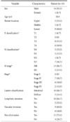
The clinicopathological features of 15 patients are presented in Table 1. Of 15 patients, 14 were male and 1 was female. The mean age was 58.9 years. The distribution of the stages according to 7th American Joint Committee on Cancer classification included seven stage II (46.7%), six stage III (40.0%), and two stage IV (13.3%).
The cytotoxic effect of the chemotherapeutic agents tested at previously published peak plasma concentrations192021 ranged from 0% to 72.7% cell death (Table 2). The highest cancer cell death rates were seen in cells treated with etoposide (44.8%), 5-FU (43.1%), and mitomycin (39.9%). The rank of each chemotherapeutic agent among 11 agents was determined according to that cell death rate in each patient. The most active chemotherapeutic agent was etoposide, with the highest chemosensitivity, 60.0% (9/15) for the tested specimens. Ten patients who underwent 5-FU chemotherapy were categorized into higher- and lower-cell death rate groups based on their median cell death rates. The overall survival of these two groups was analyzed. The 5-FU-sensitive patients showed better survival, though the difference was not statistically significant (P=0.216; Fig. 1A). Among the 15 patients, only two patients were treated with adjuvant chemotherapeutic agents as determined by ATP-CRAs (one with etoposide and one with cisplatin) that had the highest chemosensitivity results. Among the remaining 13 patients, 8 patients were treated with 5-FU based chemotherapeutic agents and 1 patient was treated with a docetaxel based chemotherapeutic agent. The other 4 patients did not received adjuvant chemotherapy. Fig. 1B shows the survival comparison of patients who were treated according to ATP-CRA results or who were treated with chemotherapeutic agents chosen by their physician (P=0.105).
The median number of cells positive for CD3, CD4, CD8, Foxp3, and Granzyme B were 156.0, 64.7, 80.7, 15.3, and 10, respectively (Table 3). Using the median values, all cases were classified into low- and high-density groups for each variable and survival rates were compared. A higher number of total T lymphocytes (CD3) was a good prognostic factor (P=0.008; Fig. 1C).
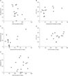
Pearson's correlation tests showed no statistically significant association between cancer cell death rates as determined by ATP-CRA for each chemotherapeutic agent and the TIL subsets from the correlating patient (Table 4). Three pairings of chemotherapeutic agents/count number of TIL subsets, docetaxel-CD4, 5-FU-Granzyme B, and methotrexate-Granzyme B had marginal associations with correlation coefficients of -0.453, 0.506, and 0.477, respectively, and P-values of 0.090, 0.054, and 0.072, respectively (Fig. 2A~C). The most commonly used chemotherapeutic agents in Korea, 5-FU and cisplatin, also showed no significant association with the CD3 subset, which showed prognostic implications in this study (Fig. 2D, E).
Accumulating evidence suggests that the immune microenvironment alters chemo-responsiveness.2223 Previous studies on chemo-responsiveness used various chemotherapeutic agents for different types of cancer. Thus, translation of the results to gastric cancer treatment is challenging. For example, high posttreatment levels of CD3 or CD8 TIL subsets in the tumor micro-environment correlates with more robust responses to paclitaxel neoadjuvant chemotherapy in breast cancer.24 Additionally, the density of CD8 TILs affects the chemo-responsiveness to 5-FU in stage III colon cancer.25 Higher densities of CD3, CD8, and Granzyme B, but not of Foxp3 TILs at invasive margins are related to improved chemo-responsiveness to irinotecan- and platinum-based chemotherapies in metastatic colorectal cancer.26 Furthermore, the level of regulatory T cells prior to chemotherapy is a predictive marker for early breast cancer.27 However, the relationship of the immune microenvironment and chemoresponsiveness in gastric cancer has never been studied. To explore whether cancer cell response to chemotherapeutic agents has any relationship with immune microenvironments, we investigated the association of cancer cell death rates in ATP-CRAs and the distribution of TIL subsets within the tumor tissue microenvironment. However, no significant associations were identified.
It is unknown whether TILs cause or enhance susceptibility to chemotherapeutic agents or are simply chemosensitivity markers. The current study suggests that TILs are more representative of susceptibility to chemotherapy rather than a marker of chemosensitivity. Our ATP-CRA and TIL analyses were separately performed using cultured cancer cells and immunohistochemical staining of tumor tissues. Thus, it may not completely and accurately represent the in situ interactive effects of chemotherapy on the tumor/host immune system. A subset of TILs in tumor microenvironments can modulate the susceptibility of chemotherapy against cancer cells.92829 Conversely, chemotherapy can enhance the efficacy of host immune functions by reducing tumor burdens and enhancing tumor cell susceptibility.30 It is also known that tumor-associated antigens are released when chemotherapeutic agents destroy tumor cells.9
Limitations of this study include the small number of the patients, emphasizing the need for further validation of these observations in additional data sets; only a small portion of the patients were treated according to ATP-CRA results; and the biological mechanism underlying these observations was not studied. Despite these limitations, we tried to assess the association of chemosensitivity test results and TIL subsets in gastric cancer. No significant association between the two suggests that current ATP-CRA has limitations for predicting cancer cell destruction, which could be affected by the immune system.
In conclusion, cancer cell death rates in response to specific chemotherapeutic agents had a poor association with the distribution of TIL subsets.
Figures and Tables
 | Fig. 1Survival analysis according to chemotherapeutic agent choice and tumor infiltrating lymphocytes (TILs). (A) Survival of patients who were treated by 5-fluorouracil (5-FU) (P=0.216). From 15 patients, 10 patients underwent 5-FU adjuvant chemotherapy. According to median cell death rates in adenosine triphosphate-based chemotherapy response assays (ATP-CRAs), patients were grouped as sensitive (n=5) and insensitive (n=5) to 5-FU. (B) Survival of patients who were treated with therapeutic agents as determined by ATP-CRAs (n=2) versus those chosen by a physician (n=8) (P=0.105). (C) Survival of high- (n=5) and low-CD3 (n=5) patients. Of the various TIL subsets examined, only high numbers of CD3 TILs (total T lymphocytes) showed favorable prognosis (P=0.008). |
 | Fig. 2Pearson's correlation scatter plots for cell death rates in response to various chemotherapeutic agents in adenosine triphosphate-based chemotherapy response assays and tumor infiltrating lymphocyte subsets for 15 patients. (A) CD4-docetaxel cell death rate (CDR). (B) Granzyme B-5-fluorouracil (5-FU) CDR. (C) Granzyme B-Methotrexate CDR. (D) CD3-5-FU CDR. (E) CD3-cisplatin CDR. For all graphs, each point represents results from a single patient. |
Table 2
Cancer cell death rates following treatment with chemotherapeutic agents in adenosine triphosphate-based chemotherapy response assays

Acknowledgments
This study was supported by a Faculty Research Grant from the Yonsei University College of Medicine for 2013 (6-2013-005).
References
1. Torre LA, Bray F, Siegel RL, Ferlay J, Lortet-Tieulent J, Jemal A. Global cancer statistics, 2012. CA Cancer J Clin. 2015; 65:87–108.
2. Macdonald JS, Smalley SR, Benedetti J, Hundahl SA, Estes NC, Stemmermann GN, et al. Chemoradiotherapy after surgery compared with surgery alone for adenocarcinoma of the stomach or gastroesophageal junction. N Engl J Med. 2001; 345:725–730.
3. Macdonald JS, Fleming TR, Peterson RF, Berenberg JL, Mc-Clure S, Chapman RA, et al. Adjuvant chemotherapy with 5-FU, adriamycin, and mitomycin-C (FAM) versus surgery alone for patients with locally advanced gastric adenocarcinoma: a Southwest Oncology Group study. Ann Surg Oncol. 1995; 2:488–494.
4. Yoo CH, Noh SH, Shin DW, Choi SH, Min JS. Recurrence following curative resection for gastric carcinoma. Br J Surg. 2000; 87:236–242.
5. Noh SH, Park SR, Yang HK, Chung HC, Chung IJ, Kim SW, et al. Adjuvant capecitabine plus oxaliplatin for gastric cancer after D2 gastrectomy (CLASSIC): 5-year follow-up of an open-label, randomised phase 3 trial. Lancet Oncol. 2014; 15:1389–1396.
6. Park S, Woo Y, Kim H, Lee YC, Choi S, Hyung WJ, et al. In Vitro adenosine triphosphate based chemotherapy response assay in gastric cancer. J Gastric Cancer. 2010; 10:155–161.
7. Park JY, Kim YS, Bang S, Hyung WJ, Noh SH, Choi SH, et al. ATP-based chemotherapy response assay in patients with unresectable gastric cancer. Oncology. 2007; 73:439–440.
8. Loi S, Sirtaine N, Piette F, Salgado R, Viale G, Van Eenoo F, et al. Prognostic and predictive value of tumor-infiltrating lymphocytes in a phase III randomized adjuvant breast cancer trial in node-positive breast cancer comparing the addition of docetaxel to doxorubicin with doxorubicin-based chemotherapy: BIG 02-98. J Clin Oncol. 2013; 31:860–867.
9. Denkert C, Loibl S, Noske A, Roller M, Müller BM, Komor M, et al. Tumor-associated lymphocytes as an independent predictor of response to neoadjuvant chemotherapy in breast cancer. J Clin Oncol. 2010; 28:105–113.
10. Yasuda K, Nirei T, Sunami E, Nagawa H, Kitayama J. Density of CD4(+) and CD8(+) T lymphocytes in biopsy samples can be a predictor of pathological response to chemoradiotherapy (CRT) for rectal cancer. Radiat Oncol. 2011; 6:49.
11. Liu H, Zhang T, Ye J, Li H, Huang J, Li X, et al. Tumor-infiltrating lymphocytes predict response to chemotherapy in patients with advance non-small cell lung cancer. Cancer Immunol Immunother. 2012; 61:1849–1856.
12. Zingg U, Montani M, Frey DM, Dirnhofer S, Went P, Oertli D. Influence of neoadjuvant radio-chemotherapy on tumor-infiltrating lymphocytes in squamous esophageal cancer. Eur J Surg Oncol. 2009; 35:1268–1272.
13. West NR, Milne K, Truong PT, Macpherson N, Nelson BH, Watson PH. Tumor-infiltrating lymphocytes predict response to anthracycline-based chemotherapy in estrogen receptor-negative breast cancer. Breast Cancer Res. 2011; 13:R126.
14. Morris M, Platell C, Iacopetta B. Tumor-infiltrating lymphocytes and perforation in colon cancer predict positive response to 5-fluorouracil chemotherapy. Clin Cancer Res. 2008; 14:1413–1417.
15. Balermpas P, Michel Y, Wagenblast J, Seitz O, Weiss C, Rödel F, et al. Tumour-infiltrating lymphocytes predict response to definitive chemoradiotherapy in head and neck cancer. Br J Cancer. 2014; 110:501–509.
16. Kim HI, Kim H, Cho HW, Kim SY, Song KJ, Hyung WJ, et al. The ratio of intra-tumoral regulatory T cells (Foxp3+)/helper T cells (CD4+) is a prognostic factor and associated with recurrence pattern in gastric cardia cancer. J Surg Oncol. 2011; 104:728–733.
17. Gooden MJ, de Bock GH, Leffers N, Daemen T, Nijman HW. The prognostic influence of tumour-infiltrating lymphocytes in cancer: a systematic review with meta-analysis. Br J Cancer. 2011; 105:93–103.
18. Minamoto T, Mai M, Watanabe K, Ooi A, Kitamura T, Takahashi Y, et al. Medullary carcinoma with lymphocytic infiltration of the stomach. Clinicopathologic study of 27 cases and immunohistochemical analysis of the subpopulations of infiltrating lymphocytes in the tumor. Cancer. 1990; 66:945–952.
19. Kang SM, Park MS, Chang J, Kim SK, Kim H, Shin DH, et al. A feasibility study of adenosine triphosphate-based chemotherapy response assay (ATP-CRA) as a chemosensitivity test for lung cancer. Cancer Res Treat. 2005; 37:223–227.
20. Weisenthal LM, Dill PL, Finklestein JZ, Duarte TE, Baker JA, Moran EM. Laboratory detection of primary and acquired drug resistance in human lymphatic neoplasms. Cancer Treat Rep. 1986; 70:1283–1295.
21. Bird MC, Bosanquet AG, Gilby ED. In vitro determination of tumour chemosensitivity in haematological malignancies. Hematol Oncol. 1985; 3:1–10.
22. Bhardwaj N. Harnessing the immune system to treat cancer. J Clin Invest. 2007; 117:1130–1136.
23. Burnette B, Weichselbaum RR. Radiation as an immune modulator. Semin Radiat Oncol. 2013; 23:273–280.
24. Demaria S, Volm MD, Shapiro RL, Yee HT, Oratz R, Formenti SC, et al. Development of tumor-infiltrating lymphocytes in breast cancer after neoadjuvant paclitaxel chemotherapy. Clin Cancer Res. 2001; 7:3025–3030.
25. Halama N, Michel S, Kloor M, Zoernig I, Pommerencke T, von Knebel Doeberitz M, et al. The localization and density of immune cells in primary tumors of human metastatic colorectal cancer shows an association with response to chemotherapy. Cancer Immun. 2009; 9:1.
26. Halama N, Michel S, Kloor M, Zoernig I, Benner A, Spille A, et al. Localization and density of immune cells in the invasive margin of human colorectal cancer liver metastases are prognostic for response to chemotherapy. Cancer Res. 2011; 71:5670–5677.
27. de Kruijf EM, van Nes JG, Sajet A, Tummers QR, Putter H, Osanto S, et al. The predictive value of HLA class I tumor cell expression and presence of intratumoral Tregs for chemotherapy in patients with early breast cancer. Clin Cancer Res. 2010; 16:1272–1280.
28. Zitvogel L, Apetoh L, Ghiringhelli F, André F, Tesniere A, Kroemer G. The anticancer immune response: indispensable for therapeutic success? J Clin Invest. 2008; 118:1991–2001.
29. Ladoire S, Arnould L, Apetoh L, Coudert B, Martin F, Chauffert B, et al. Pathologic complete response to neoadjuvant chemotherapy of breast carcinoma is associated with the disappearance of tumor-infiltrating foxp3+ regulatory T cells. Clin Cancer Res. 2008; 14:2413–2420.
30. Lake RA, Robinson BW. Immunotherapy and chemotherapy: a practical partnership. Nat Rev Cancer. 2005; 5:397–405.


 PDF
PDF ePub
ePub Citation
 Print
Print


 XML Download