

 PDF
PDF ePub
ePub Citation
 Print
Print


Introduction
Estrogens regulate the growth, differentiation, and functioning of diverse target tissues, both within and outside of the reproductive system.1 Although the stomach is not known as a direct target organ of sex hormones, both epidemiologic and animal studies suggest that gastric cancer may be estrogen-sensitive. A male dominance in the incidence of gastric cancer with a male to female ratio of about 2 : 1 sets up a hypothesis that estrogens may play a protective role against adenocarcinoma of the stomach.2 The risk of developing gastric cancer was lower among patients with prostate cancer who had been treated with estrogen than in those without such treatment.3 Studies which have shown that tamoxifen exposure has also been shown to be a risk factor for gastric cancer further support the hypothesis.4 Reduced risk of gastric cancer in a nationwide cohort study of men with a diagnosis of prostate cancer who had been treated with estrogen were reported.3 According to the experiment by Furukawa et al.,5 the carcinogenic N-methyl-N0-nitro-N-nitrosoguanidine added to drinking water induced gastric cancer in male rats but not in female, and castrated or estrogentreated male rats had a lower incidence of gastric cancer compared to untreated male rats.
Most of the actions of estrogens are exerted via the estrogen receptor (ER) of target cells, and 2 ER subtypes, ER-α and ER-β, mediate the genomic actions of estrogens. The presence of ER-α and β in gastric adenocarcinoma has been also demonstrated.5,6 The discovery of ERs has raised enormous interests in possible role of these receptors in the carcinogenesis, progression and treatment of gastric cancer.2 To date, however, only limited data regarding the detection of ERs in gastric tumors and cell lines are available, and significance of ER expression and hormone manipulation in gastric cancer is not established.
The aims of this pilot study were: 1) to describe the expression status of ER-α and -β mRNAs in five gastric carcinoma cell lines; 2) to evaluate in vitro the effects of 17β-estradiol and ER antagonists on the proliferation of the cell lines.
Materials and Methods
1. Cell lines and chemicals
Six cell lines established from human breast cancer (MCF7) and gastric adenocarcinoma (AGS, KATO III, MKN28, MKN45 and MKN74) were obtained from the Korean Cell Line Bank Center, Seoul, Korea. MCF7 cells were used for positive control for ERs. 17β-estradiol was purchased from Sigma-Aldrich, Yongin, Korea. ER-α antagonist, methyl-piperidino-pyrazole (MPP) and ER-β antagonist, pyrazolo [1,5-a] pyrimidine (PHTPP) were purchased from Tocris Bioscience, Bristol, UK. Solutions of 17β-estradiol and ER antagonists (MPP and PHTPP) were dissolved in ethanol (0.3%) and dimethylsulphoxide to 1 mM, respectively and then diluted in the medium to the final concentrations.
2. Cell culture
Cell lines were maintained in RPMI 1640 (GIBCO, Grand Island, NY, USA) without phenol red supplemented with 10% fetal bovine serum (FBS; HyClone Laboratories Inc., Logan, UT, USA), 1% penicillin/streptomycin and incubated at 37℃ in a humidified atmosphere containing 5% CO2 in air. Cultured cells were harvested at confluence by means of trypsinization and serially subcultured at a 1 : 4 split ratio.
3. Assessment of cell proliferation
The proliferative response of the cell lines was estimated by the colorimetric methylthiazol tetrazolium (MTT) test (Sigma-Aldrich, St Louis, MO, USA). After incubation with a drug, cells seeded in 96-well plates were harvested and pre-incubated with MTT (0.5 mg/ml) for 4 hours at 37℃. The culture medium was discarded, and the blue formazan crystals were solubilized with acidic isopropanol (0.1 N HCl in absolute isopropanol). MTT conversion to formazan by metabolically viable cells was monitored by spectrophotometry at 570 nm.
4. Detection of estrogen receptors
For ER analysis of cell lines, Total RNA of cells was extracted from individual cell lines using the TRIzol reagent (Invitrogen, Camarillo, CA, USA) according to the manufacturer's guidelines. Quantification of total RNA was performed by measuring absorbance at OD 260 nm. For cDNA synthesis, a reverse transcription-polymerase chain reaction (RT-PCR) technique (Promega, Madison, WI, USA) was used. One microgram RNA were reverse-transcribed in a final volume of 20 µl using the reverse transcriptase and then stored at -20℃. The semi-quantitative RT-PCR was performed in a final volume of 20 µl using G-Taq DNA polymerase (Cosmo Genetech, Seoul, Korea) according to manufacturer's instructions. To control for variations in the reaction, all the polymerase chain reaction (PCR) primers were normalized against the β-actin expression. The primer sequences were as follows: ER-α 5'-TCCTGATGATTGGTCTCGTCT-3' (forward) and 5'-ACATTTTCCCTGGTTCCTGTC-3' (reverse), ER-β 5'-AGTCTGGTCGTGTGAAGGATG-3' (forward) and 5'-ACTTCTCTGTCTCCGCACAAG-3' (reverse), and β-actin 5'-TGTTACAGGAAGTCCCTTGC-3' (forward) and 5'-AAGCAATGCTATCACCTCCC-3' (reverse). Primers were designed and synthesized by Primer 3 (http://simgene.com/Primer3). PCR was performed for 30 cycles (denaturation at 95℃ for 5 minutes, annealing at 60℃ for 1 minute and extension at 72℃ for 7 minutes). Five microliter of the reaction mixture was electrophoresed on a 2% agarose gel and visualized by ethidium bromide (MP Bio, Solon, OH, USA), applying 100-bp DNA ladder (RBC Bioscience, New Taipei, Taiwan) to estimate the band sizes.
5. Effects of 17β-estradiol and estrogen receptor antagonists on proliferation of gastric cancer cell lines
To investigate the effects of 17β-estradiol under estrogen-depleted culture conditions cell lines which expressed ER-α and ER-mRNAs were plated at a density of 1×10⁴ cells/100 ul of phenol red-free RPMI 1640 containing 10% FBS in 96 well plates (SPL Life Science, Pocheon, Korea). After 24 hours of incubation, when the cells were approximately 60% confluent, the 17β-estradiol (Sigma-Aldrich) dissolved in ethanol (0.3%) was added to the culture media at concentrations ranging from 5 nM to 32 µM. After 24 hours of treatment with 17β-estradiol the cell proliferative response was estimated by MTT test. Each experiment was repeated three times, and all results represent the means produced by three times repeated experiments.
To evaluate the effect of the ER antagonists on the proliferation of gastric cancer cell line, the cell line which expressed both ERs was chosen and treated with the ER-α antagonist MPP (50 µmol/L) and/or the ER-β antagonist PHTPP (50 µmol/L) 30 minutes before the application of 17β-estradiol (32 µM) and incubated for 72 hours.
Each experiment was repeated three times, and all results represent the means produced by three times repeated experiments.
6. Statistical analysis
The significance of the differences between the control group and each experimental group was evaluated with the Wilcoxon matched pairs test. P<0.05 were considered statistically significant. IBM SPSS Statistics ver. 20.0 for Windows (IBM Co., Armonk, NY, USA) was used for the statistical analyses.
Results
Semi-quantitive RT-PCR was performed to measure the levels of two ERs in the human gastric cancer cell lines; five cancer cell lines (AGS, KATO III, MKN28, MKN45 and MKN74) were used for study and MCF7 cells were used for controls for positive ERs. As shown in Fig. 1A, ER-α mRNA was detected in the KATO III, MKN28, and MKN45, except AGS and MKN74. ER-α/β-actin ratio of KATO III, MKN28 and MKN45 were 6.17, 0.86, and 0.04, respectively. In contrast, ER-β mRNA was detected in all the five cell lines. ER-α/β-actin ratio of AGS, KATO III, MKN28, MKN45 and MKN74 were 1.33, 1.74, 0.76, 2.06, and 1.93, respectively (Fig. 1B).
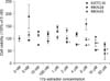
Fig. 2 shows the effects of increasing concentrations of 17β-estradiol on the proliferation of gastric cancer cell lines. The 17β-estradiol concentrations (from 5 nM to 32 M) did not significantly affect the cell viability in the MKN28 cells. In the MKN45 cells, the 17β-estradiol concentrations (from 5 nM to 4 µM) did not significantly affect the cell viability in the MKN45 cells, whereas higher concentrations (8, 16 and 32 µM) of 17β-estradiol caused a significant reduction in cell viability (-2.04%, -17.56%, -59.75%, respectively, P<0.01). Viable cells of KATO III decreased in a dose dependent manner, reaching the minimum value at a 17β-estradiol concentration of 32 µM. 17β-estradiol at concentrations lower than 1 µM did not significantly affect cell viability, whereas the higher concentrations (2, 4, 8, 16, and 32 µM) of 17β-estradiol caused a significant reduction in cell viability, -14.21%, -24.13%, -26.8%, -46.93%, and -46.23%, respectively (P<0.01).
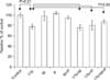
To better understand the role of ERs on the proliferation of gastric cancer cell line, KATO III cells were treated with the ER-α antagonist (MPP, 50 µmol/L) and/or the ER-β antagonist (PHTPP, 50 µmol/L) 30 minutes before the application of 17β-estradiol. Neither ER-α nor ER-β antagonist had effect on anti-proliferative effect of 17β-estradiol (Fig. 3). Proliferation rate of KATO III cells was decreased to 77.8% of control 72 hours after application of 17β-estradiol (P<0.01). The estrogenic effect was not blocked by ER-α antagonist, MPP, where the cell viability was reversed to 76.5% of 17β-estradiol treated control (P>0.05, Wilcoxon matched pairs test). ER-β antagonist, PHTPP, did not block antiproliferative effect of 17β-estradiol, with a cell viability of 80.8% of 17β-estradiol treated control (P>0.05). When treated with both MPP and PHTPP, cell proliferation was reversed to 87.5% of 17β-estradiol treated control (P>0.05).
Discussion
Estrogens regulate the growth, differentiation, and functioning of various target tissues, and their effects are mediated by the receptors, ER-α and ER-β. ER-α and ER-β have distinct cellular distributions; ER-α is predominantly expressed in female sex organs such as the breast, uterus and ovaries, and ER-β is far more widely distributed in the body than is ER-α.7,8
The oncologic importance of estrogens and ERs in cancers arising in classic target organs for estrogens such as breast and uterine endometrium have been well described.9-14 Estrogens and their receptors also have an important role in the progression of cancers of gastrointestinal tract which has been believed to be nontarget system for estrogens. In contrast to the cancer-promoting role of estrogen in breast and gynecologic cancers, it functions as a suppressor for gastrointestinal cancers.15 Current use of estrogen therapy is suggested to be associated with a decreased risk of colorectal cancer among peri- or postmenopausal women,13,16 and in comparison with tumors with high ER-β expression, tumors negative for ER-β were associated with advanced cancer stages.17 Most studies on esophageal adenocarcinoma are consistent in suggesting a detrimental effect and prognostic value for ER-β.7
Contrasting results have been obtained with regard to the expression level of ERs in gastric cancer. Some studies show that only ER-β is expressed in gastric cancer,18,19 whereas others reported that the both ERs are expressed.20-22 This prompted us to reevaluate ER status and hormonal modulation of cell growth in human gastric cancer.
Cultured gastric cancer cell line is a useful tool to study the effect of hormones in conditions that are independent of neural, hormonal, and blood flow factors.23 Past studies performed on gastric cancer cell lines showed contrasting actions of estrogens on the cell growth. Diverse effects of estradiol on the growth of six human gastric xenografts in nude mice were found, including one case of stimulation, two of inhibition and three of unchanged condition.24 Messa et al.23,25 showed that 17β-estradiol administration exerts a growth-inhibitory effect on ER-positive cell lines (HGC-27, AGS). On the other hand, Wu et al.26 found that steroid hormone administration in ER-negative human gastric carcinoma cell line NUGC-3 had a slight stimulatory effect. These studies, however, did not distinguish ER-β from ER-α. The discovery of ER-β,12 a second ER isoform, has prompted renewed investigation into the mechanism of action of estrogenic molecules. However, the biologic significance of the ERs in gastric cancer still remains to be elucidated.
In the present study, ER-α and ER-β mRNAs were expressed in three (KATO III, MKN28 and MKN45) and all of the five gastric cancer cell lines, respectively. ER-α mRNA was not expressed in AGS and MKN74 cell lines and the ER-α/β-actin ratios were very low in MKN28 and MKN45 (0.28 and 0.04, respectively). Our results indicated that ER-β mRNAs are preferentially expressed in gastric cancers, as Takano et al.20 reported, and ER-β plays more important role in both physiologic and oncologic processes in gastric tissues than ER-α.
Imbalanced ER-α/ER-β expression, i.e., an increase in ER-α/ER-β mRNA and protein ratios, is a common feature and could be a critical step of estrogen-dependent tumor progression, and ER-β seems to play a key role in the mitogenic action of estrogen by providing protection against ER-α-induced hyperproliferation.14 Our previous study indicated that The ER-β positive group was associated with lower tumor stage, Lauren's intestinal type, negative perineural invasion, and free of recurrence, and the ER-β positive group had a better 3-year survival compared with the negative group in survival analysis.19 Xu et al.27 also reported that ER-α expression and the absence of ER-β expression are associated with poor survival.
In our results, however, relations between the levels of mRNA expression of ERs and effects of 17β-estradiol on proliferation of gastric cancer cell lines were not identified. Low concentrations of 17β-estradiol stimulated cell growth of MKN28 and MKN45 cell lines, even though the values were statistically not significant. A significant decrease in the cell growth rate is obtained with 17β-estradiol concentrations equal to or higher than 16 µM. On the contrary, 17β-estradiol administration exerted a growth-inhibitory effect on the KATO III. A significant decrease in the cell growth rate is obtained with 17β-estradiol concentrations equal to or higher than 2 µM, which was much lower than those required for MKN28 or MKN45. KATO III cells display a higher expression level of ER-α than MKN28 and MKN45. This finding could be explained by the fact that ER-α mRNA expression in KATO III cell line was higher than those in MKN28 and MKN45 cell lines.
Some previous studies suggested that the ER-α pathway may have a role in the progression of gastric cancer and 17β-estradiol enhances gastric cancer cell proliferation in vitro. Based on these suggestion, a few clinical trials using an estrogen antagonist, tamoxifen, have been conducted for the management of patients with ER-α positive gastric cancer. However, the results have not been consistent. An earlier Japanese study with historical control suggested a survival benefit in tamoxifen treated patients with gastric cancer but the sample size was small and patients received chemotherapy.28 A randomized clinical study by Harrison et al showed that ER-α positive patients survived for a significantly shorter time than untreated controls if they were treated with tamoxifen. ER-α negative patients had a longer survival and tamoxifen did not significantly affect prognosis.29,30 On the contrary to these old reports, Kameda et al.31 recently reported that 17β-estradiol-induced cell proliferation was suppressed by pure anti-estrogen drug, ICI 182, 780 and suggested that the development of clinically available antagonists that can sufficiently block estrogen-induced cell proliferation is promising for management of patients with aggressive diffuse-type gastric cancer. They suggested a possibility that different anti-estrogens used in the above studies may explain the discrepancies in results.
In the present study, MPP and PHTPP were used as ER-α and ER-β antagonists, respectively. MPP is a highly selective ER-α antagonist with 200 fold greater affinity for ER-α versus ER-β,32 and PHTPP is a potent ER-β antagonist, with negligible activity with ER-α.33 The present study, however, showed that neither ER-α nor ER-β antagonist had effect on anti-proliferative effect of 17β-estradiol. In our previous study, immunohistochemical staining of ER-α showed no positivity in any of the tissue microarrays of gastric adenocarcinoma tissues, although ER-α was expressed in normal and gastric adenocarcinoma tissues on Western blot analysis.19 The expression of a particular gene mRNA may not always correspond with the expression of the functional protein.21
Our present study imply that hormone therapy rather than ER blockers may be a useful strategy for the treatment of gastric cancer in cases of ER-β positive gastric cancer. However, our study has some limitations. Our data do not show clear relationship between the effect of 17β-estradiol and ER-β. 17β-estradiol showed no effect on two ER-β positive cell line. Furthermore, ER antagonists failed to block the effect of 17β-estradiol in this study. The mechanism by which estrogens and their receptors inhibit the growth of cancer cell should be clarified by further studies.

 XML Download