

 PDF
PDF ePub
ePub Citation
 Print
Print


Introduction
The clinical outcome of pyogenic liver abscesses with malignant disease is reportedly dismal.1 Since liver abscesses can be easily detected by abdominal computed tomography (CT) or clinical symptoms, we have an opportunity to treat such diseases successfully; however, we rarely experience fetal septicemia from the occult liver abscesses which cannot be detected in advance. We hereby reported a rare case of acute septicemia unknown etiology following anti-cancer drug treatment, autopsy identified that Escherichia coli due to occult multiple liver abscesses was a trigger of these sepsis and organ failure.
Case Report
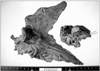
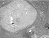
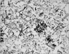
A 72-year-old man having recurrent lymph node metastases from gastric cancer was admitted to Saiseikai Sendai Hospital to receive chemotherapy for gastric cancer. He had a history of distal gastrectomy plus right hepatic lobectomy for advanced gastric cancer with liver metastases (Fig. 1). As paraaortic lymph node metastases had re-grown despite 17 courses of combination chemotherapy of paclitaxel (Bristol-Myers Squibb, NY, US) and S-1 (Taiho Pharmaceutical CO., LTD, Tokyo, Japan), the chemotherapy regime was changed to drip infusion of docetaxel (Sanofi-aventis, Paris, France). He showed no definite adverse effects at the first administration of docetaxel in short term hospitalization. This was the second treatment of docetaxel. On admission, he looked well-being and blood chemistry data showed no inflammatory disease. Before chemotherapy, abdominal CT showed neither metastasis nor abscesses in the liver. A total of 95 mg of docetaxel was infused intravenously. Before docetaxel administration, 8 mg of methyl predonine was routinely pretreated. One day after chemotherapy, the patient presented with a high-grade fever up to 39℃ and low blood pressure (80/55 mmHg). At that time, laboratory findings revealed a white blood cell count of 13,550/L, a red blood cell count of 2,420,000/L, a blood platelet count of 109,000/L, aspartate aminotransferase of 1,112 IU/L, alanine aminotransferase of 774 IU/L, and CRP of 4.6 mg/dl. Since he was diagnosed with septicemia unknown etiology, continuous dopamine infusion and broad-band antibiotics were started. The next day, blood chemistry examination revealed serious liver and renal dysfunction. Although sepsis was strongly suspected from leucocytosis and the high value of C-reactive protein, the origin of sepsis could not be identified. Despite intensive care including hemodialysis and respiratory support, the patient died 5 days against strong medical care after docetaxel administration. Blood bacterial culture revealed strongly positive E. coli. An autopsy was performed to check the cause of rapid multiple organ failure. There was no evidence of myelo-suppression by sequential chemotherapy from the result of bone marrow histology. Residual lymph node metastases from gastric cancer were histologically confirmed. Multiple occult liver abscesses which could not identified macroscopically were diagnosed by the histological examine (Fig. 2). They consisted of gram-negative rods and fungi (Fig. 3). Therefore, we concluded that fatal E. coli septicemia had been induced from multiple occult liver abscesses as a result of immuno compromised host induced by anticancer drug treatment.
Discussion
Pyogenic liver abscesses are found in 0.3~1.4% and their mortality rate is reportedly 11~13%.2,3 The majority of pyogenic liver abscesses are caused by infection originating from the biliary or intestinal tracts. E. coli infection may be introduced from the biliary tract. Patients with diabetes mellitus, immune deficiency and malignancy are at high risk for liver abscesses.4
The patient had recurrent gastric cancer and repeated anti-cancer drug administration. It seems that his occult immuno-suppressive condition has a strong association with high risk for pyogenic liver abscess.
His white blood count was preserved during chemotherapy and normal bone marrow histology was confirmed by autopsy. This fatal septic state did not directly connected with meyelo-suppression by redundant treatment of anti-cancer agents. When pyogenic liver abscess is diagnosed, the patients underwent radiologically guided percutaneous drainage of the liver abscess and they received empiric antimicrobial therapy.5 In the current case, regrettably, we could neither detect nor treat these liver abscesses before he developed fatal state. In this series, we did not perform CT examination. Considering the finding of the autopsy, we could not detect pyogenic liver abscess during the treatment.
In conclusion, we presented a rare case of fatal sepsis from multiple occult liver abscesses following redundant anti-cancer treatment. We should keep in mind the high risk of fatal sepsis from the pyogenic liver abscesses in cancer patients who had recurred tumor and history of redundant chemotherapy.
 XML Download