

 PDF
PDF ePub
ePub Citation
 Print
Print


Introduction
There have been many changes in the surgical techniques used to treat complicated duodenal ulcer disease.(1-7) Especially in the case of duodenal ulcer perforation, primary repair is one of the most common methods used by acting surgeons in emergency surgery because of the convenience of the surgical technique and advances in medical treatment. After laparoscopic repair was first introduced for duodenal ulcer perforation, many surgeons tried to perform the technique and they reported their surgical outcomes and experiences.(4,6,8,9)
With regards to surgical outcomes in laparoscopic primary repair for duodenal ulcer perforation, there are still many questions as to whether the surgeon should perform this type of surgery in an emergency. Some surgeons have suggested that laparoscopic primary repair is the best way to improve early surgical outcomes.(10-15) On the other hand, others believe that there is no significant difference between laparoscopic primary repair group and open primary repair group.(16-20)
Experience is a very important factor in improving the surgical outcome of laparoscopic surgery. Many investigators have reported that a learning period is necessary to improve the surgical outcomes of this type of surgery in the treatment of stomach cancer.(21-24) Therefore, only experienced laparoscopic surgeons participated in this study to resolve this problem. This study aims to evaluate the feasibility and safety of laparoscopic primary repair for duodenal ulcer perforation, providing detailed technical information about the procedure.
Materials and Methods
1. Patients
We enrolled 21 consecutive patients who underwent primary repair with omentopexy for duodenal ulcer perforation between March, 2011 and May, 2012 at Hanyang University Guri Hospital. We retrospectively reviewed the prospectively collected data. In this study, experienced surgeons were defined as those who had abundant experience in laparoscopic surgery and other major operations as the operating surgeon. One experienced surgeon had performed 139 laparoscopic gastrectomies to treat gastric cancer by March, 2010.(25)
There were no laparoscopic explorations during the study period and there were no contraindications to perform laparoscopic surgery. Laparoscopic primary repair was carried out regardless of time from the onset of symptoms, previous laparotomy, old age, and comorbidity. The choice of primary repair was decided by an intra-operative assessment of the perforation size. During the same period, five patients who were diagnosed with giant duodenal ulcer perforation (>2 cm) underwent laparoscopic distal gastrectomy with truncal vagotomy. These patients were excluded from the study.(18)
2. Surgical techniques
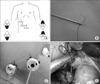
Each patient was placed in thereverse Trendelenburg position. A carbon dioxide pneumoperitoneum was formed from the umbilical port, and pressure was maintained between 12 and 15 mmHg. Two 12 mm trocars and one 5 mm trocar (ENDOPATH Xcel Bladeless Trocar) were positioned as described in Fig. 1A.
After all the trocars were inserted, the size of the perforation was measured by intra-operative assessment. If the perforation was not much bigger than 2 cm, we carried out a laparoscopic primary repair with omentopexy. To minimize spillage of gastric, biliary, and pancreatic contents from the perforation site, we closed the perforation site before peritoneal irrigation.
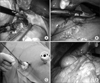
Extracorporeal knot tying was used to make it easier for the operator to perform the suturing. Our technique consists of several steps. First, undetached black silks (3-0, 26 mm, 75 cm, MERSILK, ETHICON®) were used to close the perforation site. Closure of the perforation site was performed to avoid the possibility of the detached needle dropping into the abdominal cavity. Second, the needle was prevented from dropping through the trocar during insertion and extraction by grasping the thread 2 cm below the needle (Fig. 1B). This thread was then moved into the right lower trocar (Fig. 1C). Third, the needle was held just below the swage to perform suture repair of full thickness around the perforation site (Fig. 1D, 2A). In the process of extracting the needle and thread from the suture site, a pulley principle was used to minimize injury to the inflamed tissues. Because the smooth surface of the grasper body changes the direction of the tension force in the thread and decreases the force, it was possible to extract the needle easily (Fig. 2B). Before performing extracorporeal knot tying, this thread was extracted from the right upper trocar. The detached thread was extracted again through the right upper trocar to make a knot. After tying, a knot was made just above the perforation site by pushing a knot pusher through the right upper trocar (Fig. 2C).
After primary closure of the perforation site, the contaminated abdominal cavity was irrigated with a large volume of saline (usually 1 or 2 L) to avoid the accumulation of infected fluid and other debris. Once irrigation was complete, a large volume of omentum was reinforced above the primary suture site by extracorporeal knot tying (Fig. 2D).
3. Pre- and postoperative course
A nasogastric tube was inserted in order to prevent spillage of the stomach contents into the peritoneal cavity at the time of diagnosis of the duodenal ulcer perforation. Broad spectrum antibiotics were used until all signs of peritoneal irritation disappeared.
After surgery, the nasogastric tube was maintained until it was safe to remove it, as indicated by upper gastrointestinal imaging (UGI) using gastrografin. Depending on the return of bowel activity, UGI examination was performed on postoperative day 3~5. An intravenous proton pump inhibitor was injected during the fasting period and theoral proton pump inhibitor was changed after starting a diet, and continued for 8 weeks.
During the operation, a laparoscopic biopsy of the ulcer was performed if possible. If this was not possible due to inflamed ulcer tissues, a follow-up gastrofiberscopy was performed to evaluate the state of the ulcer.
4. Clinical analysis
Clinical data obtained from medical records included patient age, gender, body mass index (BMI), and American Society of Anesthesiologist (ASA) score. Early surgical outcomes included operation time, overall postoperative complications, day of normalization of leukocytosis, day of commencement of a soft diet, number of administered analgesics, and postoperative hospital stay.
Postoperative complications were defined as any condition requiring conservative or surgical treatment. Severe postoperative complications were defined as those that required management by an endoscopic or interventional procedure or a re-operation (expanded classification, over level 3).(26)
In this study, a liquid diet was started after confirming the safety of the suture site by performing UGI between postoperative days 3 and 5. The soft diet was started when patients felt comfortable enough to consume a liquid diet twice, consecutively. Patients were discharged if they had no problems eating a soft diet, showed an absence of inflammatory conditions including leukocytosis, unstable vital signs and abrupt onset abdominal pain, and if they were generally comfortable. The final decision regarding discharge was left up to each patient.
Results
1. Patient characteristics
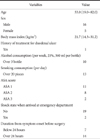
The clinical characteristics of the patients in this study are presented in Table 1. Twenty-one patients, 16 men and five women, presenting with duodenal ulcer perforation were treated with laparoscopic primary repair with omentopexy. The median age was 53 years (19~82 years). Median BMI at the time of operation was 21.7 (14.3~31.2). Ten of the 21 patients showed ASA class 2, 3. Fourteen patients underwent the operation more than 24 hours after the onset of symptoms. Two patients arrived at our institution in a state of shock.
2. Early surgical outcomes of patients who underwent primary repair with omentopexy
Details of early surgical outcomes are listed in Table 2. There was no conversion to open surgery and no postoperative mortality in any patients. Postoperative complications occurred in one patient (4.7%). The median operation time was 45.0 minutes. The median day of commencement of a soft diet was 6.0 days. The mean number of administered analgesics was 1.3 during the postoperative period. The duration of the postoperative hospital stay was 8.0 days.
3. Details of postoperative complications
A 58-year-old male patient was operated on due to a duodenal ulcer perforation. The time period between onset of symptoms and diagnosis was over 48 hours. Before being transferred to our hospital, he was hospitalized in a specialized dementia hospital because of alcohol dementia. Based on operative findings, the size of the perforation measured about 2 cm. The whole abdominal cavity had been severely contaminated by infected bowel contents. Two stitches were used to close the perforation. The operation took 1 hour from skin incision to skin closure. The first postoperative UGI series showed a contrast leakage from the suture site, although there were no clinical signs of leakage during the recovery period. Radiologic leakage disappeared by postoperative day 15. After confirming closure of the perforation, the patient started a soft diet on postoperative day 17. He was discharged from the hospital on postoperative day 18.
Discussion
After laparoscopic surgery was first described as a surgical method to repair a duodenal ulcer perforation in 1990, several investigators suggested that this surgery could be a better way to improve surgical outcomes in patients undergoing emergency surgery for duodenal ulcer perforation.(1-6,9) By contrast, several researchers suggested that laparoscopic primary repair did not improve surgical outcomes compared to open primary repair.(7-11)
Despite recent controversies, laparoscopic surgery is likely to play an increasing role in the future, as surgical techniques evolve and more surgeons are expected to apply laparoscopic techniques in the treatment of surgical emergencies such as duodenal ulcer perforation. In reality, many surgeons hesitate to perform laparoscopic emergency surgery due to their inexperience. Therefore, this study was designed to assess the feasibility of laparoscopic primary repair for duodenal ulcer perforation and to provide less experienced surgeons with detailed information about the practical procedures.
In our study, the analysis of surgical outcomes after laparoscopic primary repair yielded positive surgical outcomes, similarly to previous reports.(1-6) Particularly with consideration of the study population, we believe that our results were satisfactory. We did not have any contraindications to laparoscopic emergency surgery in our study. The interval between symptom onset and diagnosis, the grade of peritoneal contamination, and the ASA score were not taken into consideration.
In the present study, postoperative complications occurred in one patient. The leakage at the suture site was diagnosed by routine postoperative UGI. Before and after the diagnosis of this complication, there were no clinical signs of leakage. During the operation, the size of the perforation was measured at 2 cm. We could find marked wall thickening around the perforation site. In the case of a large perforation or severely inflamed ulcer tissues, we avoided creating a tight closure as it can cause substantial disruption. A large volume of omentum was placed above the suture site as possible. We believe that the leakage observed radiologically did not fall under the heading of morbidity. On balance, we are confident that laparoscopic primary repair is a good method for the surgical treatment of duodenal ulcer perforation.
With respect to surgical outcomes, we believe that there were several reasons for our findings. First, surgeons experienced in laparoscopic gastrectomy participated in this study. The operating surgeon had experience on more than 139 cases of laparoscopic gastrectomy for stomach cancer before performing this procedure. We believe that hands-on experience is a key element to successful laparoscopic emergency surgery. Therefore, the accumulation of experience in this technique is expected to help inexperienced surgeons in many ways. Second, it is important to find a way to make this procedure more convenient. For example, we did not change the operating setup, such as the positioning of the operating surgeon and patient, during laparoscopic gastrectomy for stomach cancer. In addition, we did not insist on performing intracorporeal knottying and suturing, as many surgeons are anxious to perform this procedure due to their inexperience. Therefore, extracorporeal knottying and intracorporeal suturing techniques were used successfully. Third, we tried to avoid tight suturing at the perforation site because this may lead to dehiscence of anastomosis by penetration of the thread into inflamed tissues. In our institution, we inserted and extracted the needle tip into normal tissue outside of the inflamed tissue range.
We acknowledge that there are some limitations to our study. We could not compare surgical outcomes between open and laparoscopic primary repair because open primary repair was performed by other surgeons. In addition, our sample size was relatively small.
In conclusion, surgeons should adapt to the changing needs of future generations. Especially in the case of benign diseases such as duodenal ulcer perforation, laparoscopic surgery should be performed, as the outcome of such surgery is very important in terms of quality of life. Easier and more detailed techniques are needed for less experienced surgeons to perform laparoscopic primary repair. We hope that our detailed method will help 'beginners' to perform laparoscopic primary repair easily in the case of duodenal ulcer perforation.

 XML Download