

 PDF
PDF ePub
ePub Citation
 Print
Print


Introduction
Bezoars are an unusual cause of gastric outlet obstruction and are formed in the gastrointestinal tract. Most beazoars form in the stomach. They occasionally cause gastrointestinal obstruction after passing from the stomach to the small intestine.(1,2) Several approaches for the treatment of gastric phytobezoars have been proposed such as enzymatic or drug-based dissolution, endoscopic fragmentation and extraction, and surgical treatment.(3) Large multiple gastric bezoars require surgical management, and conventional surgical management of large multiple gastric bezoars traditionally involves laparotomy. However, it is increasingly feasible to remove large bezoars through laparoscopy without the necessity of an abdominal incision.
Case Report
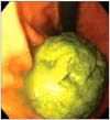
A previously healthy 52-year-old woman, without diabetes, presented with intermittent severe epigastric pain after consuming 15 persimmons at one sitting 3 weeks before admission, with a pertinent history of persimmon overconsumption since childhood. The body mass index of the patient was 25.3 kg/m2 and physical examination revealed a palpable hard mass in the upper abdomen without abdominal distension, guarding, or rigidity. Computed tomography scans demonstrated large heterogeneous masses with a mottled air pattern within the stomach. Gastroscopy showed 3 large bezoars (about 10 cm, 7 cm, 7 cm diameter) occluding almost the entire pylorus (Fig. 1). Nasogastric irrigation and endoscopic fragmentation were attempted for conservative management but found to be unsuccessful and laparoscopic removal was decided.
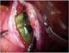
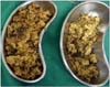
One 10-mm port, one 12-mm port, and two 5-mm ports were used. A 4 cm-sized gastrotomy incision was made on the anterior wall of the gastric body using an ultrasonic shears. A laparoscopic endo-bag was introduced and positioned next to the gastrotomy site. All gastric bezoars were retrieved and placed directly in one endo-bag without any spillage (Fig. 2). The endobag was retrieved through the subumbilical port site and the open end of the bag was exteriorized outside the abdomen. The wound of the subumbilical port was enlarged to 20 mm, and the bezoars were morcellated with ring forceps and removed in fragments (Fig. 3).
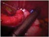
The gastrotomy site was closed using endo-GIA staplers (ETS Flex45, Ethicon Enodo-Surgery, Cincinnati, OH, USA) after the placement of stay sutures (Fig. 4). The entire small intestine and the stomach were explored carefully for retained bezoars, and a drainage tube was not inserted into the peritoneal cavity.
The total operating time was 90 minutes and there were no significant complications during surgery. The postoperative course was uneventful, with a return to soft food on the third postoperative day and discharge on the sixth postoperative day.
DISCUSSION
Bezoars are retained collections of swallowed material (either food or foreign body) that fail to clear the stomach and accumulate into masses of concretions. They are usually found in the stomach, but on occasion can be located in the small intestine and the rectum.(1,2)
Gastric bezoar formation may occur in patients with altered gastric physiology, impaired gastric emptying, and reduced acid production. So, they mainly occur in patients who have undergone gastric surgeries, including antrectomy or vagotomy with pyloroplasty. Illnesses which affect gastrointestinal motility can contribute to risk.
Bezoars can be phytobezoars (made up of vegetable material), trichobezoars (hair balls), pharmacobezoars (made up of various ingested chemical substances), and lactobezoars (undigested milk concretions). It may produce ulceration, epigastric pain and discomfort, gastrointestinal hemorrhage, perforation, and intestinal obstruction, and should always be removed from the stomach because of these risks.(3)
Many conservative and surgical approaches are used in the management of gastric bezoars. Surgical management is more commonly used for trichobezoars, but gastric phytobezoars can be treated by several conservative methods, including nasogastric lavage, enzymatic dissolution, and endoscopic fragmentation.(4) Many endoscopic techniques have been described which result in the breakage of gastric bezoars. Instruments used for breakage include biopsy forceps, polypectomy snares, electrohydraulic lithotripters, pulsed water jets, Nd:YAG lasers, needle-knife bezotomes, and endoscopic injection with enzymes.(5-7)
Gastroscopic removal of bezoars has not always been a successful mode of therapy, since conservative management is not useful in the presence of large multiple bezoars, and surgical removal by gastrotomy is the preferred clinical option. Additionally, some patients cannot tolerate the repetitive passage of the endoscope for multiple bezoars and the long manipulation times. Recent developments in minimally invasive surgery enable effective laparoscopic removal of gastric bezoars instead of the traditional surgical treatment through an abdominal incision.(8,9) Further, the simplicity of the traditional surgical removal of bezoars with a gastrotomy predicted the easy applicability of the laparoscopic approach without complex laparoscopic skills.
We minimized the possibility of bezoar spillage and wound infection in this clinical case by direct transfer of bezoars from the stomach into the laparoscopic endo-bag positioned directly next the gastrotomy site. We ensured complete removal of all bezoars from gastrointestinal tract by careful laparoscopic examination of the stomach and the remainder of the intestine. These measures are also important in open conventional surgery for bezoar removal.
Laparoscopic procedures provide superior cosmetic effects, shorter hospital stays, earlier return to normal daily activity, less postoperative pain, and a decreased incidence of adhesion formation as compared with conventional open laparotomy. Therefore, we recommend this laparoscopic approach for the patients with multiple large gastric bezoars in whom surgical management is considered as a treatment option.
 XML Download