Abstract
Objectives
To assess the correlation between symptom improvement and spinal canal dimensions in patients who underwent selective nerve root block for lumbar spinal stenosis.
Summary of Literature Review
When the canal size is relatively small, the pressure on the nerve root increases. Decompressive surgery relieves more pain in such patients.
Materials and Methods
From July 2009 to March 2011, 141 patients received selective nerve root block for 1-level central lumbar spinal stenosis in our hospital. We evaluated the patients using a visual analog scale (VAS) before the procedure and 1 hour, 1 month, and 3 months following the procedure. We measured the spinal canal using magnetic resonance imaging.
Results
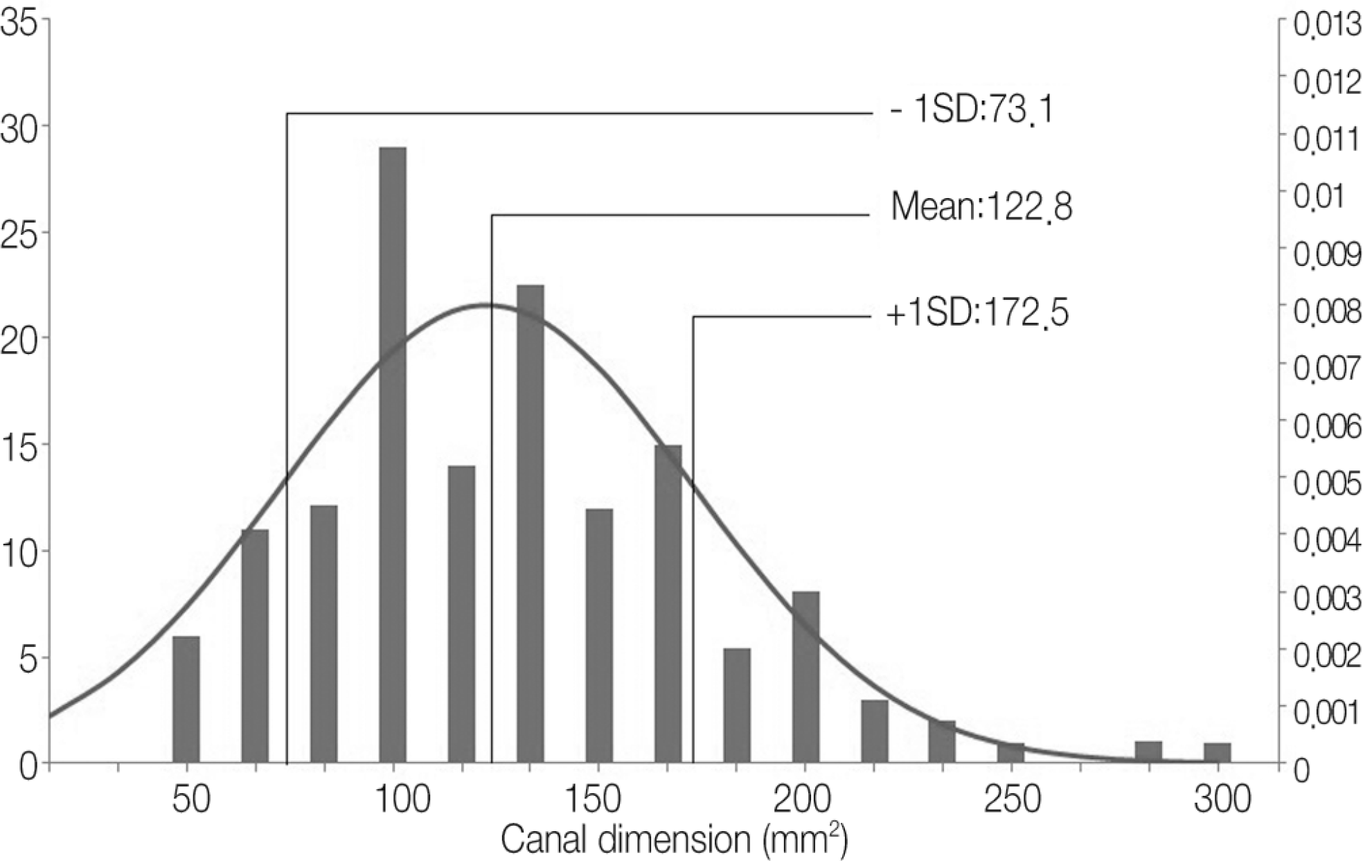
There was no significant correlation between spinal canal dimensions and the pre-procedure VAS. We divided the patients into 3 groups using the average and the standard deviation of the patients'spinal canal dimensions (<73.1 mm2, 73.1-172.5 mm2, >172.5 mm2) (p<0.01). One hour after the procedure, the VAS scores changed by 1.43±1.8, 1.62±1.7, and 1.53±1.5, respectively, with no significant differences among the 3 groups. However there were significant differences in the VAS changes 1 month and 3 months following the procedure, with results of 2.39±1.7 and 1.39±1.5, 4.65±2.1 and 4.28±2.3, and 4.97±2.2 and 6.83±1.9 (p<0.01), respectively.
REFERENCES
1. Sachs B, Fraenkel J. �rogressive ankylotic rigidity of the spine. J Nerv Ment Dis. 1900; 27:1–15.
2. Chang � Y, Chung YK, �ark WC. Spinal stenosis; �eview of 40 cases. J Korean �rthop �ssoc. 1982; 17:808–14.
3. Chae � J. Lumbar spinal stenosis. J Korean Soc Spine Surg. 1999; 6:220–7.
4. Chung SS, Lee CS, Lee SG, et al. Correlation between clinical features and M�� findings in one level lumbar spinal stenosis. J Korean �rthop �ssoc. 1999; 34:541–6.
5. Bolender NF, Schonstrom N, Sepengler D. �ole of computed tomography and myelography in the diagnosis of central spinal stenosis. J Bone Joint Surg �m. 1985; 67:240–6.
6. �rnoldi CC, Brodsky � E, Cauchoix J, et al. Lumbar spinal stenosis and nerve root entrapment syndromes. Definition and classification. Clin �rthop �elat �es. 1976; 115:4–5.
7. �mundsen T, Weber H, Lilleas F, et al. Lumbar spinal stenosis. Clinical and radiologic features. Spine (�hila �a 1976). 1995; 20:1178–86.
8. �nel D, Sari H, Donmez C. Lumbar spinal stenosis: Clini-cal/�adiologic therapeutic evaluation in 145 patients. Conservative treatment or surgical intervention? Spine (�hila �a 1976). 1993; 18:291–8.
9. Shim DM, Choi YH, Yang JH, et al. �nalysis and measurement of the lumbar spinal canal dimension using magnetic resonance imaging. J Korean �rthop �ssoc. 2008; 43:588–94.
10. Tajima T, Furukawa K, Kuramochi E. Selective lumbosacral radiculography and block. Spine (�hila �a 1976). 1980; 5:68–77.

11. Weber H. Lumbar disc herniation. � controlled, prospective study with ten years of observation. Spine. 1983; 8:131–40.
12. Saal J�, Saal JS. Nonoperative treatment of herniated lumbar intervertebral disc with radiculopathy. �n outcome study. Spine (�hila �a 1976). 1989; 14:431–7.
13. Deyo ��, Loeser JD, Bigos SJ. Herniated lumbar intervertebral disk. �nn �ntern Med. 1990; 112:598–603.

15. Lindahl D, �exed B. Histologic changes in spinal nerve roots of operated cases of sciatica. �cta �rthop Scand. 1951; 20:215–25.

16. Macnab �. Negative disc exploration. �n analysis of the causes of nerve-root involvement in sixty-eight patients. J Bone Joint Surg �m. 1971; 53:891–903.
17. Kikuchi S, Hasue M, Nishiyama K, et al. �natomic and clinical studies of radicular symptoms. Spine (�hila �a 1976). 1984; 9:23–30.
18. Derby �, Kine G, Saal J�, et al. �esponse to steroid and duration of radicular pain as predictors of surgical outcome. Spine (�hila �a 1976). 1992; 17:176–83.
19. Kim � D, �hn JC, �ark BC, et al. The effect of epidural steroid injection in low back pain. J Korean Soc Spine Surg. 1994; 1:81–6.
20. �ark BM, Han DY, �hn J�, et al. � study of the effect of epidural steroid injection for low back pain and sciatica. J Korean �rthop �ssoc. 1984; 19:454–60.
21. �oh YW, Song JE, Byun CS, et al. � clinical study of low back pain. J Korean �rthop �ssoc. 1985; 20:445–53.
22. Stephens MM, Evans JH, �'Brien J�. Lumbar intervertebral foramens. �n in vitro study of their shape in relation to intervertebral disc pathology. Spine (�hila �a 1976). 1991; 16:525–9.
23. Schonstrom N, Bolender NF, Spengler DM, et al. �ressure changes within the cauda equina following constriction of the dural sac. �n in vitro experimental study. Spine (�hila �a 1976). 1984; 9:604–7.
24. �orter � W, Wicks M, �ttewell D. Measurement of the spinal canal by diagnostic ultrasound. J Bone Joint Surg Br. 1978; 60:481–4.
25. Haughton VM. M� imaging of the spine. �adiology. 1988; 166:297–301.
26. �aushter D, Modic MT, Masaryk TJ. Magnetic resonance imaging of the spine: applications and limitations. �adiol Clin North �m. 1985; 23:551–62.
27. �oss JS, Masaryk TJ, Modic MT, et al. Lumbar spine: �ostoperative assessment with surface-coil M� imaging. �adiology. 1987; 164:851–60.
28. Berman � T, Garbarino JL Jr, Fisher SM, et al. The effects of epidural injection of local anesthetics and corticosteroids on patients with lumbosciatic pain. Clin �rthop �elat �es. 1984; 188:144–51.
29. Kadziolka �, �sztely M, Hanai K, et al. Ultrasonic measurement of the lumbar spinal canal. J Bone Joint Surg Br. 1981; 63:504–7.
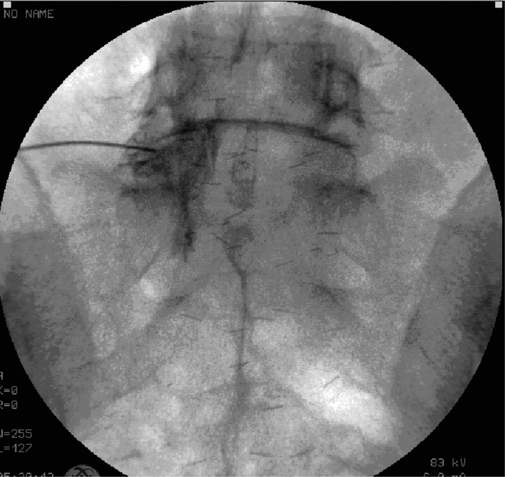
Fig. 1.
(A) We measured spinal canal dimensions using the free-line region of interest calculator in the Infinitt PACS system in axial MRI images, as shown by an inverted triangular line on the image. (B) There was no definitive disc herniation or spondylolisthesis in the sagittal MRI image. PACS, picture archiving and communication system; MRI, magnetic resonance imaging.

Table 1.
Demographics
| Age | Male | Female | Total |
|---|---|---|---|
| 30-39 | 5 | 1 | 6(4.3%) |
| 40-49 | 13 | 5 | 18(12.8%) |
| 50-59 | 32 | 10 | 42(29.8%) |
| 60-69 | 26 | 15 | 41(29.1%) |
| 70-79 | 17 | 9 | 26(18.4%) |
| 80- | 4 | 4 | 8(5.7%) |
| Total | 97(68.8%) | 44(31.2%) | 141(100%) |
Table 2.
The effect of the selective nerve root block according to age
Table 3.
The effect of the selective nerve root block according to sex
| Male | Female | p-value | |||
|---|---|---|---|---|---|
| Average | Standard deviation | Average | Standard deviation | ||
| Pre-1hr VAS | 0.85 | 0.910 | 0.97 | 1.088 | 0.468 |
| Pre-1month VAS | 2.38 | 1.317 | 2.34 | 1.292 | 0.728 |
| Pre-3month VAS | 2.24 | 1.557 | 2.34 | 1.445 | 0.893 |
Table 4.
VAS and effect of selective nerve root block by canal dimension.
Table 5.
Result of t-test


 PDF
PDF ePub
ePub Citation
 Print
Print


 XML Download