

 PDF
PDF ePub
ePub Citation
 Print
Print


Abstract
Objectives
To report the effectiveness of open reduction and internal (screw) fixation treatment performed to treat dislocation of the first coccygeal vertebra.
Summary of Literature Review
Most treatment methods for coccygeal dislocation were conservative treatment for acute coccygodynia and coccygectomy for chronic coccygodynia.
Materials and Methods
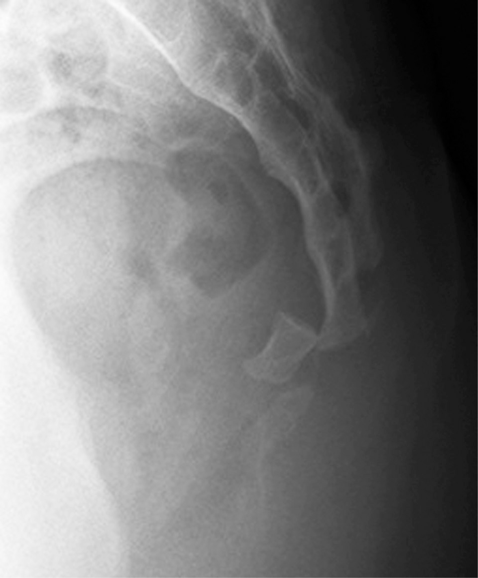
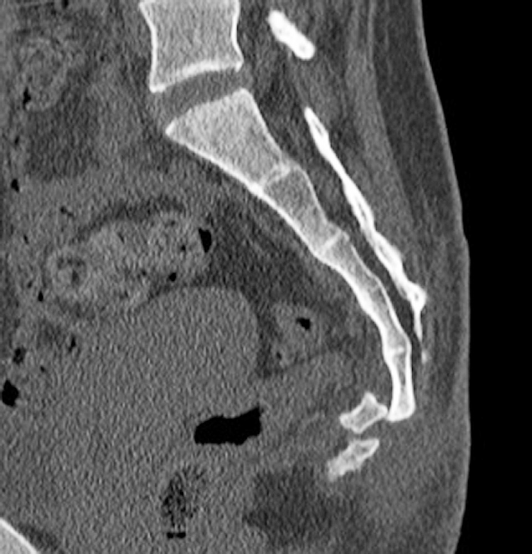
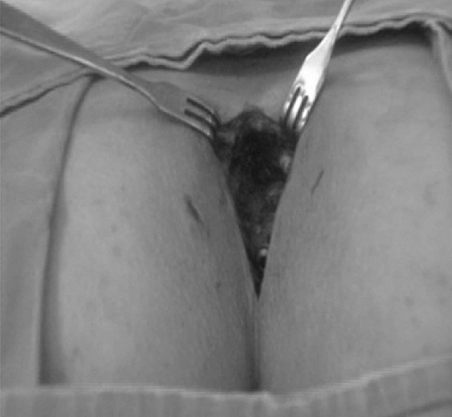
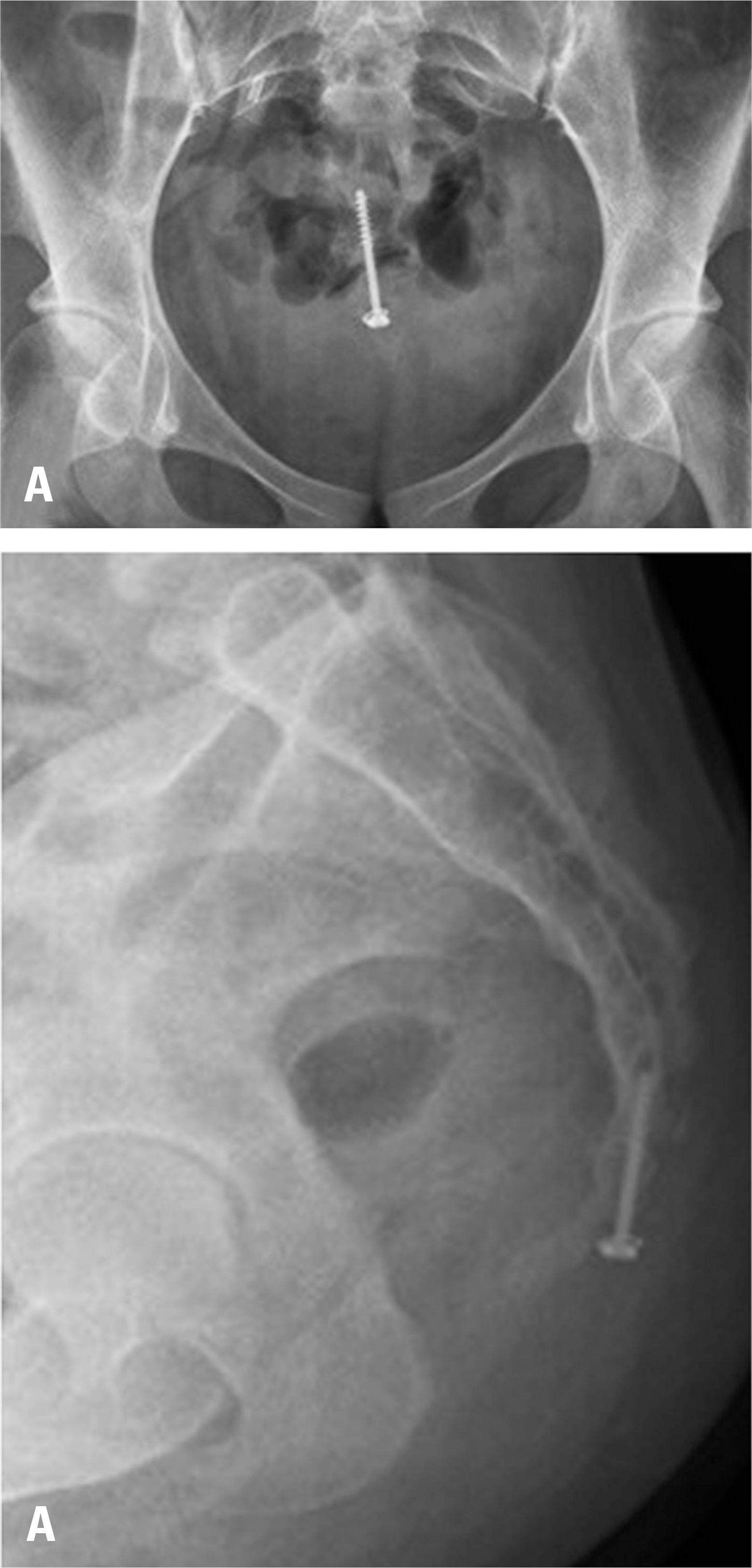
A 18-year-old female presented with severe coccygodynia due to a fall down the stairs. Computed tomography showed dislocation of the first coccygeal vertebra. We performed open reduction and internal fixation with a 4.0-mm short-thread cancellous screw with a washer, with no additional procedure for bone union.
Go to : 
REFERENCES
1. Johnson PH. Coccygodynia. J Ark Med Soc. 1981; 77:421–4.
2. Karadimas EJ, Trypsiannis G, Giannoudis PV. Surgical treatment of coccygodynia: an analytic review of the literature. Eur Spine J. 2010; 20:698–705.

3. Maigne JY, Doursounian L, Chatellier G. Causes and mechanisms of common coccydynia: role of body mass index and coccygeal trauma. Spine. 2000; 25:3072–9.
4. Perkins R, Schofferman J, Reynolds J. Coccygectomy for severe refractory sacrococcygeal joint pain. J Spinal Disord Tech. 2003; 16:100–3.
5. Key J A. Operative treatment of coccygodynia. J Bone Joint Surg. 1937; 19:759–64.
6. Ramieri A, Domenicucci M, Cellocco P, et al. Acute traumatic instability of the coccyx: results in 28 consecutive coccygectomies. Eur Spine J. 2013; 22:939–44.
7. Bergkamp AB, Verhaar JA. Dislocation of the coccyx: a case report. J Bone Joint Surg Br. 1995; 77:831–2.
8. Kim WY, Han CW, Kim YH. Joystick reduction and percutaneous pinning for an acutely anteriorly dislocated coccyx: a case report. J Orthop Trauma. 2004; 18:388–9.
Go to :
 XML Download