

 PDF
PDF ePub
ePub Citation
 Print
Print


Abstract
Summary of Literature Review
For patients who undergo traffic accidents, most cases of seat belt injury cause trauma to the lower torso. Seat belt injury is associated with variable clinical problems such as vascular injury, intestinal injury (perforation), vertebral injury (flexion-distraction injury), chest wall injury, diaphragmatic rupture/hernia, bladder rupture, lumbosacral plexopathy, and other related conditions.
Materials and Methods
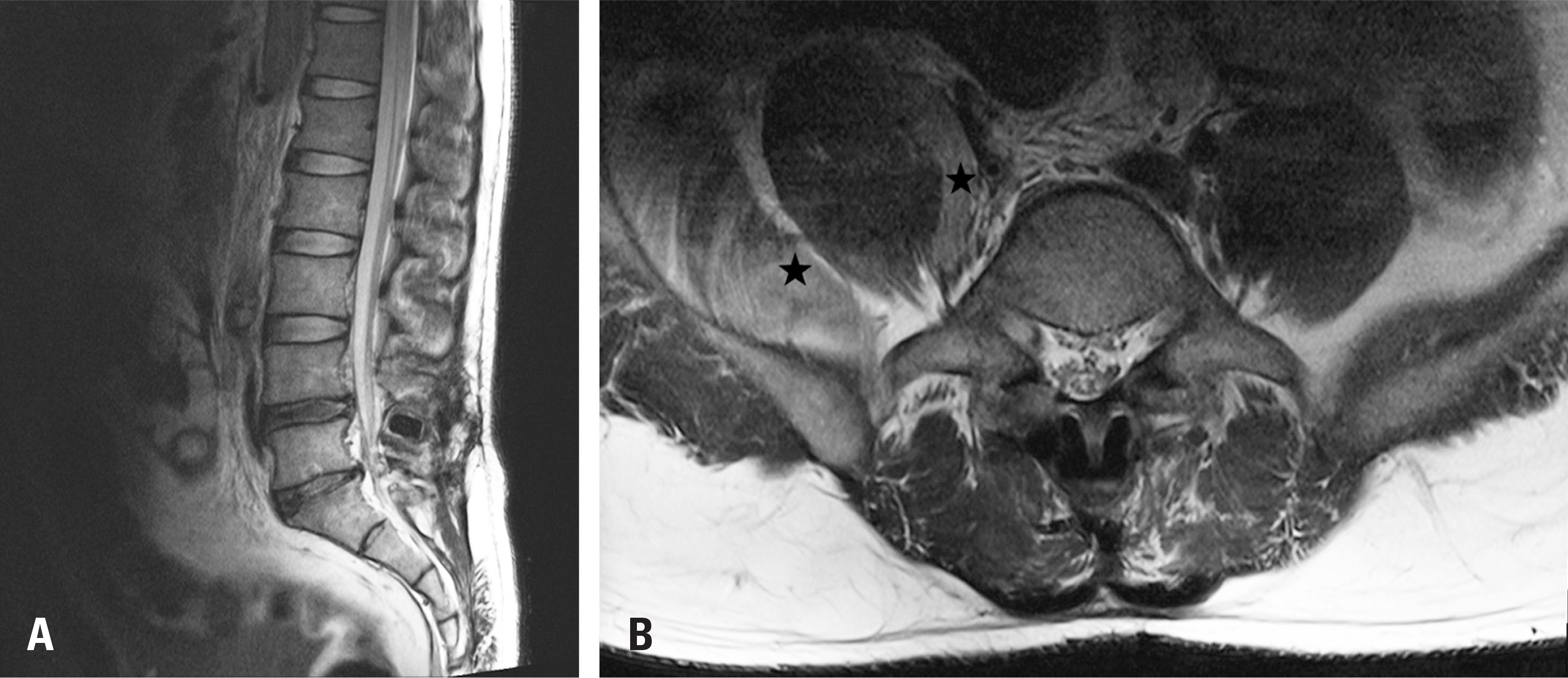
A 38-year-old male truck driver (traffic accident victim) who suffered monoplegia of his right leg due to lumbar plexus injury without spinal column involvement. Injury to a lumbar plexus and the internal vasculatures originated from direct compression to internal abdominal organs (the iliopsoas muscle and internal vasculatures anterior to the lumbar vertebrae) caused by the seat belt. We have illustrated an extremely uncommon cause of a neurologic deficit from a traffic accident through this case.
Results
Under the impression of traumatic lumbar plexopathy, we managed it conservatively, and the patient showed signs of recovery from neurologic deficit.
Conclusions
We need to review the lumbar plexus pathway, in patients with atypical motor weakness and sensory loss of the lower extremities which are not unaccompanied by demonstrable spinal lesions. Therefore, close history taking, physical examination and comprehension of injury mechanism are important in the diagnosis.
REFERENCES
1. Onu DO, Hunn AW, Bohmer RD. Seat belt syndrome with unstable Chance fracture dislocation of the second lumbar vertebra without neurological deficits. BMJ Case Rep[Internet]. 2014 Jan 8. Available form: http://caser-eports.bmj.com/content/2014/bcr-2013-202412.

2. Bushby N, Wickramasinghe SY, Wickramasinghe DN. Lumbosacral plexopathy due to a rupture of a common Iliac artery aneurysm. Emerg Med Australas. 2010; 22:351–3.
3. Freni L, Barbetta I, Mazzaccaro D, et al. Seat belt injuries of the abdominal aorta in adults–case report and literature review. Vasc Endovascular Surg. 2013; 47:138–47.
4. Campbell DK, Austin RF. Seat-belt injury: injury of the abdominal aorta. Radiology. 1969; 92:123–4.
5. Planner AC, Donaghy M, Moore NR. Causes of lumbosacral plexopathy. Clinical Radiol. 2006; 61:987–95.
6. Crosby ET, Reid DR, DiPrimio G, et al. Lumbosacral plexopathy from iliopsoas haematoma after combined gen-eral-epidural anaesthesia for abdominal aneurysmectomy. Can J Anaesth. 1998; 45:46–51.
Fig. 1.
(A) Abdomen CT scan showing hemoperitoneum (star), a short-segmental filling defect at the proximal portion of the right common iliac artery (arrow). (B) Axial view of the abdomen CT scan showing active bleeding of the superior mesenteric artery branch. (C) Abdominal angiography showing proximal ileum branch of the superior mesenteric artery (arrow). (D) 2 cm sized pseudo-aneurysm arising at right proximal common iliac artery with segmental non-visualization of right common iliac artery (star).

 XML Download