

 PDF
PDF ePub
ePub Citation
 Print
Print


Abstract
Objectives
To report a case of paraplegia in a patient with thoracic kyphosis after osteosynthesis for a fracture of the femur.
Summary of the Literature Review
There are few reports about cases of paraplegia after low extremity fracture surgery in patients with thoracic kyphosis with ankylosing spondylitis.
Materials and Methods
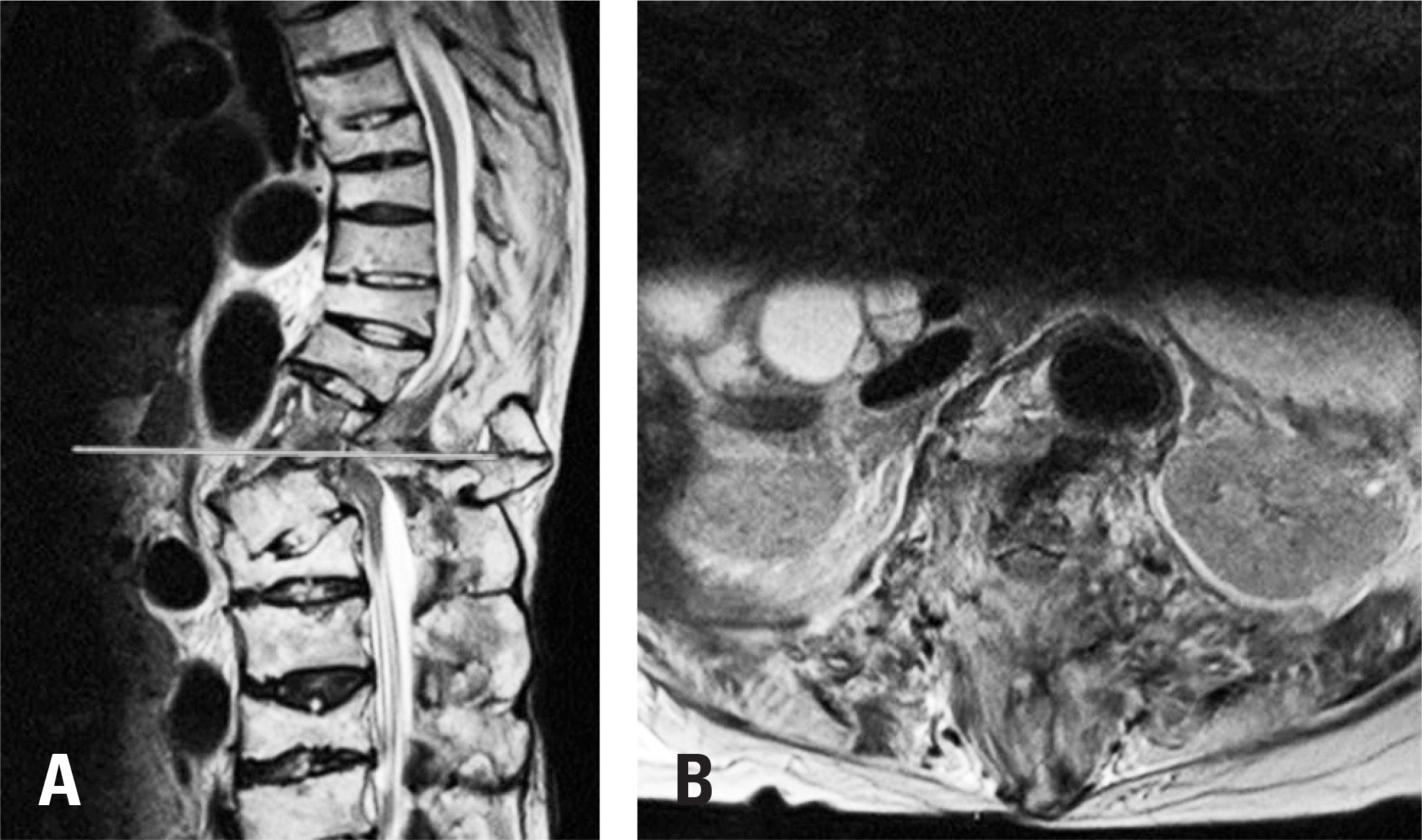
An 86-year-old female patient presented with right hip pain. She had undergone surgery for an intertrochanteric fracture of the femur in the supine position under general anesthesia. Immediately after surgery, she showed paraplegia. Postoperative thoracolumbar spine images revealed a fracture through the disc at T12 and L1. However, she did not complain of back pain or any neurologic deficits before surgery.
Results
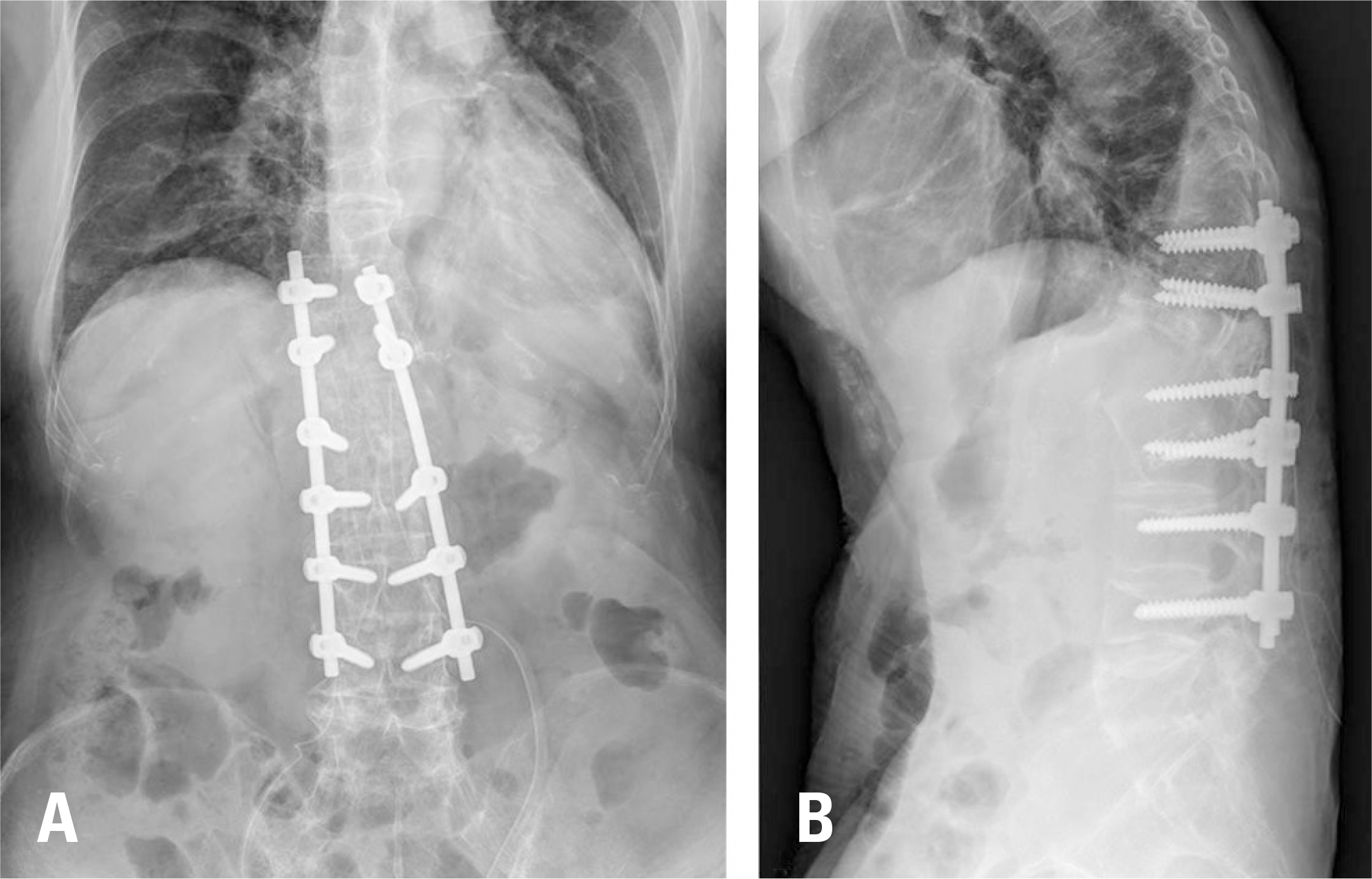
Although the patient underwent emergent posterior decompression and fusion surgery, her neurologic compromise did not improve during 1 year of follow-up.
Conclusions
It is necessary to check preoperative spine radiographs before surgery in elderly patients who have a kyphotic deformity and lower extremity fractures. Surgeons should consider changing the position of the patient and the type of anesthesia used during surgery when spine stability is in doubt.
Go to : 
REFERENCES
1. Labelle H, Roussouly P, Berthonnaud É, et al. Spondylolisthesis, pelvic incidence, and spinopelvic balance: a correlation study. Spine (Phila Pa 1976). 2004 Sep; 29(18):2049–54. DOI: 10.1097/01.brs.0000138279.53439.cc.
2. Murray GC, Persellin RH. Cervical fracture complicating ankylosing spondylitis: a report of eight cases and review of the literature. Am J Med. 1981 May; 70(5):1033–41. DOI: 10.1016/0002-9343 (81)90860-3.
3. Patel SN, Turtz A, Dixon A, et al. Neurologically intact lumbar spine displaced fracture with ankylosing spondylitis. West J Emerg Med. 2011 Feb; 12(1):142–3.
4. Chaudhary SB, Hullinger H, Vives MJ. Management of acute spinal fractures in ankylosing spondylitis. ISRN Rheumatol. 2011 Apr; 2011(2011):1–9. DOI: 10.5402/2011/150484.

5. Good AE. Nontraumatic fracture of the thoracic spine in ankylosing spondylitis. Arthritis Rheum. 1967 Oct; 10(5):467–9. DOI: 10.1002/art.1780100509.
6. Osgood CP, Abbasy M, Mathews T. Multiple spine fractures in ankylosing spondylitis. J Trauma. 1975 Feb; 15(2):163–6. DOI: 10.1097/00005373-197502000-00011.
7. Ghozlani I, Ghazi M, Nouijai A, et al. Prevalence and risk factors of osteoporosis and vertebral fractures in patients with ankylosing spondylitis. Bone. 2009 May; 44(5):772–6. DOI: 10.1016/j.bone.2008.12.028.
8. Olerud C, Frost A, Bring J. Spinal fractures in patients with ankylosing spondylitis. Eur Spine J. 1996 Jan; 5(1):51–5. DOI: 10.1007/BF00307827.
9. Danish SF, Wilden JA, Schuster J. Iatrogenic paraplegia in 2 morbidly obese patients with ankylosing spondylitis undergoing total hip arthroplasty. J Neurosurg Spine. 2008 Jan; 8(1):80–3. DOI: 10.3171/SPI-08/01/080.
Go to :
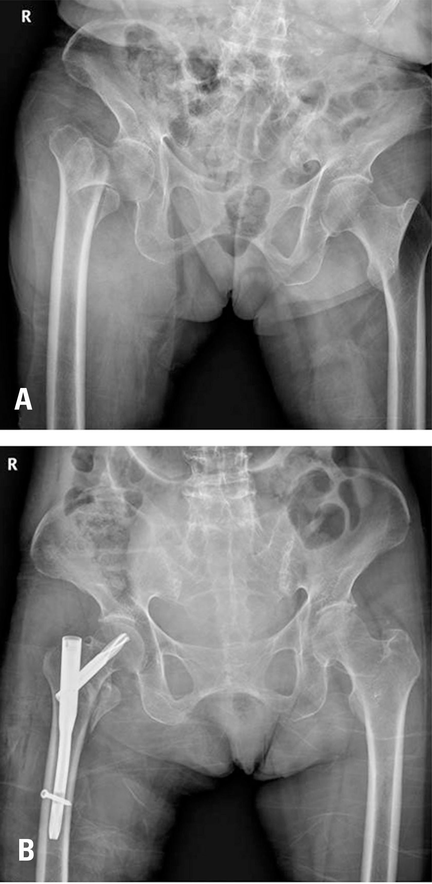 | Fig. 1.An intertrochanteric fracture of the right femur (Boyd-Griffin type II, Evans unstable) is observed on a preoperative pelvic radiograph (A). Postoperative pelvic radiographs show that the fracture was fixed by a cephalomedullary nail (B). |
 | Fig. 2.Lumbar spine radiography before hip surgery showed bamboo spine with extensive fusion across the entire spine (A). On the lateral view, there was a gap between T12 and L1 (B). Thoracolumbar lateral spine radiographs after hip surgery show a more displaced thoracolumbar fracture and deviated spine alignment (C). |
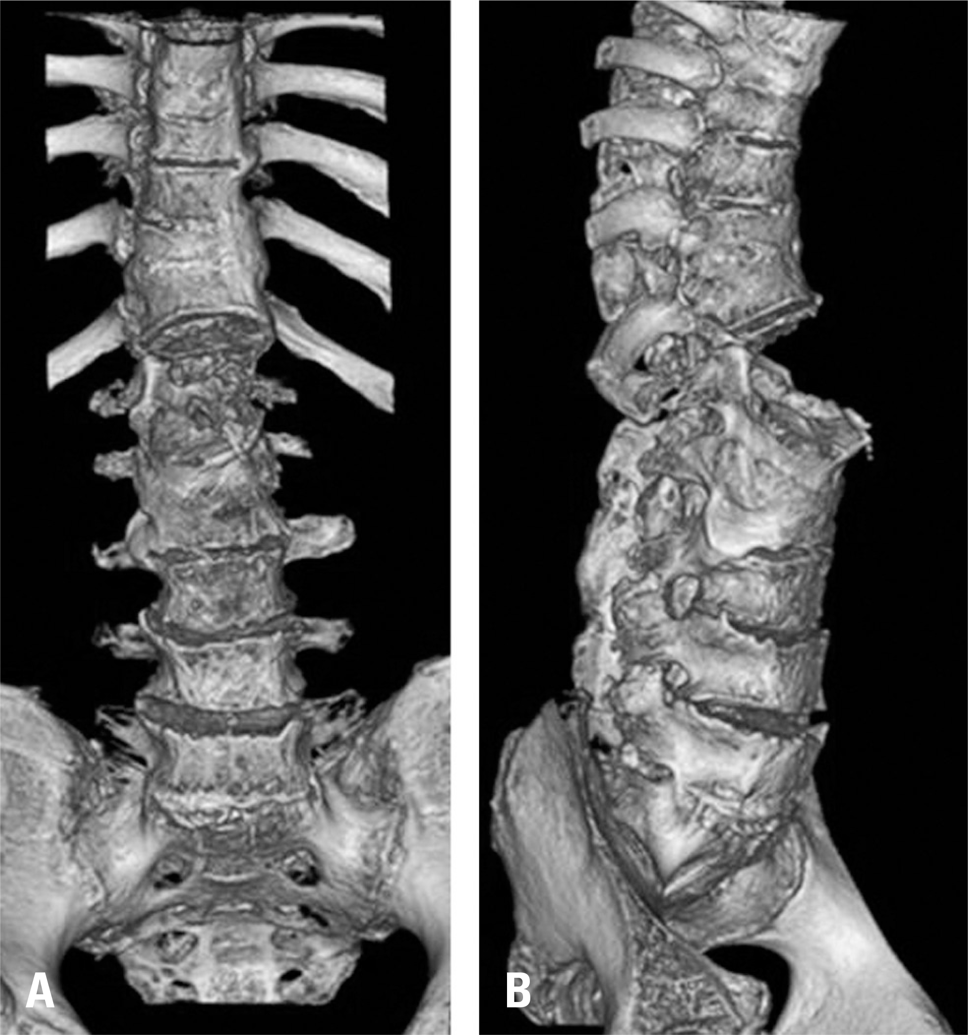 | Fig. 3.A 3-dimensional reconstructed computed tomography scan after hip surgery revealed a wide range of upper and lower vertebral fusion and worsening of the thoracolumbar fracture in the anteroposterior view (A) and lateral view (B). |
 XML Download