

 PDF
PDF ePub
ePub Citation
 Print
Print


Abstract
Objectives
To evaluate the natural history of conservatively treated lumbar degenerative kyphosis (LDK).
Summary of Literature Review
The correlations between the clinical and radiologic parameters of general adult spinal deformity (ASD) are widely known. However, in LDK, dynamic sagittal imbalance is present during ambulation, meaning that its pathogenesis and natural history are different and not widely recognized compared to those of other forms of ASD, resulting in many controversies regarding treatment. To elucidate the natural history of LDK, we analyzed the clinical and radiologic outcomes of patients, comparing their first and final follow-up visits, and evaluated correlations among clinical and radiologic parameters.
Materials and Methods
From June 2006 to January 2014, 31 patients diagnosed with LDK who underwent conservative treatment were studied. The mean age of the patients was 72.5 years, and the mean follow-up period was 59.2 months. Clinical and radiologic evaluations were conducted on the first and final follow-up visits. Clinical evaluations were done using a visual analog scale and the Oswestry disability index, and radiologic evaluations were performed using spinal and pelvic parameters over a follow-up period of at least 24 months.
Results
Patients who were diagnosed with LDK and underwent conservative treatment showed no significant differences in their clinical outcomes between the first and final follow-up. Some radiologic parameters significantly progressed. There were no significant differences between clinical and radiologic parameters at the initial and final follow-up visits.
Conclusions
During the follow-up period of patients diagnosed with LDK, some radiologic parameters progressed. However, the progress of LDK and the clinical symptoms reported by the patients did not significantly change. Decisions regarding the treatment of LDK should not be made according to radiologic parameters showing the degree of deformity, but by carefully determining the patients’ clinical symptoms and disability level.
Go to : 
REFERENCES
1. Lee C-S, Kim Y-T, Kim E. Clinical study of lumbar degenerative kyphosis. J Korean Soc Spine Surg. 1997; 4:27–35.
2. Lee CS, Lee CK, Kim YT, et al. Dynamic sagittal imbalance of the spine in degenerative flat back: significance of pelvic tilt in surgical treatment. Spine (Phila Pa 1976). 2001; 26:2029–35.
3. Lee CS, Chung SS, Chung KH, et al. Significance of pelvic incidence in the development of abnormal sagittal alignment. J Korean Orthop Assoc. 2006; 41:274–80.

4. Doherty J. Complications of fusion in lumbar scoliosis. J Bone Joint Surg Am. 1973; 55:438–49.
5. Takemitsu Y, Harada Y, Iwahara T, et al. Lumbar degenerative kyphosis. Clinical, radiological and epidemiological studies. Spine (Phila Pa 1976). 1988; 13:1317–26.
6. Kim E-H, Han S-K, Kim H-J. A clinical analysis of surgical treatment of lumbar degenerative kyphosis. J Korean Soc Spine Surg. 2001; 8:210–8.
7. Carlsson AM. Assessment of chronic pain. I. Aspects of the reliability and validity of the visual analogue scale. Pain. 1983; 16:87–101.
8. Fairbank JC, Pynsent PB. The Oswestry disability index. Spine (Phila Pa 1976). 2000; 25:2940–53.
9. O'Brien M, Kuklo T, Blanke K, et al. Radiographic Mea-surement Manual. Spinal Deformity Study Group (SDSG) Medtronic Sofamor Danek USA: Inc;2008.
10. Kim MS, Chung SW, Hwang C, et al. A radiographic analysis of sagittal spinal alignment for the standardization of standing lateral position. J Korean Orthop Assoc. 2005; 40:861–7.
11. Kim W-J. Optimal standing radiographic positioning in patients with sagittal imbalance. J Korean Orthop Assoc. 2010; 17:198–204.
12. Jackson RP, McManus AC. Radiographic analysis of sagittal plane alignment and balance in standing volunteers and patients with low back pain matched for age, sex, and size. A prospective controlled clinical study. Spine (Phila Pa 1976). 1994; 19:1611–8.
13. Schwab F, Ungar B, Blondel B, et al. Scoliosis research soci-ety—Schwab adult spinal deformity classification: a validation study. Spine (Phila Pa 1976). 2012; 37:1077–82.
14. Moe J, Denis F. The iatrogenic loss of lumbar lordosis. Orthop Trans. 1977; 1:131.
15. Smith JS, Lafage V, Shaffrey CI, et al. Outcomes of operative and nonoperative treatment for adult spinal deformity: a prospective, multicenter, propensity-matched cohort assessment with minimum 2-year follow-up. Neurosurgery. 2016; 78:851–61.
16. McCarthy I, O'Brien M, Ames C, et al. Incremental cost-effectiveness of adult spinal deformity surgery: observed quality-adjusted life years with surgery compared with predicted quality-adjusted life years without surgery. Neurosurg Focus. 2014; 36:E3.
17. Sciubba DM, Scheer JK, Yurter A, et al. Patients with spinal deformity over the age of 75: a retrospective analysis of operative versus nonoperative management. Eur Spine J. 2015. 1–9.
18. Kim WJ, Kang J-W, Kim H-Y, et al. Change of pelvic tilt before and after gait in patients with lumbar degenerative kyphosis. J Korean Soc Spine Surg. 2009; 16:95–103.
19. Garbossa D, Pejrona M, Damilano M, et al. Pelvic parameters and global spine balance for spine degenerative disease: the importance of containing for the well being of content. Eur Spine J. 2014; 23:616–27.
20. Benoist M. The natural history of lumbar degenerative spinal stenosis. Joint Bone Spine. 2002; 69:450–7.
21. Marty-Poumarat C, Scattin L, Marpeau M, et al. Natural history of progressive adult scoliosis. Spine (Phila Pa 1976). 2007; 32:1227–34.
22. Silva FE, Lenke LG. Adult degenerative scoliosis: evaluation and management. Neurosurg Focus. 2010; 28:E1.
23. Benoist M. Natural history of the aging spine. Eur Spine J. 2003; 12(Suppl):86–9.
24. Kim WJ, Song DG, Lee JW, et al. Proximal junctional problems in surgical treatment of lumbar degenerative sagittal imbalance patients and relevant risk factors. J Korean Soc Spine Surg. 2013; 20:156–62.
25. Lee JC, Soh J-W, Jo J-H, et al. Comparative analysis of surgical options in the treatment of lumbar degenerative kyphosis. J Korean Soc Spine Surg. 2009; 16:8–16.
26. Kim K-T, Lee SH, Suk K-S, et al. Loss of sagittal balance and clinical outcomes following corrective osteotomy for lumbar degenerative kyphosis. J Korean Orthop Assoc. 2009; 44:83–92.
27. Kim WJ, Kang JW, Kang SI, et al. Factors affecting clinical results after corrective osteotomy for lumbar degenerative kyphosis. Asian Spine J. 2010; 4:7–14.
Go to :
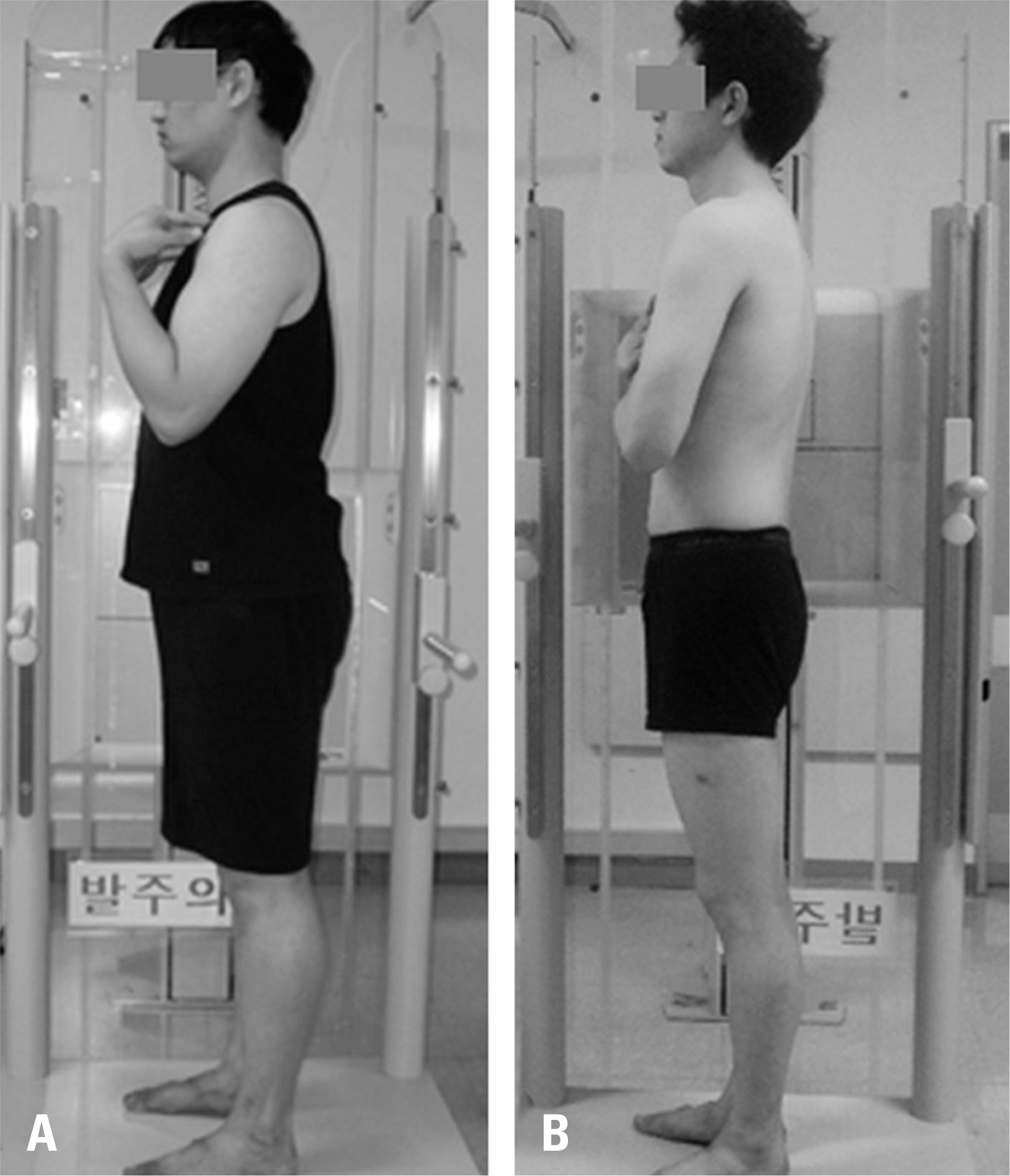 | Fig.1.The fists-on-clavicle (A) or cross-arm position (B) is recommended with an extended hip and knee while taking radiographs. |
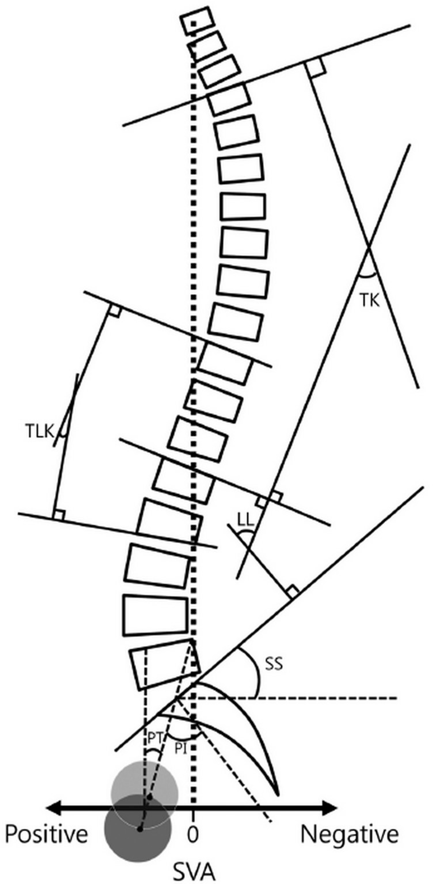 | Fig. 2.Schema displaying Cobb's method for thoracic kyphosis, thoracolumbar kyphosis, lumbar lordosis, and the sagittal vertical axis. The pelvic parameters (pelvic tilt, sacral slope, and pelvic incidence) are also indicated for the lateral whole spine. LL, lumbar lordosis; TK, thoracic kyphosis; TLK, thoracolumbar kyphosis; SVA, sagittal vertical axis; PI, pelvic incidence; SS, sacral slope; PT, pelvic tilt. |
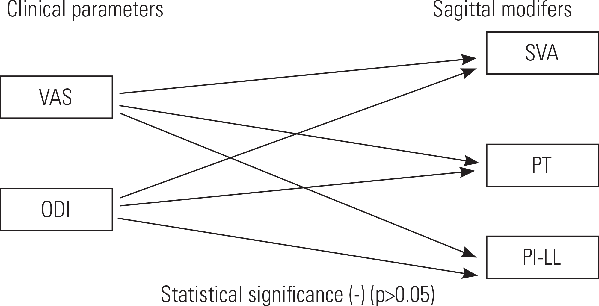 | Fig. 3.Schematic diagram relating the clinical and radiological param-eters at the initial and final follow-up visits. |
 | Fig. 4.Differential diagnosis of sagittal imbalance. (A) Initial radiograph of a 65-year-old man shows L3-on-L4 spondylolisthesis with severe pain and disability. (B) Magnetic resonance imaging shows L3-L4 spinal stenosis. (C) The patient underwent L4 nerve root block. (D) Two months later, the pain subsided and follow-up radiography revealed no sagittal imbalance. |
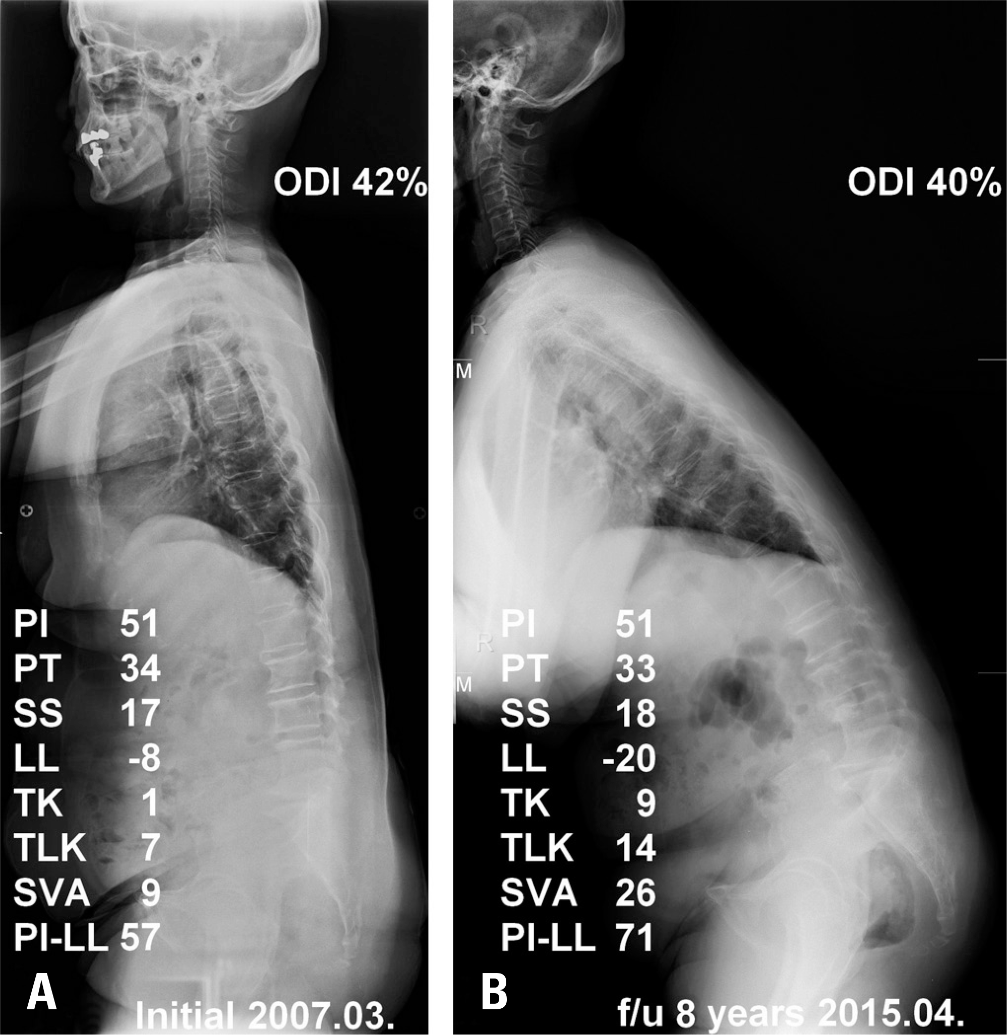 | Fig. 5.A 64-year-old woman with lumbar degenerative kyphosis with sagittal imbalance. (A) Initial radiograph showing sagittal imbalance and an Oswestry disability index (ODI) score of 42%. (B) Sagittal imbalance developed 97 months after conservative treatment was initiated, and her ODI score (40%) slightly decreased. |
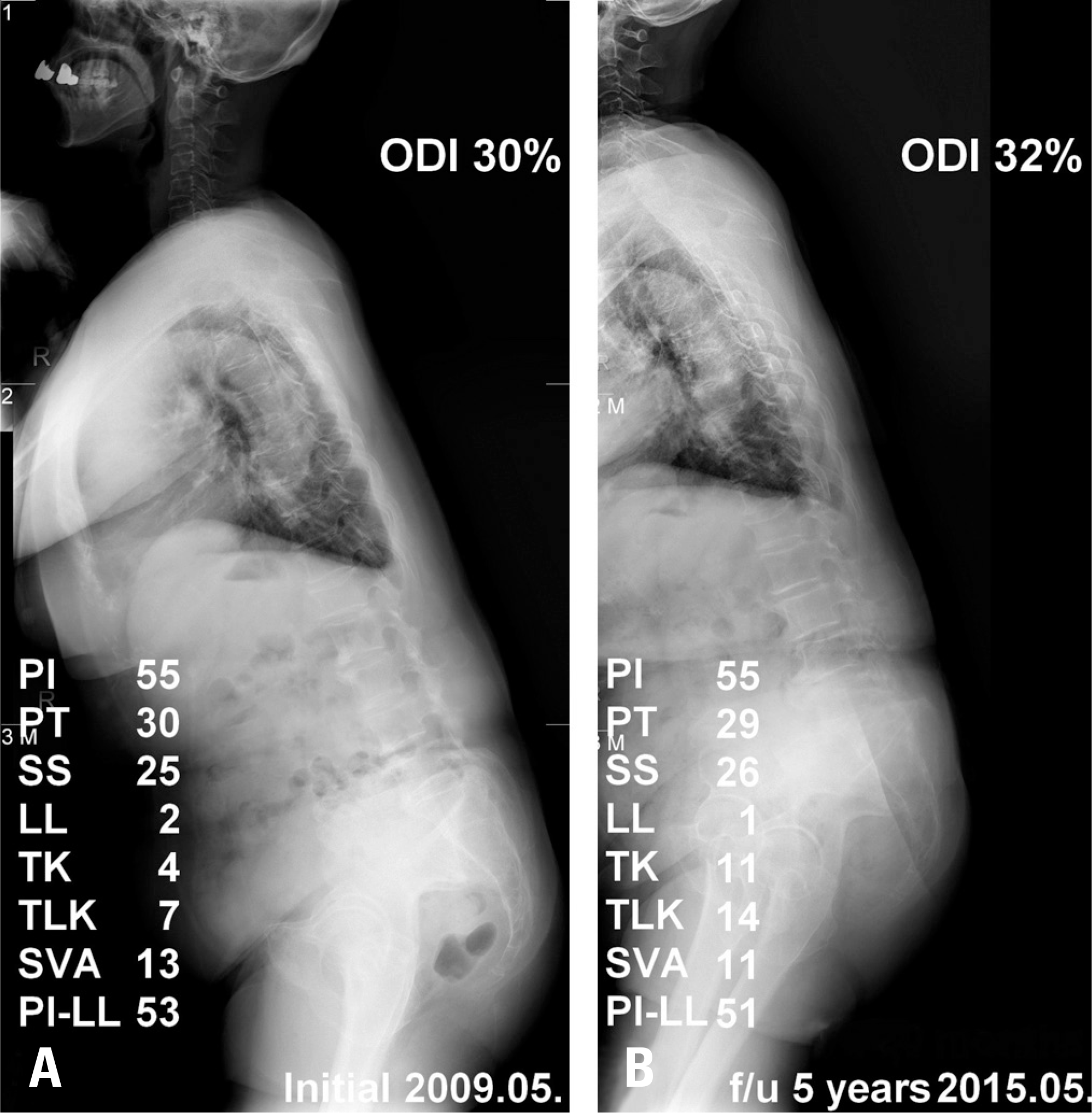 | Fig. 6.An 84-year-old woman with lumbar degenerative kyphosis with sagittal imbalance. (A) Initial radiograph showing sagittal imbalance and an Oswestry disability score (ODI) of 30%. (B) Sagittal imbalance had not developed 60 months after conservative treatment, but the ODI score (32%) slightly increased. |
Table 1.
Average results of clinical index in initial and last follow-up
| Initial | Last follow-up | p-value | |
|---|---|---|---|
| VAS∗ | 3.3 (2∼6) | 3.4 (2∼5) | 0.763 |
| ODI† | 31.9 (19∼58) | 34.7 (20∼52) | 0.102 |
Table 2.
Average values of radiologic parameters in initial and last follow-up
| Initial | Last follow-up | p-value | |
|---|---|---|---|
| LL (°)∗ | 6.2(−15.6∼24.9) | −5.4(−20.3∼8.4) | 0.001†† |
| TK (°)† | 8.1(1.3∼13.4) | 8.4(1.2∼22) | 0.841 |
| TLK (°)‡ | 15.9(1.4∼31.6) | 19.5(2.3∼37.8) | <0.001†† |
| SVA (cm)§ | 8.5(5.5∼24.3) | 17.0(5.8∼45.8) | 0.003†† |
| PI (°)|| | 57.5(36.3∼85) | 59.8(38.3∼80.9) | 0.188 |
| SS (°)¶ | 18.4(1.1∼28.4) | 19.4(2.1∼32.7) | 0.482 |
| PT (°)∗∗ | 39.2(22.5∼65.2) | 40.4(29.6∼60.8) | 0.403 |
| PI-LL (°) | 51.1(35.4∼76.1) | 67.4(39.7∼101) | <0.001†† |
 XML Download