Abstract
Objectives
To report an unusual case of an abscess of the cervical spine caused by Klebsiella peumoniae accompanied by an acute compressive flexion injury.
Summary of Literature Review
Spondylitis caused by Klebsiella peumoniae is very rare, and an unrecognized epidural abscess complicated with spinal cord compression can lead to severe neurologic deficits.
Materials and Methods
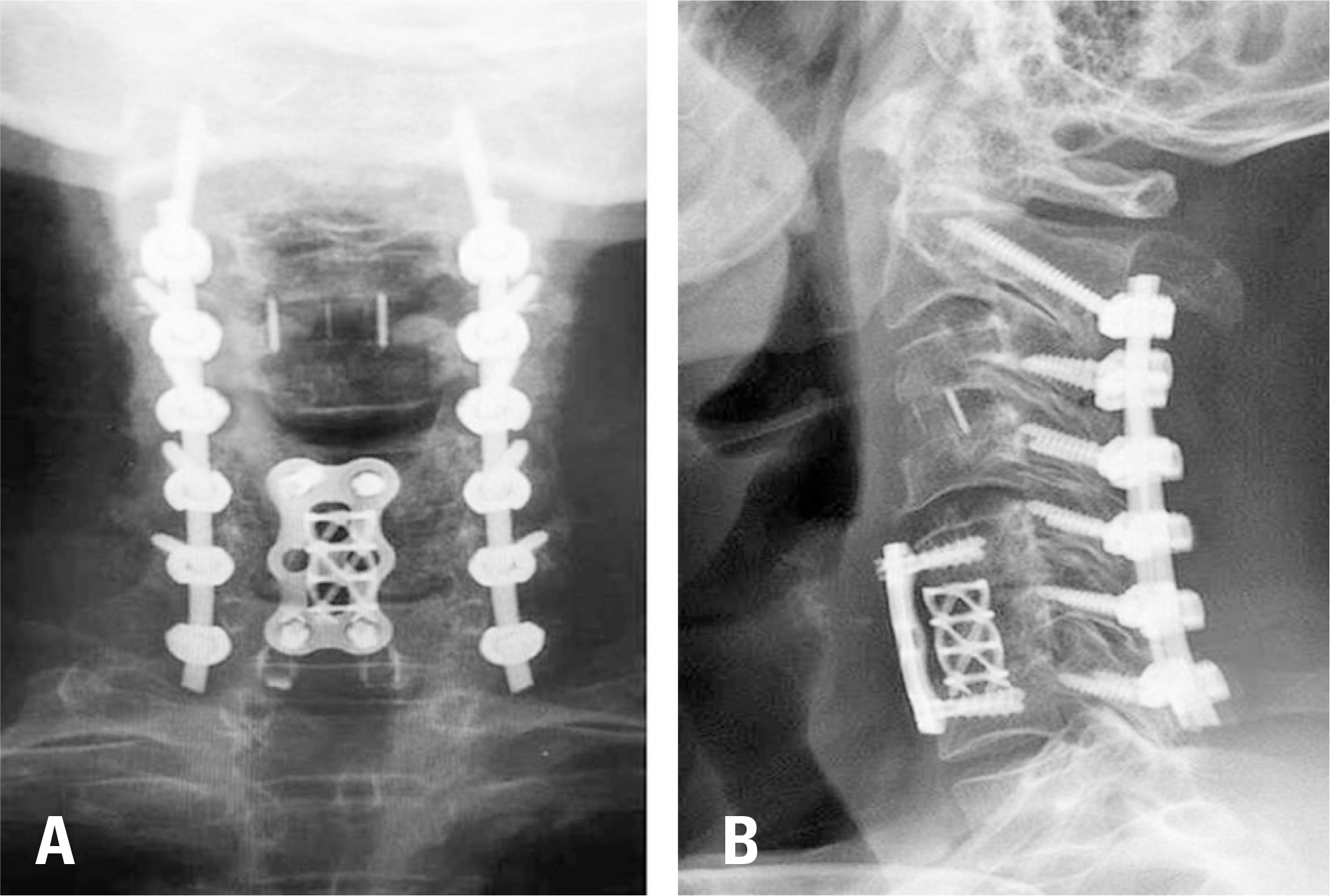
A 66-year-old male patient diagnosed with a liver abscess caused by Klebsiella peumoniae was referred from the internal medicine department to our department due to abrupt posterior neck pain and limitation of motion after a fall from the bed. He showed persistent fever and progressive dysphagia. We diagnosed the condition as a massive cervical abscess caused by Klebsiella peumoniae accompanied by an acute compressive flexion injury. We performed drainage of the massive abscess, anterior fusion to treat the loss of the intervertebral discs at the C3/4 level, and corpectomy for a compression fracture of the C6 vertebral body using a cage and plate via an anterior approach. Subsequently, we performed posterior laminectomy with drainage at the C3-6 level and posterior instrumentation of C2-7 via a posterior approach.
REFERENCES
1. Bluman EM, Palumbo MA, Lucas PR. Spinal epidural abscess in adults. J Am Acad Orthop Surg. 2004 May-Jun; 12(3):155–63. DOI: 10.5435/00124635-200405000-00003.

2. Joshi SM, Hatfield RH, Martin J, et al. Spinal epidural abscess: a diagnostic challenge. Br J Neurosurg. 2003 Apr; 17(2):160–3. DOI: 10.1080/0268869031000108918.

3. Cho CH, Min WK, Park BC. Spondylodiscitis with epidural abscess caused by Klebsiella pneumoniae J Korean Orthop Assoc. 2011 Dec; 46(6):528–32. DOI: 10.4055/jkoa.2011.46.6.528.
4. Kuramochi G, Takei SI, Sato M, et al. Klebsiella pneumoniae liver abscess associated with septic spinal epidural abscess. Hepatol Res. 2005 Jan; 31(1):48–52. DOI: 10.1016/j.hepres.2004.09.006.
5. Joo YE, Choi SK, et al. Klebsiella pneumoniae septic arthritis in a cirrhotic patient with hepatocellular carcinoma. J Korean Med Sci. 2004 Aug; 19(4):608–10. DOI: 10.3346/jkms.2004.19.4.608.

6. Chew LC. Septic monoarthritis and osteomyelitis in an elderly man following Klebsiella pneumoniae genitourinary infection: case report. Ann Acad Med Singapore. 2006 Feb; 35(2):100–3.
7. Hsieh MJ, Lu TC, Ma MH, et al. Unrecognized cervical spinal epidural abscess associated with metastatic Klebsiella pneumoniae bacteremia and liver abscess in nondiabetic patients. Diagn Microbiol Infect Dis. 2009 Sep; 65(1):65–8. DOI: 10.1016/j.diagmicrobio.2009.05.013.

8. Murphy MJ, Ogden JA, Southwick WO. Spinal stabilization in acute spinal injuries. Ortho Clin North Am. 1980 Oct; 60(5):1035–47. DOI: 10.1016/s0039-6109 (16)42231-0.

9. Stauffer ES, Kelly EG. Fracture-dislocations of the cervical spine: Instability and recurrent deformity following treatment by anterior interbody fusion. J Bone Joint Surg Am. 1977 Jan; 59(1):45–8. DOI: 10.2106/00004623-197759010-00006.

10. Kadam A, Millhouse PW, Kepler CK, et al. Bone substitutes and expanders in spine Surgery: A review of their fusion efficacies. Int J Spine Surg. 2016 Sep 22; 10:33. DOI: 10.14444/3033.

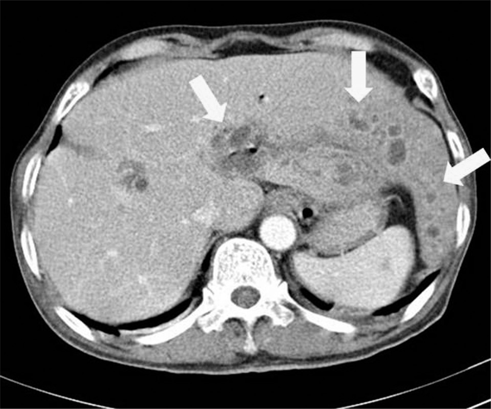
Fig. 1.
Enhanced abdominal computed tomography shows a large immature hepatic abscess and multiple small abscesses.

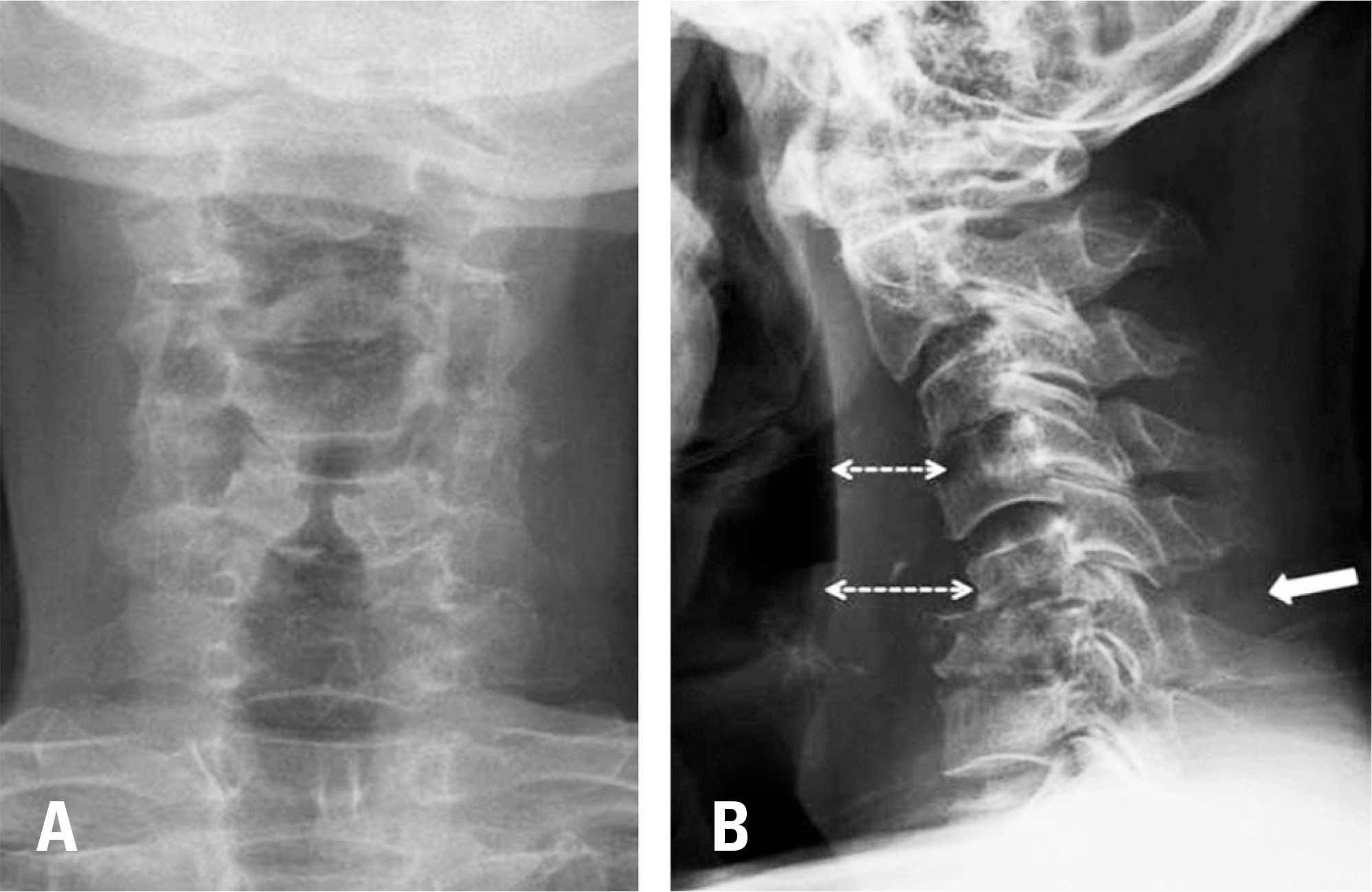
Fig. 2.
Anteroposterior (A) and lateral (B) views of the cervical spine show soft tissue thickening (dotted line) on the anterior aspect of the cervical spine with osteolysis of C4. A compression fracture of the C6 vertebral body is seen, with a widened interspinous space at C5-6 (arrow).

Fig. 3.
A cervical T2-weighted sagittal magnetic resonance (MR) image shows a massive abscess collection (A). Cervical axial T2-weighted MR images show the spinal canal compressed by the abscess at C3-4 (B). High signal enhancement of the C5-6 interspinous ligament was seen on a T2 fat-suppression sagittal MR image (C). Axial computed tomography image shows a comminuted compression fracture at C6 (D).


 PDF
PDF ePub
ePub Citation
 Print
Print


 XML Download