

 PDF
PDF ePub
ePub Citation
 Print
Print


Abstract
Objectives
To evaluate the outcomes of dual growing rod treatment over a follow-up period of at least 2 years in patients with progressive pediatric spinal deformity.
Summary of Literature Review
The dual growing rod treatment is safe and effective in curve correction and maintenance in patients with progressive pediatric spinal deformity.
Materials and Methods
Between 2009 to 2017, 14 patients who underwent dual growing rod treatment were followed up for more than 2 years. We analyzed their demographic and radiologic data, including age at surgery, sex, diagnosis, instrumented levels, number of total operations, number of lengthening procedures, interval of lengthening, Cobb angle, thoracic kyphosis angle, lumbar lordosis angle, T1-S1 length, and complications.
Results
The mean age of the patients was 11.0±2.9 years old. There were 10 male and 4 female patients, including 8 cases of neuromuscular scoliosis, 3 cases of idiopathic scoliosis, 2 cases of spondyloepiphyseal dysplasia, and 1 case of congenital scoliosis. The mean follow-up period was 42.4±14.0 months. The total number of operations was 6.6±2.6. The average number of lengthening procedures was 4.3±2.3 at an interval of 6.9±2.1 months. The Cobb angle improved from 60.4°±27.9° to 33.5°±19.7° after the initial treatment and 29.1°±16.4° after the last follow-up or final fusion. The T1-S1 length increased from 328.2±57.5 mm to 388.0±64.9 mm after the initial treatment and 424.9±64.4 mm after the last follow-up or final spinal fusion. The average growth rate was 11.5 mm/year. Six patients experienced 11 complications, of which 4 were Implant-related, and 7 were Infections.
Go to : 
REFERENCES
1. Goldberg CJ, Gillic I, Connaughton O, et al. Respiratory function and cosmesis at maturity in infantile-onset scoliosis. Spine (Phila Pa 1976). 2003; 28:2397–406.

2. Fernandes P, Weinstein SL. Natural history of early onset scoliosis. J Bone Joint Surg Am. 2007; 89(Suppl):21–33.
3. Emery JL, Mithal A. The number of alveoli in the terminal respiratory unit of man during late intrauterine life and childhood. Arch Dis Child. 1960; 35:544–7.
4. Davies G, Reid L. Effect of scoliosis on growth of alveoli and pulmonary arteries and on right ventricle. Arch Dis Child. 1971; 46:623–32.
5. Moe JH, Kharrat K, Winter RB, et al. Harrington instrumentation without fusion plus external orthotic support for the treatment of difficult curvature problems in young children. Clin Orthop Relat Res. 1984; 185:35–45.
6. Kim HB, Chong HS, Moon ES, et al. Results of Dual Growing Rods Treatment for Progressive Pediatric Spinal Deformity. J Korean Soc Spine Surg. 2013; 20:8–15.
7. Harrington PR. Treatment of scoliosis. Correction and internal fixation by spine instrumentation. J Bone Joint Surg Am. 1962; 44:591–610.
8. Vitale MG, Gomez JA, Matsumoto H, et al. Variability of expert opinion in treatment of early-onset scoliosis. Clin Orthop Relat Res. 2011; 469:1317–22.
9. Akbarnia BA, Marks DS, Boachie-Adjei O, et al. Dual growing rod technique for the treatment of progressive early-onset scoliosis: a multicenter study. Spine (Phila Pa 1976). 2005; 30(Suppl):46–57.
10. Akbarnia BA, Breakwell LM, Marks DS, et al. Dual growing rod technique followed for three to eleven years until final fusion: the effect of frequency of lengthening. Spine (Phila Pa 1976). 2008; 33:984–90.
11. Murphy RF, Emans JB, Troy M, et al. Sagittal plane parameters in growing rod patients following final fusion. J Pediatr Orthop B. 2017 Mar 23. [Epub ahead of print].
12. Shah SA, Karatas AF, Dhawale AA, et al. The effect of serial growing rod lengthening on the sagittal profile and pelvic parameters in early-onset scoliosis. Spine (Phila Pa 1976). 2014; 39:E1311–7.
13. Li WJ, Sun ZJ, Guo SG, et al. The effect of growing Rod treatment on coronal balance during serial lengthening surgeries in early onset scoliosis. BMC Musculoskelet Disord. 2016; 17:158.
13. Bess S, Akbarnia BA, Thompson GH, et al. Complications of growing-rod treatment for early-onset scoliosis: analysis of one hundred and forty patients. J Bone Joint Surg Am. 2010; 92:2533–43.
14. Cahill PJ, Marvil S, Cuddihy L, et al. Autofusion in the immature spine treated with growing rods. Spine (Phila Pa 1976). 2010; 35:E1199–203.
15. Jain A, Sponseller PD, Flynn JM, et al. Avoidance of “Final” Surgical Fusion After Growing-Rod Treatment for Early-Onset Scoliosis. J Bone Joint Surg Am. 2016; 98:1073–8.
Go to :
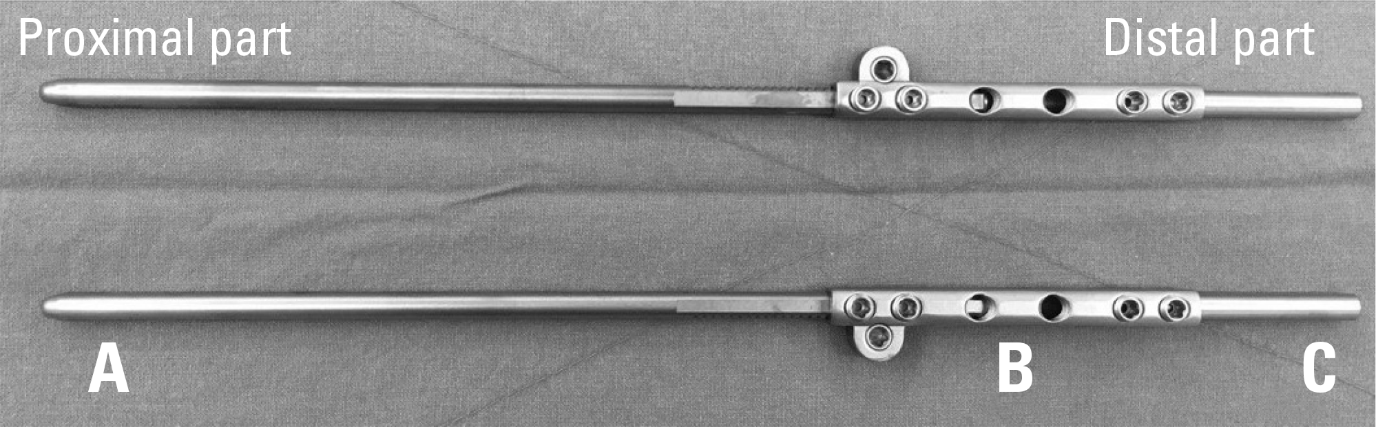 | Fig. 1.Three-part composition of the growing rod system. (A) Growing rod for thoracolumbar contouring. (B) Growing connector. (C) Conventional rod for lumbar contouring. |
 | Fig. 2.Serial radiographs of an 11-year-old male patient with idiopathic scoliosis who underwent 9 corrective operations using growing rod lengthening. (A) Preoperative whole spine posteroanterior (PA) X-ray showing a Cobb angle of 109.5°. (B) Postoperative whole spine PA view after the first operation, where the curve was corrected to a Cobb angle of 60.1°. (C) Postoperative whole-spine PA view of the fourth operation after 3 lengthening procedures. (D) Postoperative whole-spine PA view of the fifth operation after rod exchange and lengthening. (E) Postoperative whole-spine PA view of the eighth operation after final lengthening. (F) Postoperative whole-spine PA view of the final fusion operation where the curve was corrected to a Cobb angle of 46.5°. |
Table 1.
Characteristics of 14 patients treated with dual growing rods with or without fusion surgery–demographics, surgical data and complications
| No. | Sex | Age at Surgery (year) | Diagnosis | Total Treatment Period (month) | Instrumented Level | No. of Total Surgeries | No. of lengthenings | Average Lengthening Interval (month) | Final Fusion Surgery | Complication (11∗) | |
|---|---|---|---|---|---|---|---|---|---|---|---|
| Implant related (4) | Infection (7) | ||||||||||
| 1 | M | 12.5 | Congenital | 31 | T1-L1 | 7 | 3 | 6.7 | Yes | Rod breakage (1) | Deep wound infection (2) |
| 2 | M | 12.8 | Idiopathic | 55 | T1-L5 | 10 | 7 | 5.9 | Yes | Deep wound infection (1) | |
| 3 | F | 12.4 | Idiopathic | 48 | T1-ilium | 5 | 4 | 8.5 | No | ||
| 4 | M | 13.4 | Neuromuscular | 47 | T1-L4 | 4 | 2 | 6.0 | Yes | ||
| 5 | M | 14.9 | Neuromuscular | 25 | T3-L4 | 5 | 3 | 5.0 | Yes | ||
| 6 | M | 12.9 | Neuromuscular | 39 | T2-L2 | 8 | 4 | 5.8 | Yes | Deep wound infection (2) | |
| 7 | M | 10.8 | Spondyloepiphyseal Dysplasia | 64 | T4-ilium | 12 | 9 | 6.4 | Autofusion | Rod breakage (1) | |
| 8 | M | 7.0 | Neuromuscular | 39 | T2-ilium | 9 | 5 | 6.6 | Autofusion | Rod breakage (2) | Deep wound infection (1) |
| 9 | F | 7.5 | Neuromuscular | 58 | T1-L2 | 9 | 8 | 7.3 | No | ||
| 10 | F | 6.3 | Neuromuscular | 36 | T1-ilium | 6 | 5 | 7.2 | No | ||
| 11 | M | 11.6 | Neuromuscular | 35 | T1-ilium | 6 | 3 | 11.0 | Yes | Superficial wound infection (1) | |
| 12 | M | 6.3 | Neuromuscular | 27 | T1-ilium | 3 | 2 | 11.5 | No | ||
| 13 | F | 11.4 | Spondyloepiphyseal Dysplasia | 65 | T1-S1 | 6 | 2 | 4.0 | Yes | ||
| 14 | M | 13.9 | Idiopathic | 24 | T4-L5 | 4 | 3 | 5.0 | No | ||
Table 2.
Spinal curvature and length before and after implantation of growing rod
 XML Download