Abstract
Objectives
To evaluate changes in the adjacent segment after posterior instrumentation and fusion in thoracolumbar spinal fractures.
Summary of Literature Review
The incidence of adjacent-segment disease is increasing as spinal surgery becomes more common. Many studies have been conducted on the risk factors for adjacent-segment changes in the lumbar spine, but few articles have been published on this topic in the thoracolumbar spine.
Material and Methods
The records of 50 patients who received treatment from 2000 to 2013 were reviewed retrospectively. They underwent posterior instrumentation and fusion due to thoracolumbar fracture and were followed up for more than 2 years. To evaluate changes in the adjacent segment, immediate postoperative and last follow-up values of the sagittal angle, disc height, and disc angle were compared between groups divided by age (more or less than 50 years), laminectomy, and fusion levels. The Pfirrmann grade of the discs proximal and distal to the fusion level was also measured using preoperative magnetic resonance imaging.
Results
Thirty-six patients were male and 14 were female. The average age of the 50 patients was 45.6 years, and the mean follow-up period was 4.3 years. There were no cases of adjacent-segment disease. The mean kyphotic sagittal angle progression was 6.8° (range, −11° to 28.5°, p=0.000). The mean change of disc height of the proximal adjacent segment was 0.3 mm (range, −1.6 to 3.4 mm, p=0.013) and 0.6 mm (range, −4.1 to 5.8 mm, p=0.013) in the distal adjacent segment. Laminectomy did not make a significant difference. In the group below 50 years of age, the angle of the adjacent segment discs increased by 0.8° (range, −3.1° to 5.1°, p=0.004) at the proximal adjacent segment and by 0.5°(range, −4.8° to 2.9°, p=0.016) at the distal adjacent segment. Proximal adjacent disc height decreased as the fusion levels increased. As the preoperative Pfirrmann grade increased, degenerative changes in the proximal adjacent segment disc tended to accelerate.
Conclusions
Adjacent-segment disease after lumbar fusion surgery was not found in adjacent segments of the thoracolumbar spine. This seems to be due to the anatomical characteristics of the lumbar spine, which is more flexible than the thoracolumbar vertebra. The mobile segments of the lumbar spine may account for this difference, rather than the instrumentation and fusion procedure itself.
Go to : 
REFERENCES
1. Albee FH. Transplantation of a portion of the tibia into the spine for pott's disease: A preliminary report. J Am Med Assoc. 1911; 57:885–6.
2. Ghiselli G, Wang JC, Bhatia NN, et al. Adjacent segment degeneration in the lumbar spine. J Bone Joint Surg Am. 2004; 86:1497–503.

3. Lee CK, Langrana NA. Lumbosacral spinal fusion. A biomechanical study. Spine (Phila Pa 1976). 1984; 9:574–81.

4. Booth KC, Bridwell KH, Eisenberg BA, et al. Minimum 5-year results of degenerative spondylolisthesis treated with decompression and instrumented posterior fusion. Spine (Phila Pa 1976). 1999; 24:1721–7.

5. Lehmann TR, Spratt KF, Tozzi JE, et al. Long-term followup of lower lumbar fusion patients. Spine (Phila Pa 1976). 1987; 12:97–104.

6. Penta M, Sandhu A, Fraser RD. Magnetic resonance imaging assessment of disc degeneration 10 years after anterior lumbar interbody fusion. Spine (Phila Pa 1976). 1995; 20:743–7.

7. Cheh G, Bridwell KH, Lenke LG, et al. Adjacent segment disease followinglumbar/thoracolumbar fusion with pedicle screw instrumentation: a minimum 5-year follow-up. Spine (Phila Pa 1976). 2007; 32:2253–7.
8. Charles Malveaux WMS, Sharan AD. Adjacent Segment Disease After Lumbar Spinal Fusion: A Systematic Review of the Current Literature. Semin Spine Surg. 2011; 23:266–74.

9. Nagata H, Schendel MJ, Transfeldt EE, et al. The effects of immobilization of long segments of the spine on the adjacent and distal facet force and lumbosacral motion. Spine (Phila Pa 1976). 1993; 18:2471–9.

10. Cunningham BW, Kotani Y, McNulty PS, et al. The effect of spinal destabilization and instrumentation on lumbar intradiscal pressure: an in vitro biomechanical analysis. Spine (Phila Pa 1976). 1997; 22:2655–63.
11. Aota Y, Kumano K, Hirabayashi S. Postfusion instability at the adjacent segments after rigid pedicle screw fixation for degenerative lumbar spinal disorders. J Spinal Disord. 1995; 8:464–73.

12. Etebar S, Cahill DW. Risk factors for adjacent-segment failure following lumbar fixation with rigid instrumentation for degenerative instability. J Neurosurg. 1999; 90:163–9.

13. Kumar MN, Jacquot F, Hall H. Long-term follow-up of functional outcomes and radiographic changes at adjacent levels following lumbar spine fusion for degenerative disc disease. Eur Spine J. 2001; 10:309–13.

14. Schlegel JD, Smith JA, Schleusener RL. Lumbar motion segment pathology adjacent to thoracolumbar, lumbar, and lumbosacral fusions. Spine (Phila Pa 1976). 1996; 21:970–81.

15. Wiltse LL, Radecki SE, Biel HM, et al. Comparative study of the incidence and severity of degenerative change in the transition zones after instrumented versus noninstrumented fusions of the lumbar spine. J Spinal Disord. 1999; 12:27–33.

16. Cho KJ, Park SL, Kim MG, et al. Proximal Adjacent Segment Disease following Posterior Instrumentation and Fusion for Degenerative Lumbar Scoliosis. J Korean Orthop Assoc. 2009; 44:109–17.

17. Shufflebarger H, Suk SI, Mardjetko S. Debate: determining the upper instrumented vertebra in the management of adult degenerative scoliosis: stopping at T10 versus L1. Spine (Phila Pa 1976). 2006; 31(Suppl):185–94.
Go to : 
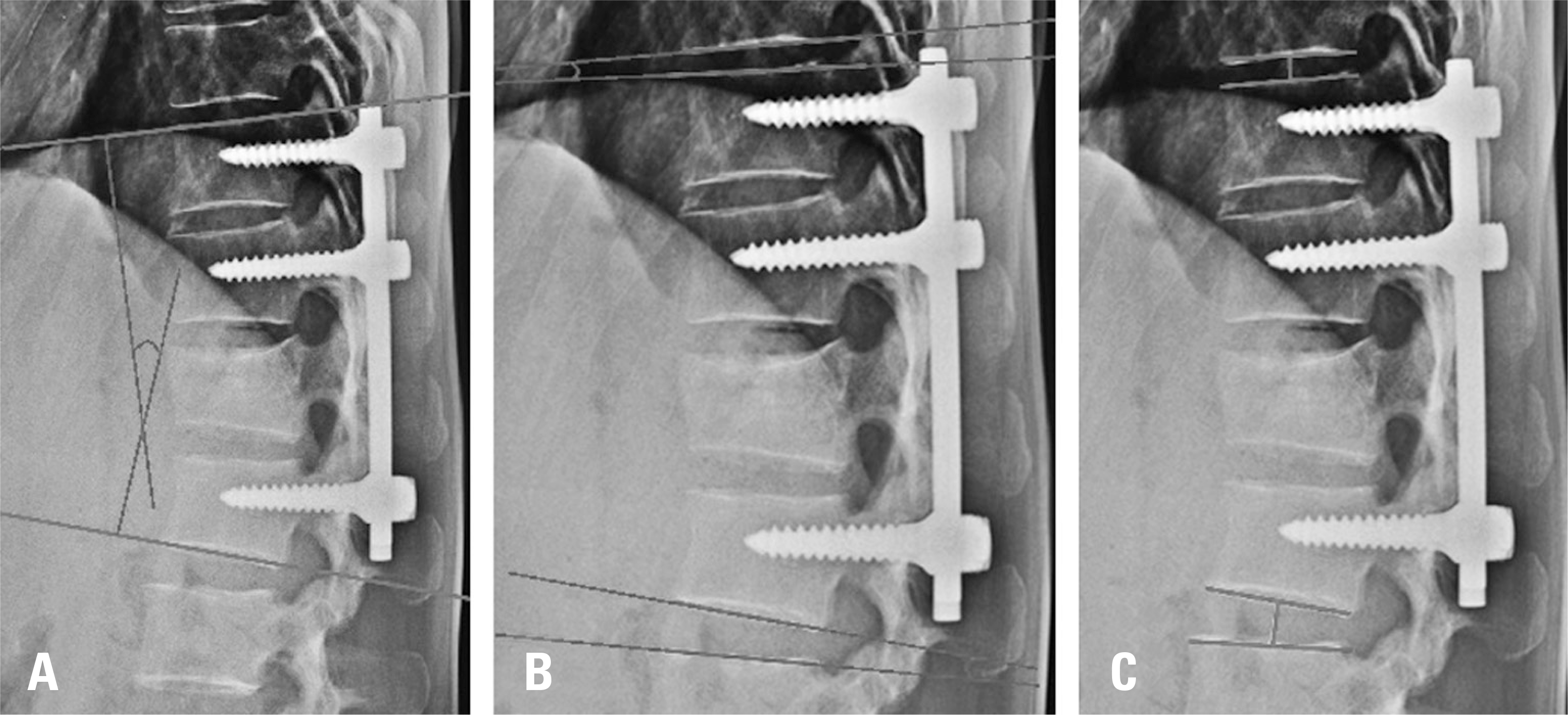
 | Fig. 1.Radiologic measurements of the adjacent segments. (A) Measurement of the segmental sagittal angle. (B) Measurement of the disc angle in the proximal and distal adjacent segments. (C) Measurement of the disc height from the midpoint of the proximal segment to the midpoint of the distal adjacent segment. |
Table 1.
Baseline data of patients
| Variable | Value |
|---|---|
| Gender (Male:Female) | 36:14 |
| Age | |
| <50 (n) | 27 |
| >50 (n) | 23 |
| Mean age (years) | 45.6 |
| Mean follow up (years) | 4.3 |
| Fusion levels | |
| 2 levels (n) | 17 |
| > 2 levels (n) | 33 |
Table 2.
Surgical level analysis through fracture site
Table 3.
Intraclass correlation coefficient of variables
Table 4.
Differences of radiologic parameters between postoperative and last follow up
Table 5.
Correlation between radiologic parameter and Pfirrmann grade
| Pearson correlation | p-value | |
|---|---|---|
| Difference of proximal adjacent disc height | −0.308 | 0.029 |
| Difference of proximal adjacent disc angle | −0.313 | 0.027 |
Table 6.
Mean differences of radiologic parameters between postoperative and last follow up according to the age.
Table 7.
Mean differences of radiologic parameters between postoperative and last follow up according to the fusion levels.


 PDF
PDF ePub
ePub Citation
 Print
Print


 XML Download