

 PDF
PDF ePub
ePub Citation
 Print
Print


Abstract
Study design
A literature review regarding the correlation between a kyphotic neck and its clinical outcomes.
Objectives
This review examines normal cervical alignment, methods for assessing alignment, a specific correlation between kyphotic neck and clinical outcomes, and indications and methods of surgical treatment.
Summary of Literature Review
Cervical kyphotic deformity is problematic in terms of HRQOL due to nerve damage or loss of horizontal gaze.
Results
Cervical kyphosis can be caused by postlaminectomy, degenerative disc disease, and trauma, and the symptoms exhibit diverse clinical progression including compensatory mechanisms, adjacent segment disease, changes in quality of life, and cervical myelopathy. Given the serious complications of cervical surgery, we need a deep understanding of spine anatomy, preoperative planning, and correction methods.
Go to : 
REFERENCES
1. Ames CP, Blondel B, Scheer JK, et al. Cervical radiographical alignment: comprehensive assessment techniques and potential importance in cervical myelopathy. Spine (Phila Pa 1976). 2013; 38(Suppl):149–60.
2. Suk KS, Kim KT, Lee JH, et al. Sagittal Alignment of the Cervical Spine After the Laminoplasty. Spine (Phila Pa 1976). 2007; 32:E656–60.

3. Tang JA, Scheer JK, Smith JS, et al. The impact of standing regional cervical sagittal alignment on outcomes in posterior cervical fusion surgery. Neurosurgery. 2012; 71:662–9.
4. Smith JS, Lafage V, Ryan DJ, et al. Association of myelopathy scores with cervical sagittal balance and normalized spinal cord volume: analysis of 56 preoperative cases from the AOSpine North America Myelopathy study. Spine (Phila Pa 1976). 2013; 38(Suppl):161–70.
5. Ryan DJ, Protopsaltis TS, Ames CP, et al. T1 pelvic angle (TPA) effectively evaluates sagittal deformity and assesses radiographical surgical outcomes longitudinally. Spine (Phila Pa 1976). 2014; 39:1203–10.
6. Three-dimensional analysis of the scoliotic deformity. in. Weinstein SL, editor. (ed):. The Pediatric Spine: Principles and Practice. New York, NY: Raven Press;1994. pp. p. 479–96.
7. Scheer JK, Tang JA, Smith JS, et al. Cervical spine alignment, sagittal deformity, and clinical implications: a review. J Neurosurg Spine. 2013; 19:141–59.
8. Hardacker JW, Shuford RF, Capicotto PN, Pryor PW. Radiographic standing cervical segmental alignment in adult volunteers without neck symptoms. Spine. 1997; 22(13):1472–80. discussion 80. Epub 1997/07/01. PubMed PMID: 9231966.
9. Harrison DE, Bula JM, Gore DR. Roentgenographic findings in the cervical spine in asymptomatic persons: A 10-year follow-up. Spine (Phila Pa 1976). 2002; 27:1249–50.
10. Albert TJ, Vacarro A. Postlaminectomy kyphosis. Spine (Phila Pa 1976). 1998; 23:2738–45.
11. Butler JC, Whitecloud TS 3rd. Postlaminectomy kyphosis. Causes and surgical management. Orthop Clin North Am. 1992; 23:505–11.
12. Belanger TA, Milam RAt, Roh JS, et al. Cervicothoracic ex-tension osteotomy for chin-on-chest deformity in ankylosing spondylitis. J Bone Joint Surg Am. 2005; 87:1732–8.
13. Sharan AD, Kaye D, Charles Malveaux WM, et al. Dropped head syndrome: etiology and management. J Am Acad Orthop Surg. 2012; 20(12):766–74.
14. Simmons EH, Graziano GP, Heffner R Jr. Muscle disease as a cause of kyphotic deformity in ankylosing spondylitis. Spine (Phila Pa 1976). 1991; 16(8 Suppl):S351–60.
15. Graziano GP, Hensinger R, Patel CK. The use of traction methods to correct severe cervical deformity in rheumatoid arthritis patients: a report of five cases. Spine (Phila Pa 1976). 2001; 26(9):1076–81.
16. He Z, Liu Y, Xue F, et al. Surgical management of congeni-tal cervical kyphosis. Orthopedics. 2012; 35(9):
17. Harrison DE, Harrison DD, Cailliet R, et al. Cobb method or Harrison posterior tangent method: which to choose for lateral cervical radiographic analysis. Spine (Phila Pa 1976). 2000; 25:2072–8.
18. El Fegoun AB, Schwab F, Gamez L, et al. Center of gravity and radiographic posture analysis: a preliminary review of adult volunteers and adult patients affected by scoliosis. Spine (Phila Pa 1976). 2005; 30(13):1535–40.
19. Polly DW Jr., Kilkelly FX, McHale KA, et al. Measurement of lumbar lordosis. Evaluation of intraobserver, in-terobserver, and technique variability. Spine (Phila Pa 1976). 1996; 21:1530–5.
20. Singer KP, Jones TJ, Breidahl PD. A comparison of radiographic and computer-assisted measurements of thoracic and thoracolumbar sagittal curvature. Skeletal Radiol. 1990; 19:21–6.
21. Mac-Thiong JM, Transfeldt EE, Mehbod AA, et al. Can c7 plumbline and gravity line predict health related quality of life in adult scoliosis? Spine (Phila Pa 1976). 2009; 34:E519–27.
22. Zheng X, Chaudhari R, Wu C, et al. Repeatability test of C7 plumb line and gravity line on asymptomatic volunteers using an optical measurement technique. Spine (Phila Pa 1976). 2010; 35(18):E889–94.
23. Suk KS, Kim KT, Lee SH, et al. Significance of chin-brow vertical angle in correction of kyphotic deformity of ankylosing spondylitis patients. Spine (Phila Pa 1976). 2003; 28:2001–5.
24. Lee SH, Kim KT, Seo EM, et al. The influence of thoracic inlet alignment on the craniocervical sagittal balance in asymptomatic adults. J Spinal Disord. 2012; 25:E41–7.
25. Kim TH, Lee SY, Kim YC, et al. T1 slope as a predic-tor of kyphotic alignment change after laminoplasty in patients with cervical myelopathy. Spine (Phila Pa 1976). 2013; 38:E992–7.
26. Lee SH, Son ES, Seo EM, et al. Factors determining cervical spine sagittal balance in asymptomatic adults: correlation with spinopelvic balance and thoracic inlet alignment. Spine J. 2015; 15:705–12.
27. Ferch RD, Shad A, Cadoux-Hudson TA, et al. Anterior correction of cervical kyphotic deformity: effects on myelopathy, neck pain, and sagittal alignment. J Neurosurg Spine. 2004; 100(1 Suppl Spine):13–9.
28. Ames CP, Smith JS, Scheer JK, et al. A standardized no-menclature for cervical spine soft-tissue release and osteotomy for deformity correction: clinical article. J Neurosurg Spine. 2013; 19:269–78.
29. Katsuura A, Hukuda S, Saruhashi Y, et al. Kyphotic malalignment after anterior cervical fusion is one of the factors promoting the degenerative process in adjacent interverte-bral levels. Eur Spine J. 2001; 10:320–4.
30. Okada E, Matsumoto M, Ichihara D, et al. Does the sagittal alignment of the cervical spine have an impact on disk de-generation? Minimum 10-year follow-up of asymptomatic volunteers. Eur Spine J. 2009; 18:1644–51.
31. Pal GP, Sherk HH. The vertical stability of the cervical spine. Spine (Phila Pa 1976). 1988; 13(5):447–9.
32. Katsuura A, Hukuda S, Saruhashi Y, et al. Kyphotic malalignment after anterior cervical fusion is one of the factors promoting the degenerative process in adjacent interverte-bral levels. Eur Spine J. 2001; 10:320–4.
33. Bridwell KH, Baldus C, Berven S, et al. Changes in radiographic and clinical outcomes with primary treatment adult spinal deformity surgeries from two years to three- to five-years follow-up. Spine (Phila Pa 1976). 2010; 35:1849–54.
34. Deutsch H, Haid RW, Rodts GE, et al. Postlaminectomy cervical deformity. Neurosurg Focus. 2003; 15:E5.
35. Shimizu K, Nakamura M, Nishikawa Y, et al. Spinal kyphosis causes demyelination and neuronal loss in the spinal cord: a new model of kyphotic deformity using ju-venile Japanese small game fowls. Spine (Phila Pa 1976). 2005; 30:2388–92.
36. Klineberg E. Cervical spondylotic myelopathy: a review of the evidence. Orthop Clin North Am. 2010; 41(2):193–202.
37. Kim HJ, Piyaskulkaew C, Riew KD. Comparison of Smith-Petersen osteotomy versus pedicle subtraction osteotomy versus anterior-posterior osteotomy types for the correction of cervical spine deformities. Spine (Phila Pa 1976). 2015; 40:143–6.
38. Lee SH, Kim KT, Suk KS, et al. A sterile-freehand reduction technique for corrective osteotomy of fixed cervical kyphosis. Spine (Phila Pa 1976). 2012; 37:2145–50.
Go to :
 | Fig. 1.Sagittal radiographs showing 3 different methods used to determine cervical lordosis. (A) The 4-line method for measuring cervical Cobb angles. This method includes drawing a line either parallel to the inferior endplate of C-2 or extending from the anterior tubercle of C-1 to the posterior margin of the spinous process, and another line parallel to the inferior endplate of C-7. Perpendicular lines are then drawn from each of the 2 lines noted above, and the angle subtended between the crossing of the perpendicular lines is the cervical curvature angle. (B) The Jackson physiological stress lines method for measuring cervical curvature. The method requires drawing 2 lines, both parallel to the posterior surface of the C-7 and C-2 vertebral bodies and measuring the angle between them. (C) The Harrison method for measuring cervical curvature. The Harrison posterior tangent method involves drawing lines that are parallel to the posterior surfaces of all cervical vertebral bodies from C-2 to C-7 and then summing the segmental angles for an overall cervical curvature angle. Adapted from Sheer J.K et al; J Neurosurg Spine. 2013;19(2):141-7)
|
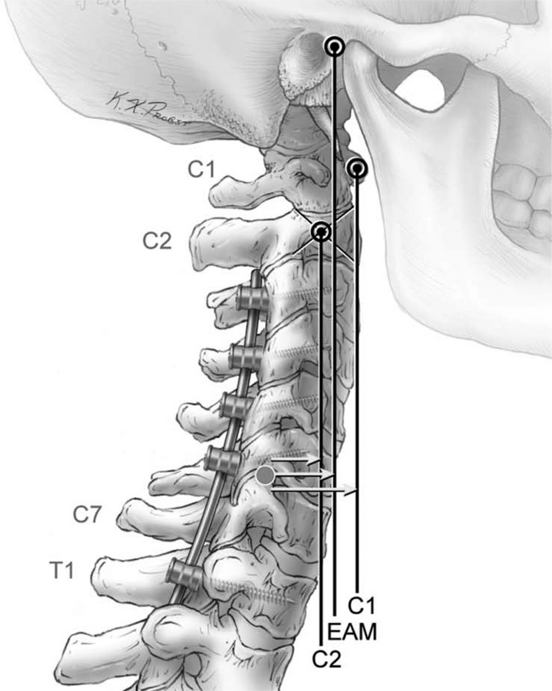 | Fig. 2.Visual representation of the technique used to measure cervical SVA. The arrows represent the distance between the posterior superior corner of C7 and a plumb line dropped from C2 (C2-C7 SVA), the external auditory meatus (EAM; CGH-C7 SVA), and C1 (C1-C7 SVA). SVA, sagittal vertical axis; CGH, center of gravity of head. Adapted from Tang JA et al; Neurosurgery. 2012;71(3):662-9.3)
|
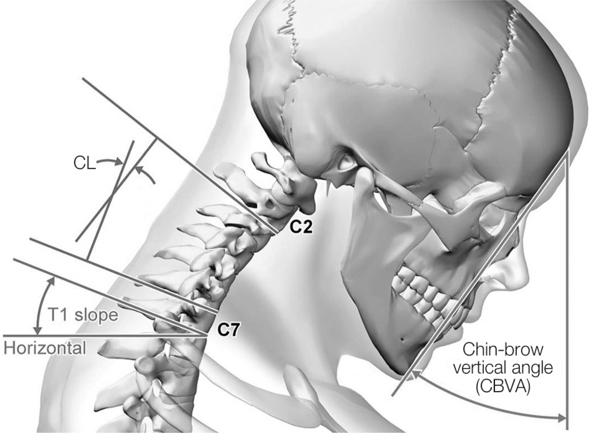 | Fig. 3.Artist's illustration of the following spinal measurements Chin-brow vertical angle(CBVA) T1 slope. Adapted from Ames CP et al; Spine (Phila Pa 1976). 2013;38(22 Suppl 1):S149-60.1)
|
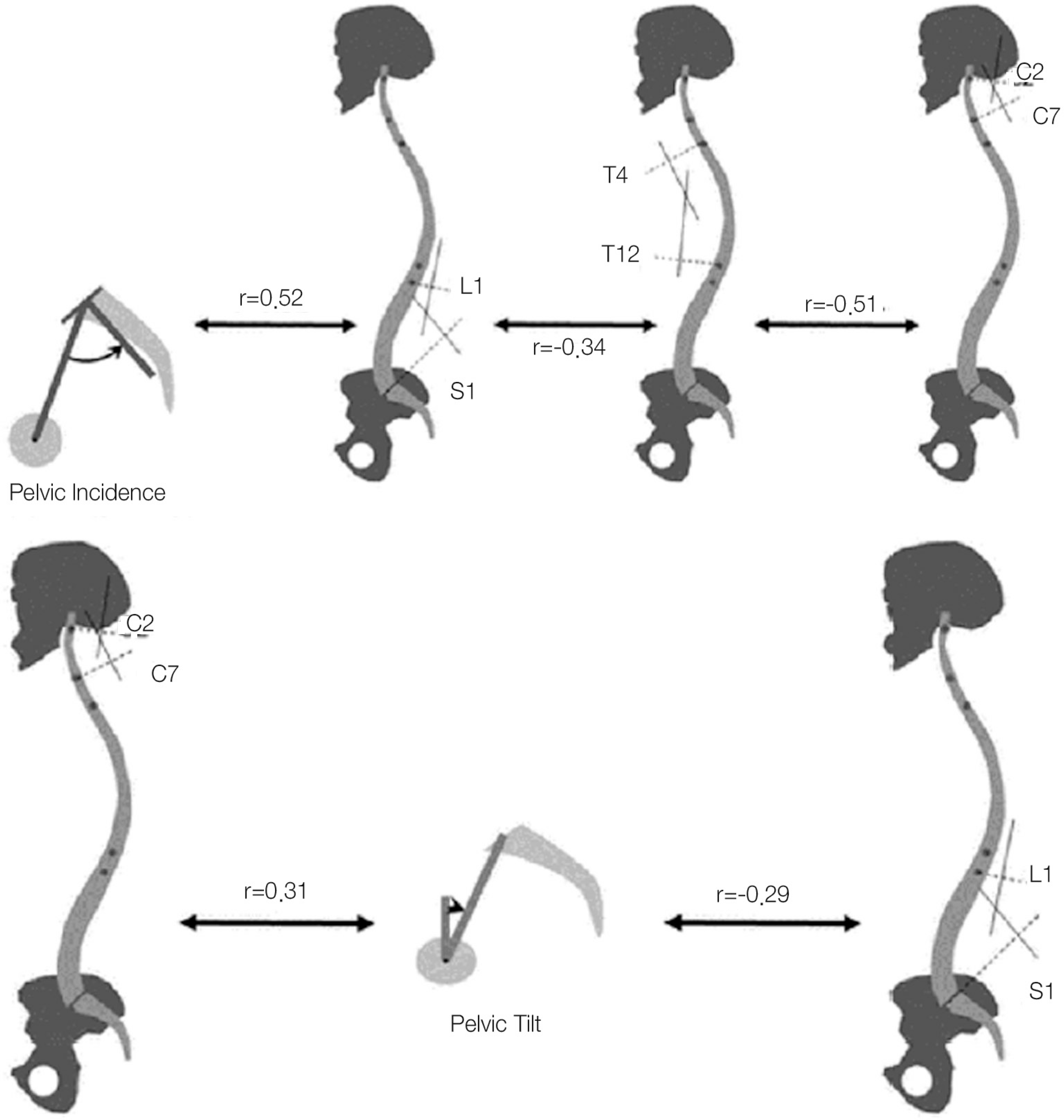 | Fig. 4.Upper: Chain of correlation between pelvic incidence and regional sagittal parameters with the corresponding Pearson coefficient (r) values. A large pelvic incidence requires a large lumbar lordosis (r=0.52). An increase of lumbar lordosis is correlated with an increased thoracic kyphosis (r=-0.34), which is correlated with an increased cervical lordosis (r= -0.51). Lower:Correlation between pelvic tilt and lumbar/ cervical lordosis. A loss of lumbar lordosis is correlated with a pelvic retroversion acting as compensatory mechanisms (r=-0.29). Pelvic retro-version is also correlated with an increased cervical lordosis (r=0.31). Adapted from Blondel et al., unpublished data, 2012. |
 | Fig. 5.Comparison of effects of positive sagittal alignment on NDI and PCS scores. Left, patient with C2-C7 SVA of 20.9mm exhibiting PCS score of 55.1 and NDI score of 3 (no disability). Right, patient with C2-C7 SVA of 59.2 mm exhibiting PCS score of 28 and NDI score of 37 (severe disability). SVA, sagittal vertical axis; NDI, neck disability index; PCS, physical component score Adapted from Tang JA et a l; Neurosurgery. 2012;71(3):662-9.3)
|
Table 1.
Normal segmental cervical angles in asymptomatic adults from literature∗
| Level | Angle(°) |
|---|---|
| C0-C1 | 2.1 ± 5.0 |
| C1-C2 | −32.2 ± 7.0 |
| C2-C3 | −1.9 ± 5.2 |
| C3-C4 | −1.5 ± 5.0 |
| C4-C5 | −0.6 ± 4.4 |
| C5-C6 | −1.1± 5.1 |
| C6-C7 | −4.5 ± 4.3 |
| C2-C7 | −9.6 |
| Total(C1-C7) | −41.8 |
 XML Download