Abstract
Objectives
To report a rare case of cauda equina syndrome due to lumbar ossification of the posterior longitudinal ligament (OPLL).
Materials and Methods
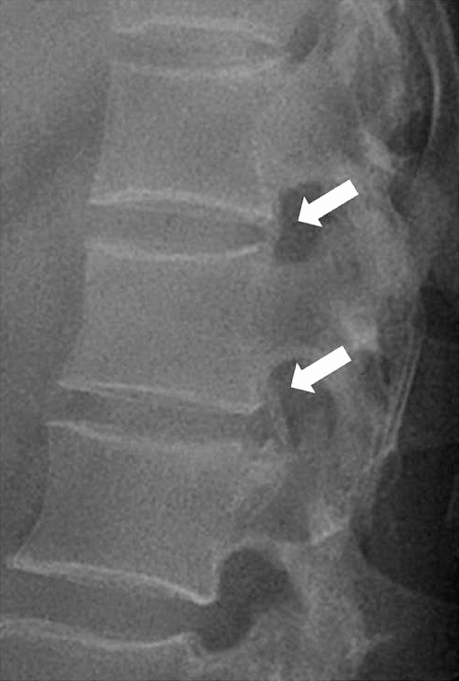
A 49-year-old female had experienced weakness in both lower extremities and radiating pain for 1 day prior to presentation. Simple radiography and CT showed OPLL at the L1-L2 level. We performed a total laminectomy and posterolateral fusion at the L1-L2 level using a posterior approach.
REFERENCES
1. Okada S, Maeda T, Saiwai H, et al. Ossification of the posterior longitudinal ligament of the lumbar spine: A Case series. Neurosurgery. 2010; 67:1311–8.

2. Albisinni U, Chianura G, Merlini L, et al. Ossification of the posterior longitudinal ligament of the lumbar spine. Radiol Med. 1988; 75:482–5.
3. Tamura M, Machida M, Aikawa �, et al. Surgical treatment of lumbar ossification of the posterior longitudinal ligament. Report of two cases and description of surgical technique. J Neurosurg Spine. 2005; 3:230–3.
4. Sakamoto R, Ikata T, Murase M, et al. Comparative study between magnetic resonance imaging and histopathologic findings in ossification or calcification of ligament. Spine (Phila Pa 1976). 1991; 16:1253–61.
5. Kurihara A, Tanaka Y, Tsumura N, et al. Hyperostotic lumbar spinal stenosis. A review of 12 surgically treated cases with roentgenographic survey of ossification of the yellow ligament at the lumbar spine. Spine (Phila Pa 1976). 1988; 13:1308–16.
6. Izumi S, Hayashi Y, Uemura M, et al. A clinical study on ossification of the posterior longitudinal ligament of the lumbar spine. Orthop Surg. 1991; 42:893–8.
Fig. 1.
A plain lateral radiograph of the lumbar spine shows ossification of the posterior longitudinal ligament (OPLL) (arrows) at the T12-L1 and L1-L2 levels.


 PDF
PDF ePub
ePub Citation
 Print
Print


 XML Download