Abstract
Objectives
To assess radiological follow-up results, including progression of bone cement augmented vertebrae, of patients who underwent percutaneous vertebroplasty (PVP).
Summary of Literature Review
There are few studies of radiological follow-up results that include progression of bone cement augmented vertebrae after PVP, regardless of good clinical results.
Materials and Methods
Between January 2000 and August 2007, 253 patients were treated with PVP for osteoporotic compression fracture. Among them, 81 patients died during follow-up and 101 patients (157 vertebrae) were available for follow-up over 7 years. We analyzed the radiologic outcomes, focusing on augmented bone cement feature and progressive change with adjacent vertebrae.
Results
The mean follow-up period was 7.9 years. Anterior body height in the last follow-up was improved about 0.3 mm compared with the preprocedural value, but this improvement was not statistically significant. The focal kyphotic angle was reduced from 12.3° at the preprocedural state to 11.7° at the postprocedural state but this change was also not statistically significant (p>0.05). Out of the 101 cases, we observed 7 cases of radiolucent line with decreased bone density in the adjacent area of bone cement and 5 cases of bone cement cracks accompanied with vertebral collapse were observed. Eleven patients (10.8%) had a solid spontaneous fusion, and 8 patients (7.9%) had partially fused with adjacent vertebrae.
REFERENCES
1. Jung �W, �ark JY, K�� KJ, et a�. Conservat�ve treat�ent o� co�press�on an� stab�e burst �ractures �n the thoraco�u�bar junct�on: ear�y a�bu�at�on Vs. �ate a�bu�at�on. J Korean Orthop. 2002; 37:483–8.
2. �uk �I, Lee CK, Kang ��, et a�. Vertebra� �racture �n Osteo-poros�s. J Korean Orthop. 1993; 28:980–7.
3. We�nste�n JN, Co��a�to �, Leh�an TR. Thoraco�u�bar burst �ractures treate� conservat�ve�y a �ong-ter� �o��owup. �p�ne (�h��a �a 1976). 1988; 13:33–8.
4. Jensen ME, Evans AJ, Math�s JM, et a�. �ercutaneous po�y�ethy��ethacry�ate vertebrop�asty �n the treat�ent o� osteoporot�c vertebra� bo�y co�press�on �ractures: techn�ca� aspects. AJNR A� J Neurora��o�ogy. 1997; 18:1897–904.
5. Layton KF, Th�e�en KR, Koch CA, et a�. Vertebrop�asty,��rst 1000 �eve�s o� a s�ng�e center: eva�uat�on o� the outco�es an� co�p��cat�ons. AJNR AM J Neurora��o�ogy. 2007; 28:683–9.
6. Ka���es DF, Co�stock BA, �eagerty �J, et a�. A Ran�o�-�ze� Tr�a� o� Vertebrop�asty �or Osteoporot�c �p�na� Fractures. N Eng� J Me�. 2009; 361:569–79.
7. �erez-��gueras A, A�varez L, Ross� RE, Qu�nones D, A�-Ass�r I �ercutaneous vertebrop�asty. �ong-ter� c��n�ca� an� ra��o�og�ca� outco�e. Neurora��o�ogy. 2002; 44:950–4.
8. K�� J�, Yoo ��, K�� J�. Long-ter� Fo��ow-up o� �er-cutaneous Vertebrop�asty �n Osteoporot�c Co�press�on Fracture: M�n��u� o� 5 Years Fo��ow-up. As�an �p�ne J. 2012; 6:6–14.
9. Roth�an R�. ���eone FA. The surg�ca� teat�ent o� spon-�y�o��sthes�s �n a�u�t �n the sp�ne 3r� e�. �h��a�ep�h�a. WB �aun�ers Co;1992. 964-5.
10. Upp�n AA, ��rsch JA, Centenera LV. Occurrence o� new vertebra� bo�y �racture a�ter percutaneous vertebrop�asty �n pat�ents w�th osteoporos�s. Ra��o�ogy. 2003; 226:119–24.
11. Tsa� TT, Chen WJ, La� �L, et a�. �o�y�ethy��ethacry�ate ce�ent ��s�o�ge�ent �o��ow�ng percutaneous vertebrop�as-ty: a case report. �p�ne (�h��a �a 1976). 2003; 28:457–60.
12. Togawa D, Bauer TW, L�eber�an I�, et a�. ��sto�og�c eva�uat�on 1 o� hu�an vertebra� bo��es a�ter vertebra� aug-�entat�on w�th po�y�ethy��ethacry�ate. �p�ne (�h��a �a 1976). 2003; 28:1521–7.
13. Cun�n G, Bo�ssonnet �, �et�te �, et a�. E�per��enta� ver-tebrop�asty us�ng ostecon�uct�ve granu�ar �ater�a�. �p�ne (�h��a �a 1976). 2000; 25:1070–6.
14. Bostro� M�, Lane JM. Aug�entat�on o� osteoporot�c vertebra� bo��es: Future ��rect�ons. �p�ne (�h��a �a 1976). 1997; 22(�upp�):38–42.
15. Cotten A, Duquesnoy B. Vertebrop�asty: Current �ata an� �uture potent�a�. Rev Rhu� Eng� E�. 1997; 64:645–9.
16. Bae �, �atten �� Jr, L�nov�tz R, et a�. A prospect�ve ran-�o��ze� FDA-IDE tr�a� co�par�ng Cortoss w�th �MMA �or vertebrop�asty: a co�parat�ve e��ect�veness research stu�y w�th 24-�onth �o��ow-up. �p�ne (�h��a �a 1976). 2012; 37:544–50.
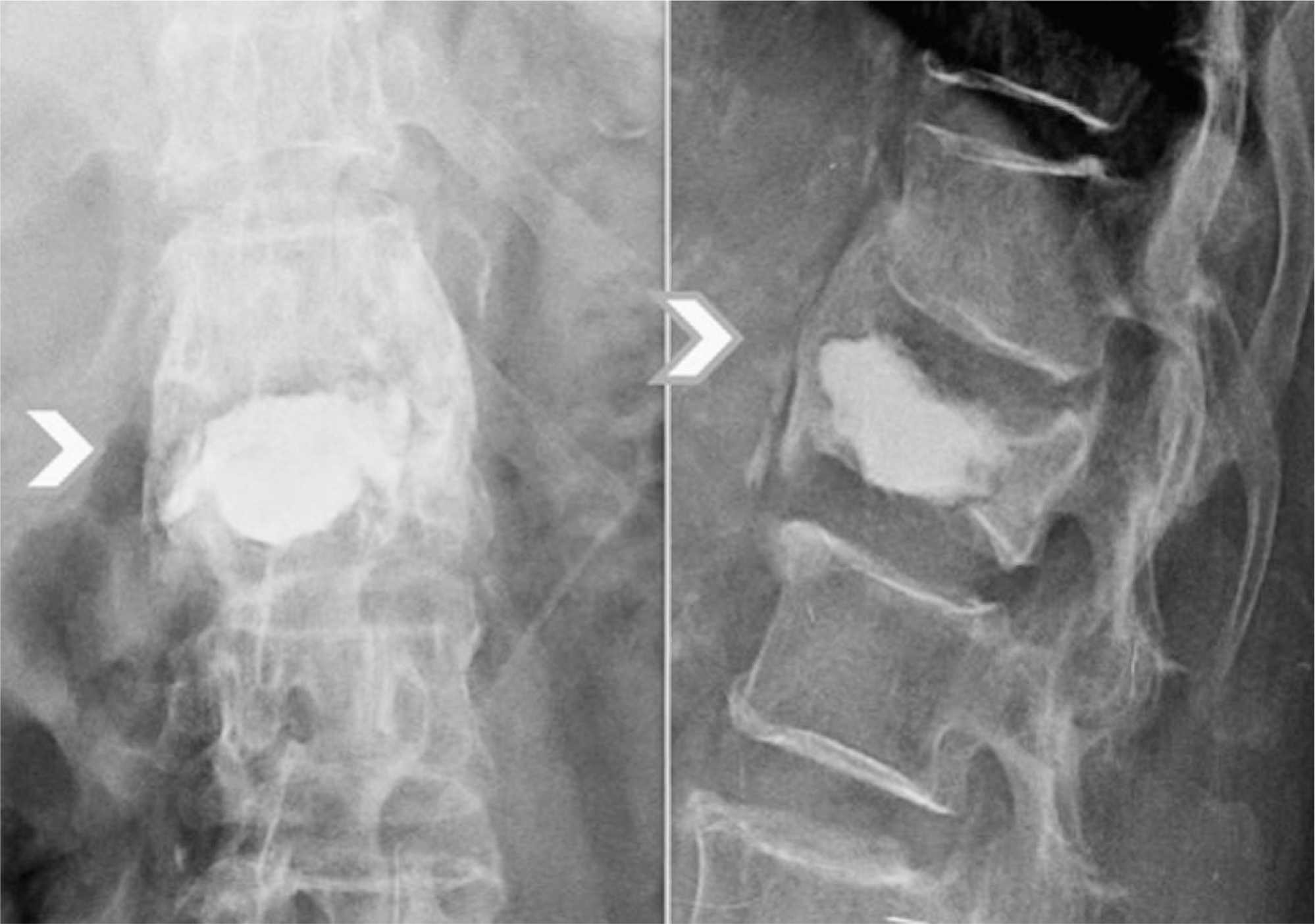
Fig. 1.
A solid fusion group defined fractured vertebrae in which continuity was achieved in at least 3 adjacent cortical vertebrae. The white ar-row shows cortical continuity between vertebrae.

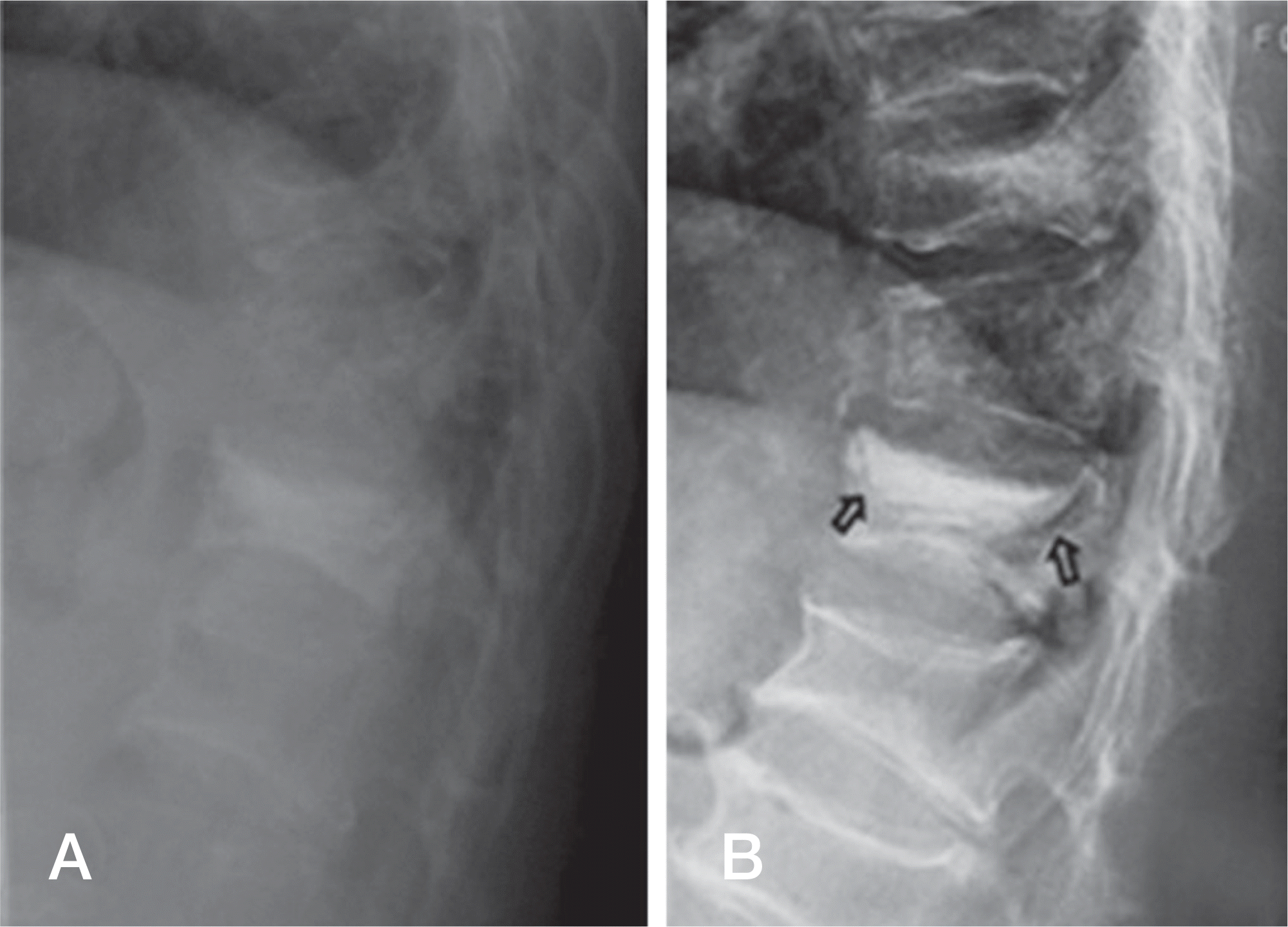
Fig. 2.
(A) A 71-year-old women had a vertebroplasty for T12 osteoporotic compression fracture. (B) The eight-year follow-up revealed a radiolucent area (arrow) between cement and bone on plain image.

Fig. 3.
(A) Seventy years old woman had osteoporotic compression fracture of L1 and L2. (B) She underwent percutaneous vertebroplasty on both L1 and L2, (C) During the two-year postoperative follow-up, an X-ray showed increased focal kyphotic angle between L1 and L2, (D) During the six-year postoperative follow-up, an X-ray showed a marked increased kyphotic angle and cemented vertebrae breakage at L1 and L2.


 PDF
PDF ePub
ePub Citation
 Print
Print


 XML Download