Abstract
Objectives
The aim of this study was to present updated information on the basic pelvic parameters associated with lumbar degenerative disease.
Summary of Literature Review
Sagittal imbalance has been known to be related to a poor prognosis in almost all adult spine problems, including lumbar degenerative disease.
Results
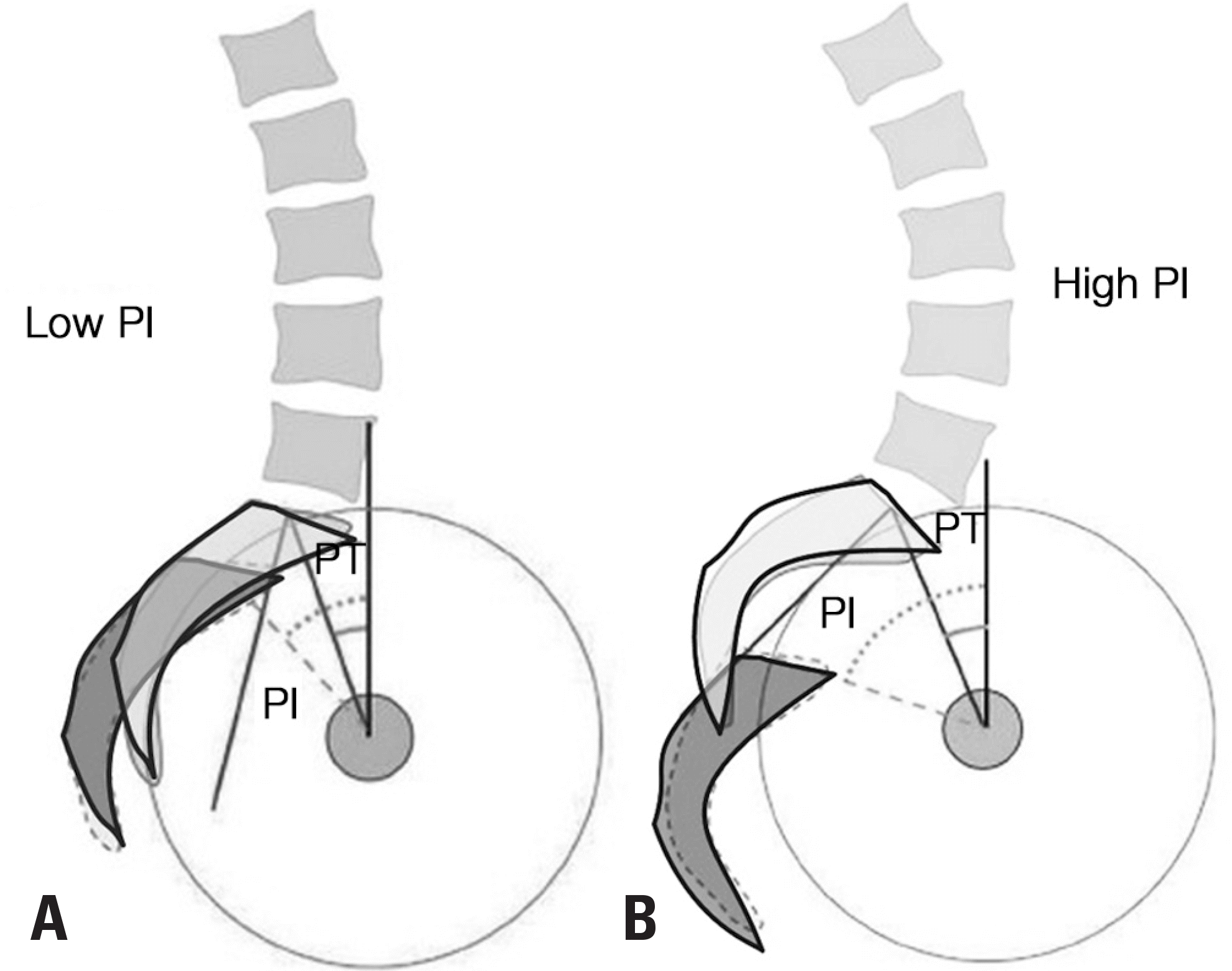
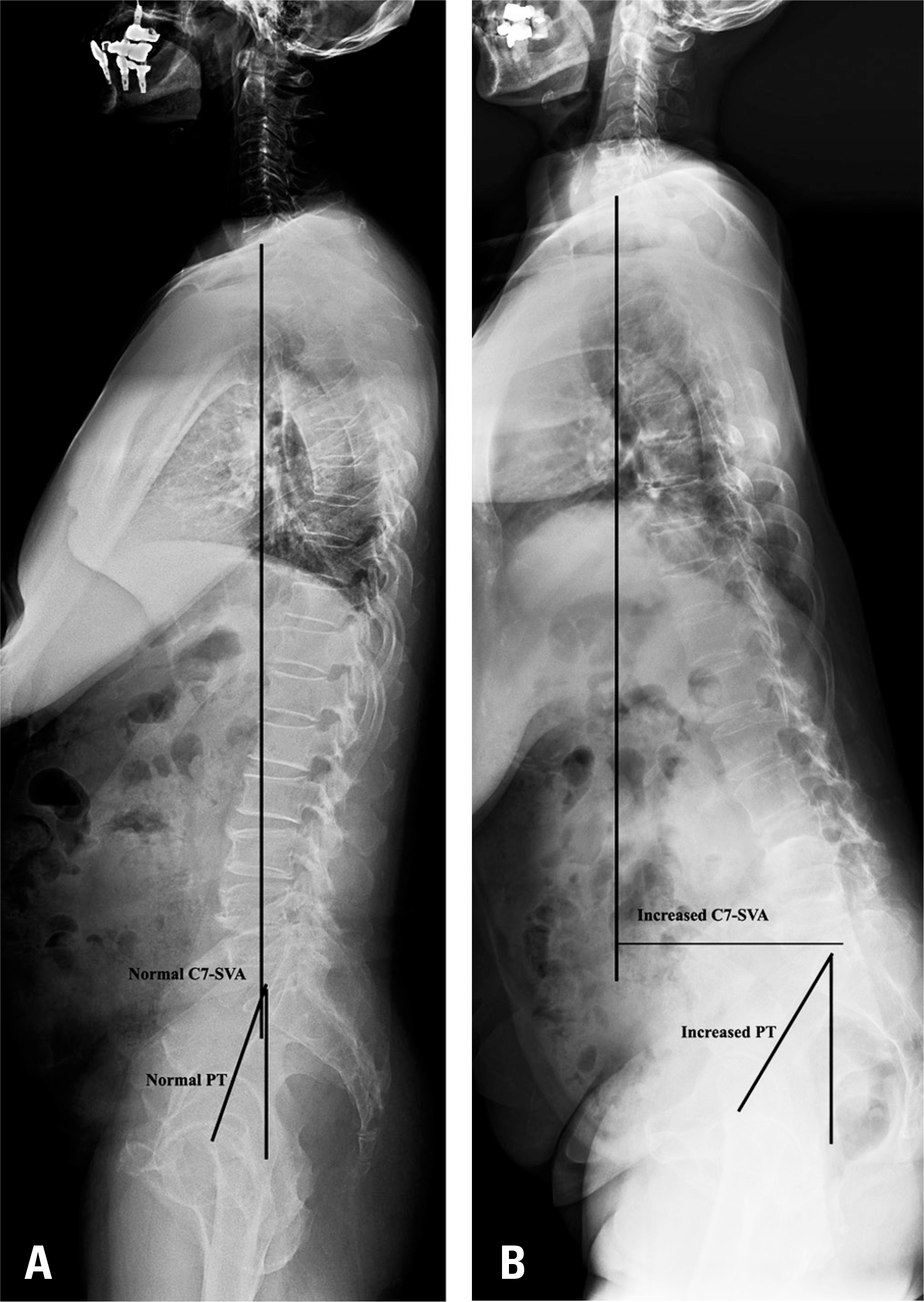
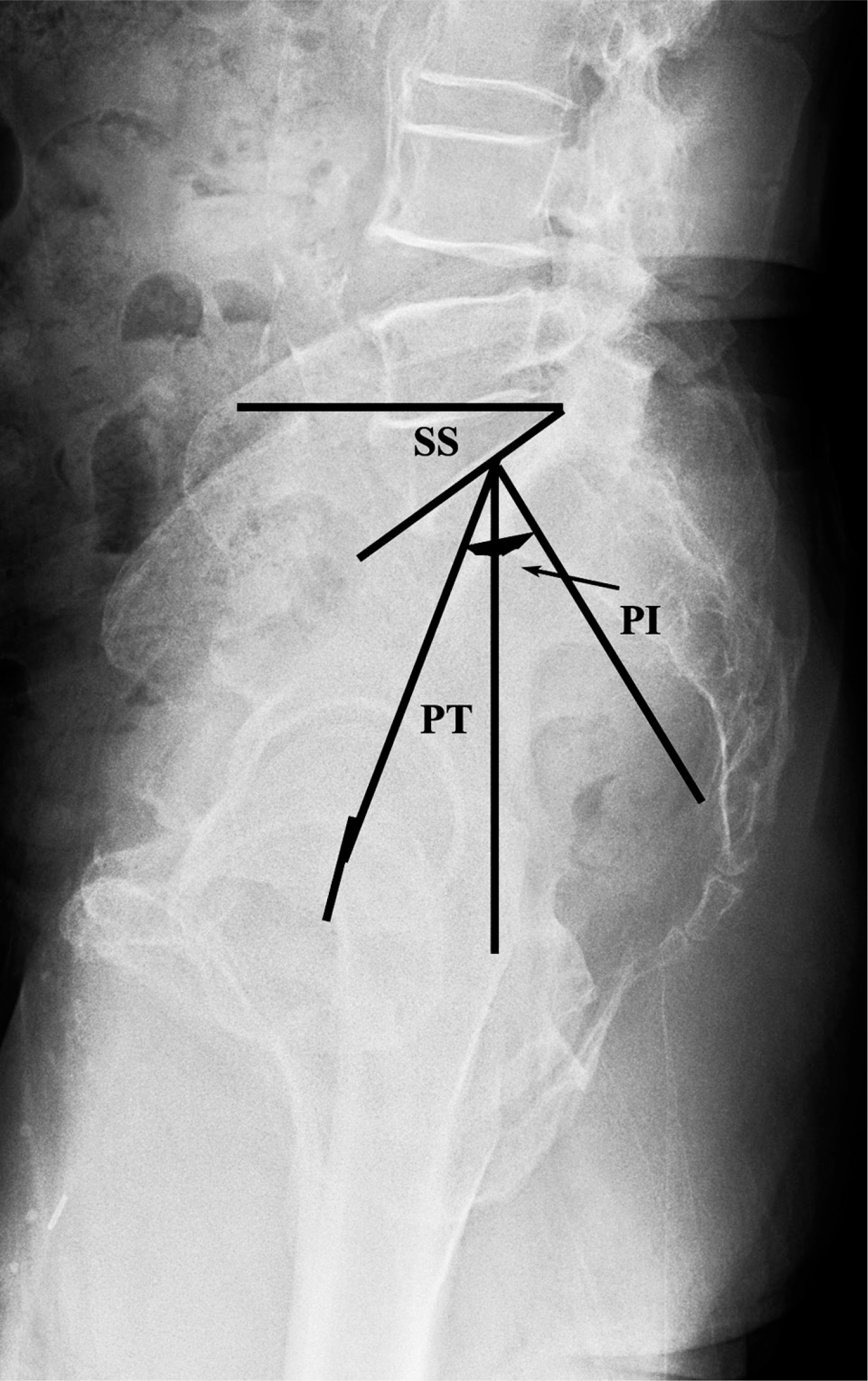
Pelvic incidence is a morphologic parameter of the pelvis. It influences lumbar lordosis and thoracic kyphosis, and determines the limitations of pelvic retroversion in sagittal imbalance. Pelvic tilt is a positional parameter of the pelvis, indicating the degree of compensation for sagittal imbalance. A C7-sagittal vertical axis >5 cm, pelvic tilt >20°, and pelvic incidence-lumbar lordosis mismatch are known to be independent factors predictive of poor outcomes.
Go to : 
REFERENCES
1. Than KD, Park P, Fu KM, et al. Clinical and radiographic parameters associated with best versus worst clinical outcomes in minimally invasive spinal deformity surgery. J Neurosurg Spine. 2016; 25:21–5.

2. Weinberg DS, Morris WZ, Gebhart JJ, et al. Pelvic incidence: an anatomic investigation of 880 cadaveric specimens. Eur Spine J. 2016; 25:3589–95.
3. Smith MW, Annis P, Lawrence BD, et al. Acute proximal junctional failure in patients with preoperative sagittal imbalance. Spine J. 2015; 15:2142–8.

4. Lin B, Zhang WB, Cai TY, et al. Analysis of sagittal balance using spinopelvic parameters in ankylosing spondylitis patients treated with vertebral column decancellation surgery. Acta Orthop Belg. 2015; 81:538–45.
5. Kim KT, Lee SH, Huh DS, et al. Restoration of Lumbar Lordosis in Flat Back Deformity: Optimal Degree of Correction. Asian Spine J. 2015; 9:352–60.

6. Jiang L, Qiu Y, Xu L, et al. Sagittal spinopelvic alignment in adolescents associated with Scheuermann's kyphosis: a comparison with normal population. Eur Spine J. 2014; 23:1420–6.

7. Dong YL, Zhao H, Zhang JG, et al. Effects of pedicle subtraction osteotomy on spinopelvic parameters of ankylosing spondylitis. Zhonghua Yi Xue Za Zhi. 2013; 93:1138–41.
8. During J, Goudfrooij H, Keessen W, et al. Toward stan-dards for posture. Postural characteristics of the lower back system in normal and pathologic conditions. Spine (Phila Pa 1976). 1985; 10:83–7.
9. Duval-Beaupere G, Schmidt C, Cosson P. A Barycentre-metric study of the sagittal shape of spine and pelvis: the conditions required for an economic standing position. Ann Biomed Eng. 1992; 20:451–62.

10. Roussouly P, Pinheiro-Franco JL. Biomechanical analysis of the spino-pelvic organization and adaptation in pathology. Eur Spine J. 2011; 20(Suppl):609–18.

11. Liu H, Shrivastava SR, Zheng ZM, et al. Correlation of lumbar disc degeneration and spinal-pelvic sagittal balance. Zhonghua Yi Xue Za Zhi. 2013; 93:1123–8.
12. Toy JO, Tinley JC, Eubanks JD, Qureshi SA, Ahn NU. Correlation of sacropelvic geometry with disc degeneration in spondylolytic cadaver specimens. Spine (Phila Pa 1976). 2012; 37:E10–5.

13. Rose PS, Bridwell KH, Lenke LG, et al. Role of pelvic incidence, thoracic kyphosis, and patient factors on sagittal plane correction following pedicle subtraction osteotomy. Spine (Phila Pa 1976). 2009; 34:785–91.

14. Bourghli A, Aunoble S, Reebye O, et al. Correlation of clinical outcome and spinopelvic sagittal alignment after surgical treatment of low-grade isthmic spondylolisthesis. Eur Spine J. 2011; 20(Suppl):663–8.

15. Schwab F, Lafage V, Patel A, et al. Sagittal plane considerations and the pelvis in the adult patient. Spine (Phila Pa 1976). 2009; 34:1828–33.

16. Schwab F, Patel A, Ungar B, et al. Adult spinal deformity-postoperative standing imbalance: how much can you tolerate? An overview of key parameters in assessing alignment and planning corrective surgery. Spine (Phila Pa 1976). 2010; 35:2224–31.
17. Legaye J, Duval-Beaupere G, Hecquet J, et al. Pelvic incidence: a fundamental pelvic parameter for three-dimensional regulation of spinal sagittal curves. Eur Spine J. 1998; 7:99–103.

18. Vialle R, Levassor N, Rillardon L, et al. Radiographic analysis of the sagittal alignment and balance of the spine in asymptomatic subjects. J Bone Joint Surg Am. 2005; 87:260–7.

19. Kim YB, Kim YJ, Ahn YJ, et al. A comparative analysis of sagittal spinopelvic alignment between young and old men without localized disc degeneration. Eur Spine J. 2014; 23:1400–6.

20. Lamartina C, Berjano P, Petruzzi M, et al. Criteria to restore the sagittal balance in deformity and degenerative spondylolisthesis. Eur Spine J. 2012; 21(Suppl):27–31.

21. Bae JS, Jang JS, Lee SH, et al. Radiological analysis of lumbar degenerative kyphosis in relation to pelvic incidence. Spine J. 2012; 12:1045–51.

22. Bae J, Lee SH, Shin SH, et al. Radiological analysis of upper lumbar disc herniation and spinopelvic sagittal alignment. Eur Spine J. 2016; 25:1382–8.

23. Le Huec JC, Aunoble S, Philippe L, et al. Pelvic parameters: origin and significance. Eur Spine J. 2011; 20(Suppl):564–71.

24. Zhu Z, Xu L, Zhu F, et al. Sagittal alignment of spine and pelvis in asymptomatic adults: norms in Chinese populations. Spine (Phila Pa 1976). 2014; 39:E1–6.
25. Jiang WY, Xu RM, Ma WH, et al. Effect of reduction on spino-pelvic parameters in treating high-grade lumbar spondylolisthesis. Zhongguo Gu Shang. 2014; 27:726–9.
26. Araujo F, Lucas R, Alegrete N, et al. Sagittal standing posture, back pain, and quality of life among adults from the general population: a sex-specific association. Spine (Phila Pa 1976). 2014; 39:E782–94.
27. Beyer F, Geier F, Bredow J, et al. Influence of spinopelvic parameters on non-operative treatment of lumbar spinal stenosis. Technol Health Care. 2015; 23:871–9.

28. Gottfried ON, Daubs MD, Patel AA, et al. Spinopelvic parameters in postfusion flatback deformity patients. Spine J. 2009; 9:639–47.

29. Hey D, Teo AQ, Tan KA, et al. How the spine differs in standing and in sitting - important considerations for cor-rection of spinal deformity. Spine J. 2016. [Epub ahead of print].
Go to : 
 | Fig. 2골반 입사각에 따른 골반의 회전(retroversion) 차이. 작은 골반 입사각에 따른 적은 골반 회전 능력(A), 큰 골반 입사각에 따른 많은 골반 회전 능력(B). |
 | Fig. 3.시상면상 균형 상태(A). 시상면상 제 7경추에서 내린 수선이 제 1천추 후상방에서 5 cm 이상 전방을 통과할 경우 시상면상 불균형이 있음을 의미(B). |
 | Fig. 43년전 타병원에서 L5-S1 유합술 받고 현재 양 하지 방사통이 심해서 내원한 66세 여자 환자. 전신 측면 기립 방사선 사진상 C7-SVA 4.5 cm, 골반 경사 16도로 비교적 균형이 잘 맞는 상태로(A), MRI 시상면 검사상 제 3-4-5요추간 협착증 소견 확인되었음(B). 제 3-4-5요추체간 유합술 이후 C7-SVA는 3 cm, 요추 전만은 46도로 호전되었으며, 시상면상 균형이 잘 유지되고 증상도 호전되었음(C). |
 | Fig. 512년전 타병원에서 L4-5-S1 유합술 받고, 현재 양쪽 엉치 통증이 심해서 내원한 67세 여자 환자. 전신 측면 기립 방사선 검사상 C7-SVA 13 cm, 골반 경사 28도로 시상면상 불균형 있었던 상태이며(A), 굴곡 측면 방사선 검사상 제 3-4요추간 불안정성 있는 상태이고(B), MRI 시상면 검사상 제 3-4요추간 협착증 소견 있었음(C). 이전 기기를 제거하고, 제 3-4요추체간 유합술을 하였으나, C7-SVA가 12 cm, 골반 경사가 26도로 거의 호전이 없어, 제 3-4요추체간 불안정성은 없지만, 불편한 증상이 계속되었음(D). |


 PDF
PDF Citation
 Print
Print


 XML Download