

 PDF
PDF Citation
 Print
Print


Abstract
Objectives
To discuss how to evaluate, interpret, and utilize measurements of spino-pelvic alignment before and after spinal surgery in patients with lumbar degenerative disease.
Summary of Literature Review
Various spino-pelvic parameters are currently utilized in the evaluation of spinal patients; however, interpretation of these parameters is not easy.
Materials and Methods
Each spino-pelvic parameter and factors affecting its value, and how to interpret and utilize the spino-pelvic parameters before and after spinal surgery were discussed for patients with lumbar degenerative disease with and without sagittal spinal deformity.
Results
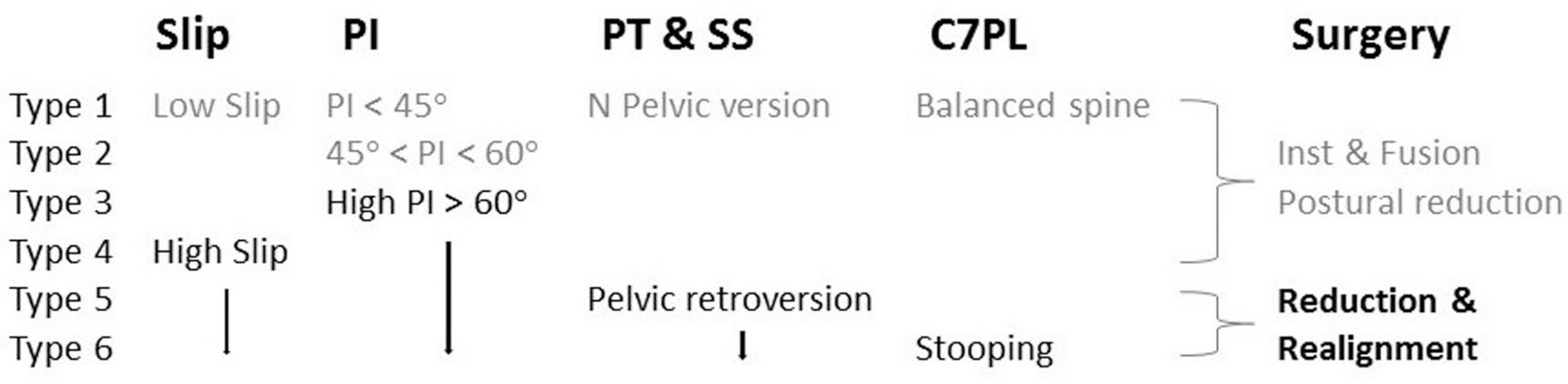
Sagittal modifiers in the SRS-Schwab classification including pelvic incidence minus lumbar lordosis (PI-LL), sagittal vertical axis (SVA), and pelvic tilt (PT) are widely accepted in the evaluation of lumbar degenerative disease with sagittal deformity. Surgery for sagittal realignment is meant to restore both the SVA and PT by restoring the LL in reference to the PI. However, patients with an extremely high SVA and PT or those with a high SVA and low PT can end up with postoperative residual malalignment. In patients without deformity, PI-LL mismatch (> 10°) should be highlighted and should be actively corrected by restoring the lordosis of the pathologic segment.
Conclusions
Sagittal modifiers are beneficial for their simplicity and comprehensibility; however, they are insufficient for evaluating sub-regional spinal deformity. Spino-pelvic parameters can be useful for evaluating spinal patients in a clinical setting, but the measurements are greatly affected by confounding factors such as poor patient posture, unqualified testers, and manual measurement techniques.
Go to : 
REFERENCES
1. Dimar JR 2nd, Carreon LY, Labelle H, et al. Intra- and inter-observer reliability of determining radiographic sagittal parameters of the spine and pelvis using a manual and a computer-assisted methods. Eur Spine J. 2008; 17:1373–9.

2. Schwab F, Ungar B, Blondel B, et al. Scoliosis Research Society-Schwab adult spinal deformity classification: a validation study. Spine (Phila Pa 1976). 2012; 37:1077–82.
3. Smith JS, Klineberg E, Schwab F, et al. Change in classification grade by the SRS-Schwab Adult Spinal Deformity Classification predicts impact on health-related quality of life measures: prospective analysis of operative and nonop-erative treatment. Spine (Phila Pa 1976). 2013; 38:1663–71.
4. Rothenfluh DA, Mueller DA, Rothenfluh E, et al. Pelvic incidence-lumbar lordosis mismatch predisposes to adjacent segment disease after lumbar spinal fusion. Eur Spine J. 2015; 24:1251–8.
5. Mac-Thiong JM, Roussouly P, Berthonnaud E, et al. Sagittal parameters of global spinal balance: normative values from a prospective cohort of seven hundred nine Caucasian asymptomatic adults. Spine (Phila Pa 1976). 2010; 35:E1193–8.
6. Protopsaltis T, Schwab F, Bronsard N, et al. TheT1 pelvic angle, a novel radiographic measure of global sagittal deformity, accounts for both spinal inclination and pelvic tilt and correlates with health-related quality of life. J Bone Joint Surg Am. 2014; 96:1631–40.
7. Lafage V, Schwab F, Patel A, et al. Pelvic tilt and truncal inclination: two key radiographic parameters in the set-ting of adults with spinal deformity. Spine (Phila Pa 1976). 2009; 34:E599–606.
8. Diebo BG, Henry J, Lafage V, et al. Sagittal deformities of the spine: factors influencing the outcomes and complications. Eur Spine J. 2015; 24(Suppl):3–15.
9. Gill JB, Levin A, Burd T, et al. Corrective osteotomies in spine surgery. J Bone Joint Surg Am. 2008; 90:2509–20.
10. Legaye J, Duval-Beaupere G. Sagittal plane alignment of the spine and gravity: a radiological and clinical evaluation. Acta Orthop Belg. 2005; 71:213–20.
11. Rose PS, Bridwell KH, Lenke LG, et al. Role of pelvic incidence, thoracic kyphosis, and patient factors on sagittal plane correction following pedicle subtraction osteotomy. Spine (Phila Pa 1976). 2009; 34:785–91.
12. Glassman SD, Bridwell K, Dimar JR, et al. The impact of positive sagittal balance in adult spinal deformity. Spine (Phila Pa 1976). 2005; 30:2024–9.
13. Berjano P, Langella F, Ismael MF, et al. Successful correction of sagittal imbalance can be calculated on the basis of pelvic incidence and age. Eur Spine J. 2014; 23(Suppl):587–96.
14. Bess S, Schwab F, Lafage V, et al. Classifications for adult spinal deformity and use of the Scoliosis Research Society-Schwab Adult Spinal Deformity Classification. Neurosurg Clin N Am. 2013; 24:185–93.
15. Ferrero E, Vira S, Ames CP, et al. Analysis of an unexplored group of sagittal deformity patients: low pelvic tilt despite positive sagittal malalignment. Eur Spine J. 2015 May 31. [Epub ahead of print].
16. Lamartina C, Berjano P. Classification of sagittal imbalance based on spinal alignment and compensatory mechanisms. Eur Spine J. 2014; 23:1177–89.
17. Lafage V, Schwab F, Vira S, et al. Does vertebral level of pedicle subtraction osteotomy correlate with degree of spinopelvic parameter correction? J Neurosurg Spine. 2011; 14:184–91.
18. Jang J-S, Lee S-H, Min J-H, et al. Changes in sagit-tal alignment after restoration of lower lumbar lordosis in patients with degenerative flat back syndrome. J Neurosurg Spine. 2007; 7:387–92.
19. Barrey C, Jund J, Perrin G, et al. Spinopelvic alignment of patients with degenerative spondylolisthesis. Neurosurgery. 2007; 61:981–6.
20. Labelle H, Mac-Thiong JM, Roussouly P. Spino-pelvic sagittal balance of spondylolisthesis: a review and classification. Eur Spine J. 2011; 20(Suppl 5):641–6.
21. Kong LD, Zhang YZ, Wang F, et al. Radiographic Restoration of Sagittal Spinopelvic Alignment After Posterior Lumbar Interbody Fusion in Degenerative Spondylolisthesis. Clin Spine Surg. 2016; 29:E87–92.
22. Jeon CH, Lee HD, Lee YS, et al. Change in sagittal profiles after decompressive laminectomy in patients with lumbar spinal canal stenosis: a 2-year preliminary report. Spine (Phila Pa 1976). 2015; 40:E279–85.
23. Marks M, Stanford C, Newton P. Which lateral radiographic positioning technique provides the most reliable and functional representation of a patient's sagittal balance? Spine (Phila Pa 1976). 2009; 34:949–54.
24. Tyrakowski M, Wojtera-Tyrakowska D, Siemionow K. In-fluence of pelvic rotation on pelvic incidence, pelvic tilt, and sacral slope. Spine (Phila Pa 1976). 2014; 39:E1276–83.
Go to :
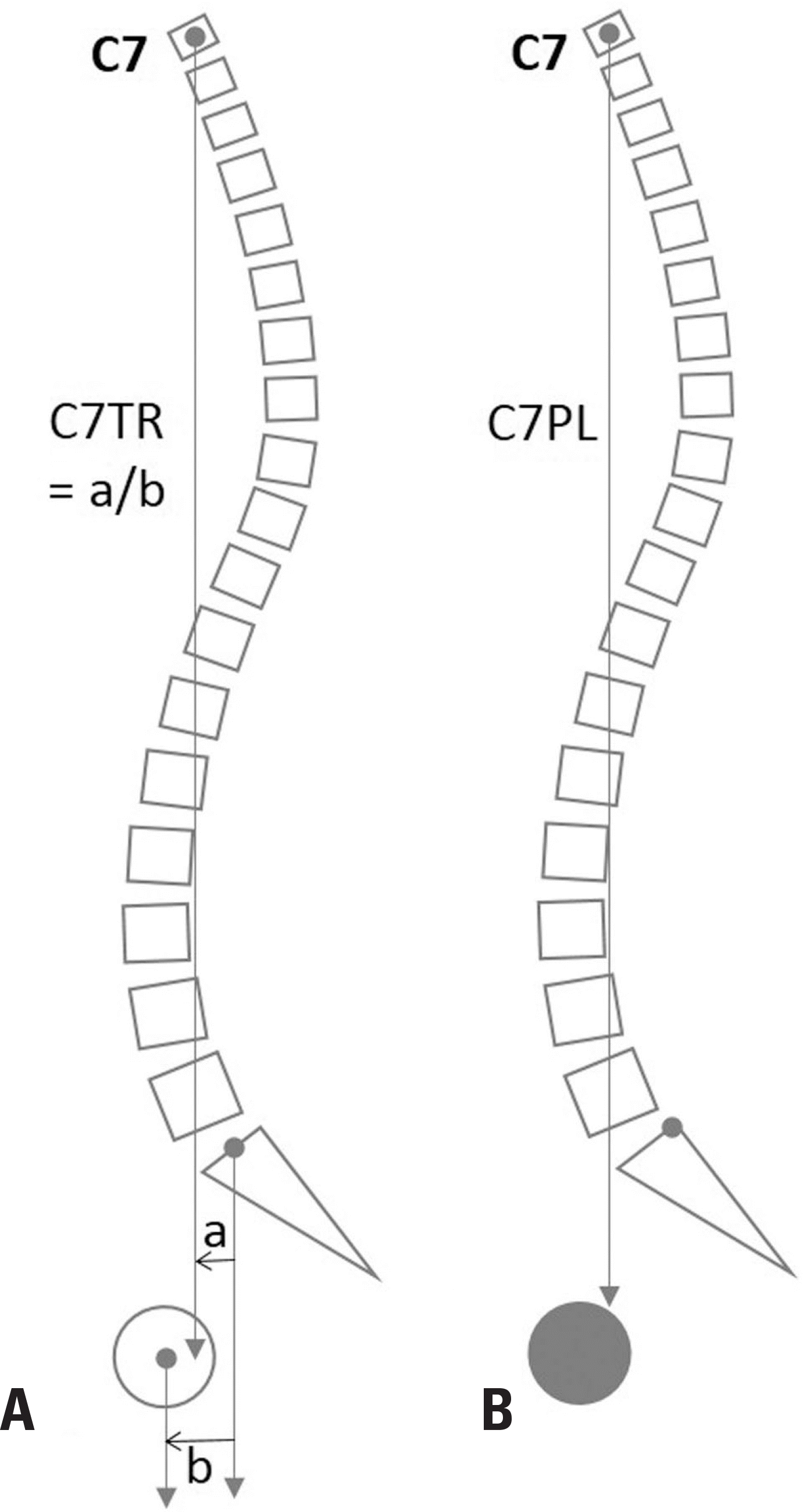 | Fig. 1.The C7 translation ratio (A) and the horizontal position of C7PL (B) between the hip joint and the posterior end of the sacral endplate. |
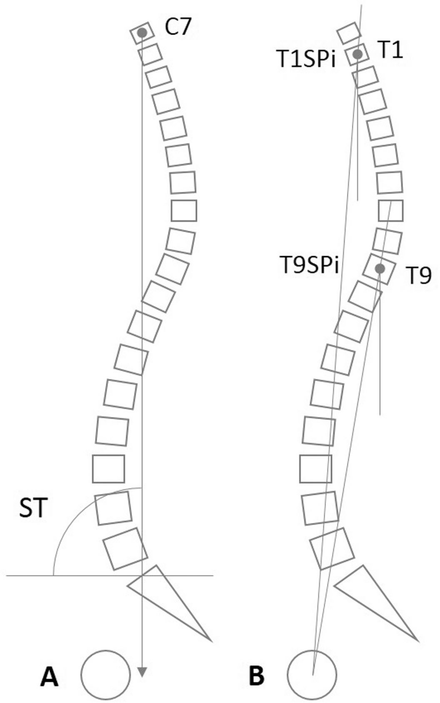 | Fig. 2.The spinal tilt (ST) (A) and the T1 (T9) spino-pelvic inclination (T1SPi) (T9SPi) (B). |
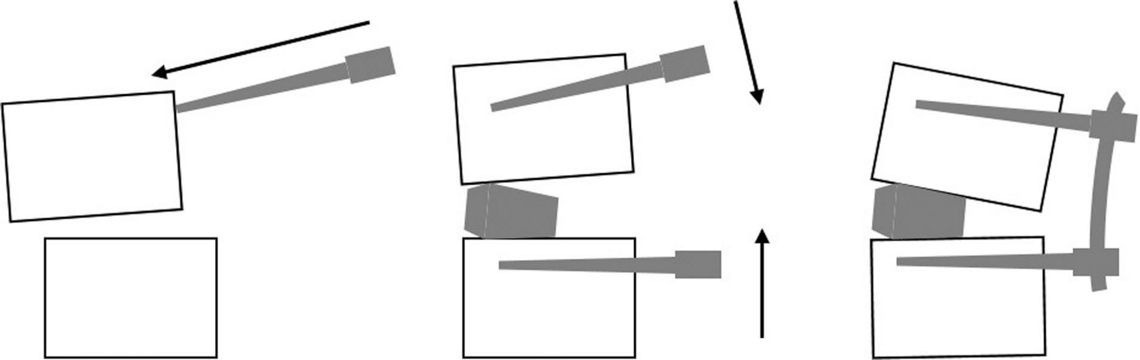
 | Fig. 3.The sagittal vertical axis can be greatly affected by a forward bending (A), neutral standing (B), or backward bending (C) body posture compensating for the posture of the lower extremity joints in patients with long spinal fusion, because the compensation mechanism of the spinal segments has been lost by long segment fusion.6)
|
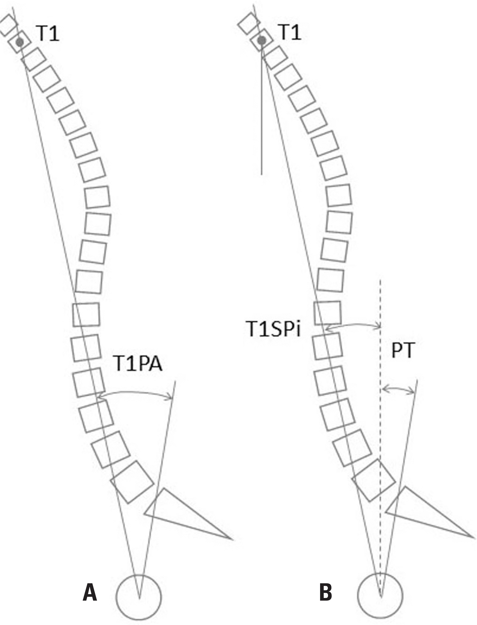 | Fig. 4.The T1 pelvic angle (T1PA) (A) is the morphologic parameter for global spinal deformity that equals T1SPi + PT (B). |
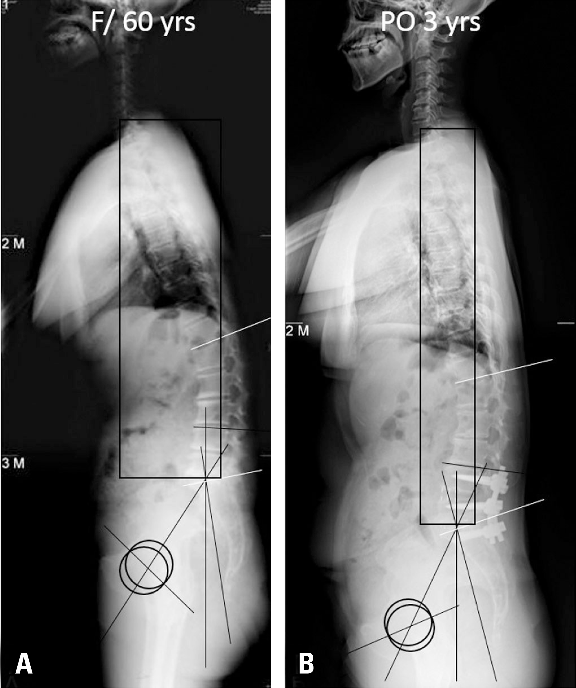 | Fig. 5.Most of the spino-pelvic parameters improved following selective fusion of the lower lumbar region in patients with lumbar degenerative kyphosis. From preoperative (A) to postoperative (B) views: PI-LL: 51° to 33°, SVA: 78 to 35, PT: 33 to 25, lower LL (L4-S1): -9 to -26. |
 XML Download