

 PDF
PDF Citation
 Print
Print


Abstract
Objectives
To report a case of video-assisted thoracoscopic (VAT) minimally invasive anterior interbody fusion of the T11-T12 level using direct lateral interbody fusion (DLIF) devices.
Summary of Literature Review
Interbody fusion of the thoracolumbar junction (especially T11-T12) is technically challenging from anterior, lateral, or posterior approaches. A VAT anterior interbody fusion approach using DLIF devices is a safe, minimally invasive alternative approach to the thoracolumbar spine.
Materials and Methods
A 37-year-old male pedestrian was struck by a car sustaining fracture-dislocation at the T11-T12 level. The accident resulted in complete paraplegia of both lower extremities and multiple lower extremity fractures. A classical instrumented posterolateral fusion from T8 to L3 and staged VAT anterior interbody fusion at the T11-T12 level were performed.
Go to : 
REFERENCES
1. Lee CG, Choi JS, Kim YC, et al. Survival Analysis of Posterior Short Fusion in Thoracolumbar Fracture-Significance of Load-Sharing Score and Bone Mineral Density. J Korean Soc Spine Surg. 2001; 8:113–20.
2. Wang X-B, Yang M, Li J, et al. Thoracolumbar fracture dislocations treated by posterior reduction, interbody fusion and segmental instrumentation. Indian J Orthop. 2014; 48:568–73.

3. Wood KB, Li W, Lebl DS, et al. Management of thoracolumbar spine fractures. Spine J. 2014; 14:145–64.
4. Xia Q, Xu Bs, Zhang Jd, et al. Simultaneous combined anterior and posterior surgery for severe thoracolumbar fracture dislocations. Orthop Surg. 2009; 1:28–33.
5. Machino M, Yukawa Y, Ito K, et al. Posterior/anterior combined surgery for thoracolumbar burst fractures— posterior instrumentation with pedicle screws and laminar hooks, anterior decompression and strut grafting. Spinal cord. 2011; 49:573–9.
6. Yu S-W, Fang K-F, Tseng I-C, et al. Surgical Outcomes of Short-Segment Fixation for Thoracolumbar Fracture Dis-location. Chang Gung med J. 2002; 25:253–9.
7. Rosenthal D. Endoscopic approaches to the thoracic spine. Eur Spine J. 2000; 9(Suppl):8–16.
8. Karikari IO, Nimjee SM, Hardin CA, et al. Extreme lateral interbody fusion approach for isolated thoracic and thoracolumbar spine diseases: initial clinical experience and early outcomes. J Spinal Disord Tech. 2011; 24:368–75.
9. Meredith DS, Kepler CK, Huang RC, et al. Extreme lateral interbody fusion (XLIF) in the thoracic and thoracolumbar spine: technical report and early outcomes. HSS J. 2013; 9:25–31.
10. Levin R, Matusz D, Hasharoni A, et al. Mini-open tho-racoscopically assisted thoracotomy versus video-assisted thoracoscopic surgery for anterior release in thoracic scoliosis and kyphosis: a comparison of operative and radiographic results. Spine J. 2005; 5:632–8.
Go to :
 | Fig. 1.The plain radiographs of 37-year-old male pedestrian, who was struck by a car, show fracture-dislocation injury at the T11-T12 level (A, B). The accident resulted in complete paraplegia of both lower extremities. |
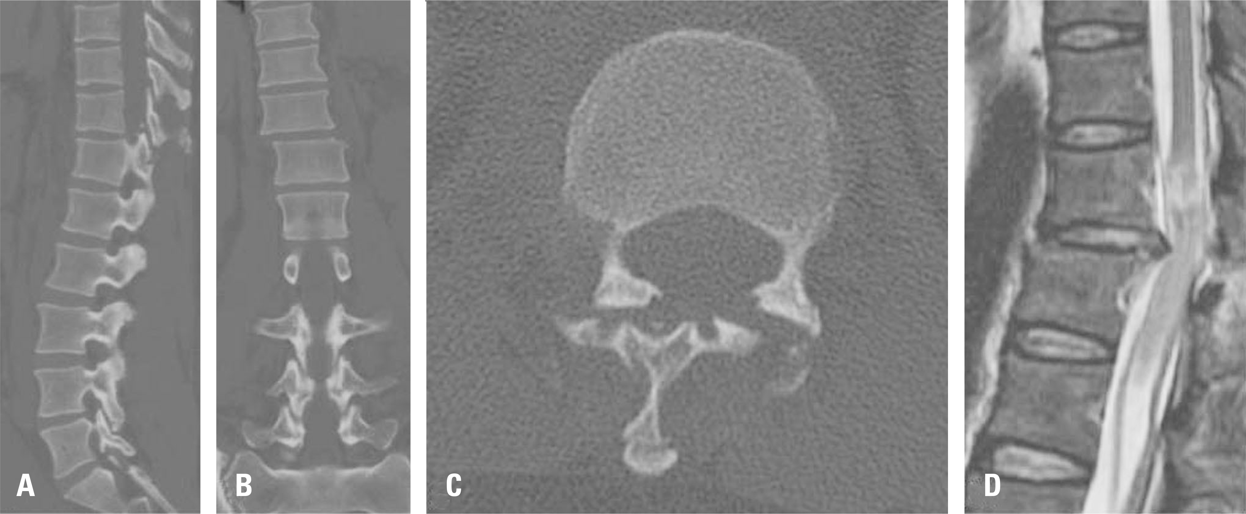 | Fig. 2.Sagittal (A), coronal (B), and axial (C) computed tomography scans and magnetic resonance imaging (D) show fracture-dislocation injury at the T11-T12 level. A transection cord injury at the T11 body level, lateral displacement with intervertebral subluxation, and fracture at both superior articular processes of the T12 vertebra occurred. |
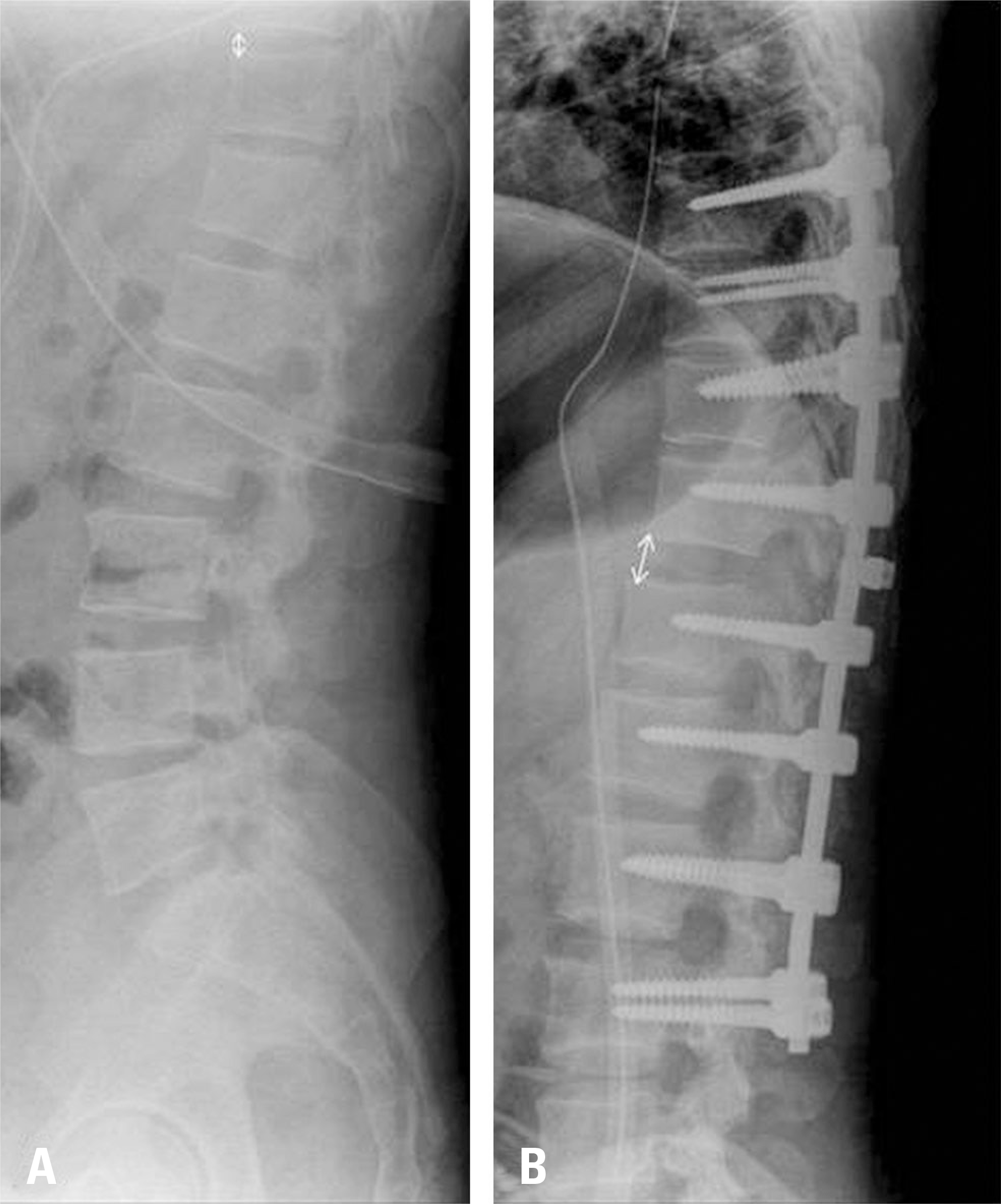 | Fig. 3.Lateral radiographs of the surgical site preoperatively (A) and after posterior operation (B) are shown. After the posterior operation, the T11-T12 interbody space was widened from 5 mm to 12 mm and facet joint subluxation still existed. |
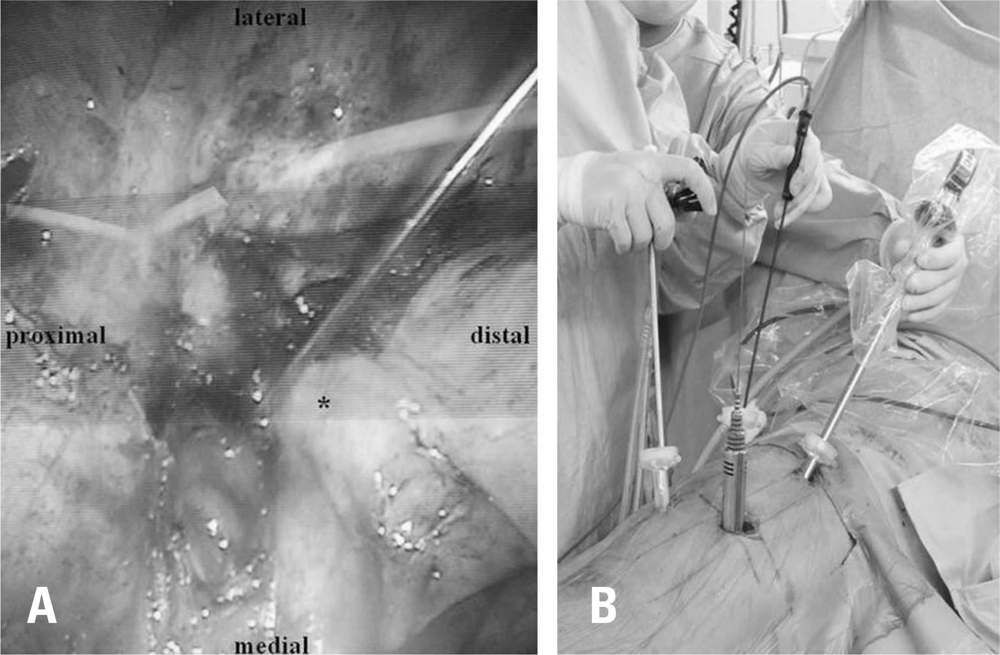 | Fig. 4.A thoracoscopic image shows the T11-T12 level (A). The dia-phragm (asterisk) was detached from the chest wall to expose the T11-T12 interbody disc space. A guide pin was inserted at the T11-T12 interbody disc space. An intraoperative image is shown (B). Serial dila-tors were inserted directly lateral to the T11-T12 interbody disc space. A viewing camera and instruments for thoracoscopy were inserted via trocars. |

 XML Download