

 PDF
PDF ePub
ePub Citation
 Print
Print


Abstract
Objectives
To present updated information on the relationship of the pelvis and lumbar degenerative disease (LDD) patients and to emphasize the importance of the pelvis in sagittal alignment of LDD patients.
Summary of Literature Review
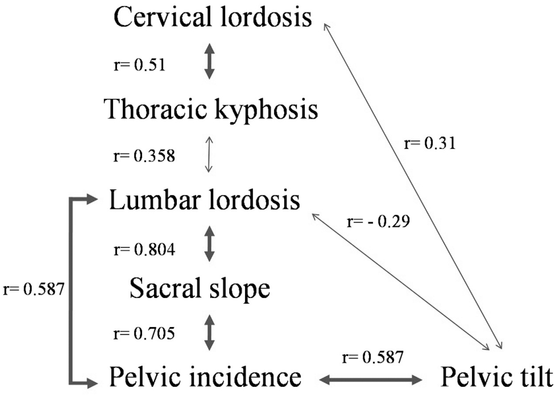
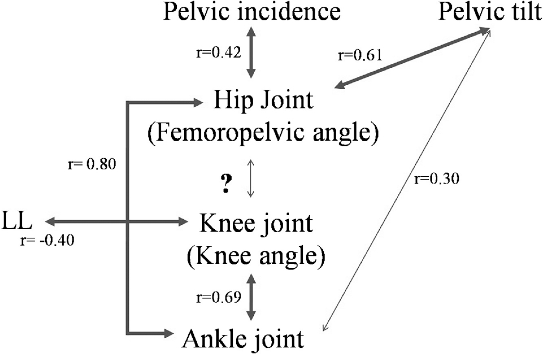
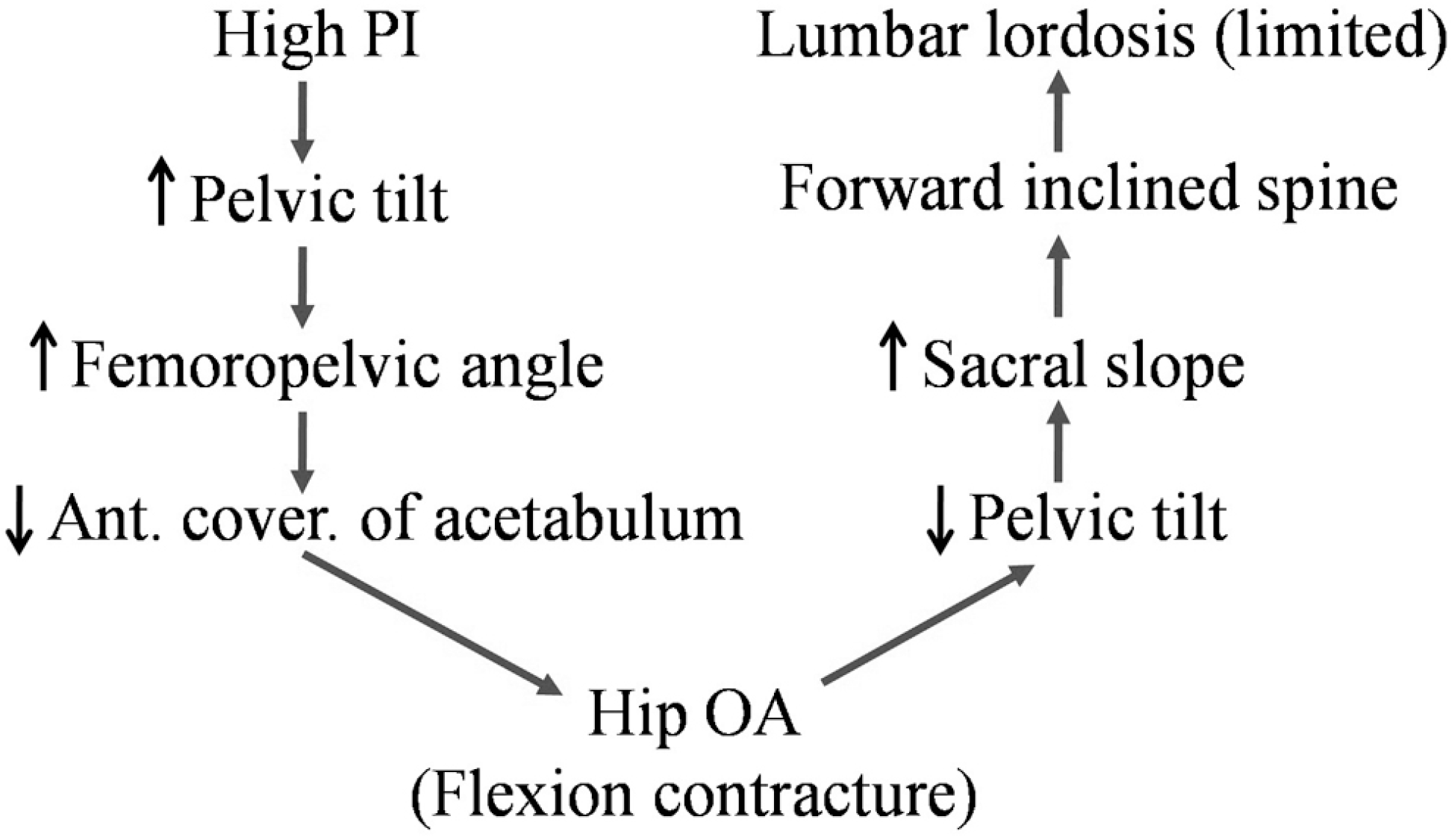
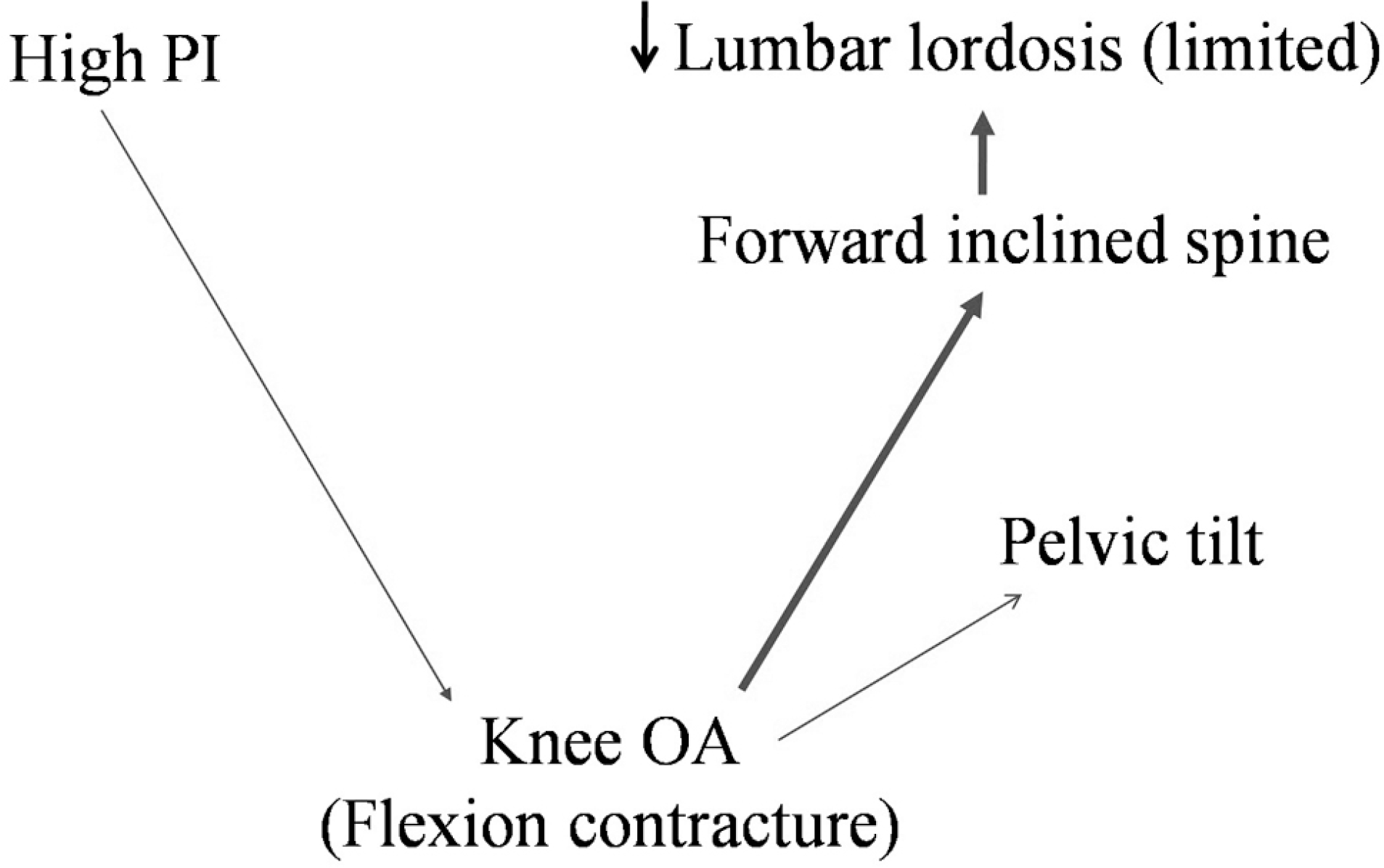
Although the relationship of the pelvis and sagittal alignment of LDD patients is controversial, many authors have reported a significant impact of the pelvis on LDD sagittal alignment.
Materials and Methods
The authors identified references through a literature search on the pelvis and LDD and continuous monitoring of the literature during the past 30 years.
REFERENCES
1. Lee CS, Chung SS, Kang KC, et al. Normal Patterns of Sagittal Alignment of the Spine in Young Adults Radiological Analysis in a Korean Population. SPINE(Phila Pa 1976). 2001; 36:E1648–54.

2. Barrey C, Jund J, Noseda O, et al. Sagittal balance of the pelvis-spine complex and lumbar degenerative dis-eases. A comparative study about 85 cases. Eur Spine J. 2007; 16:1459–67.
3. Le Huec JC, Aunoble S, Philippe L, et al. Pelvic parameters: origin and significance. Eur Spine J. 2011; 20(Suppl):564–71.
4. Suk SI. Textbook of Spinal Surgery Third Edition. Korea. Newest Medical Publishing Company;2013. p. 797–801.
5. Guillot M, Eournier J. Methods of measuring the mechanical stresses on the human lumbar spine and their results. Rev Rhum Mal Osteoartic. 1988; 55:351–9.
6. Swanson RL. Biotensigrity: a unifying theory of biological architecture with applications to osteopathic practice, edu-cation, and research. J Am Osteopath Assoc. 2013; 113:34–52.
7. Pierre Roussouly, Pinheiro-Franco JL. Sagittal parameters of the spine: biomechanical approach. Eur Spine J. 2011; 20(Suppl):578–85.
8. Lee CS, Chung SS, Park SJ, et al. The Association of Lumbosacral Sagittal Alignments and the Patterns of Lumbar Disc Degeneration. J Korean Soc Spine Surg. 2012; 19:145–51.
9. Bao H, He S. �ill Immediate Postoperative Imbalance Im-prove in Patients �ith ThoracolumbarLumbar Degenerative Kyphoscoliosis. Spine(Phila Pa 1976). 2015; 40:E293–300.
10. Boulay C, Tardieu C, Hecquet J, et al. Sagittal alignment of spine and pelvis regulated by pelvic incidence: standard val-ues and prediction of lordosis. Eur Spine J. 2006; 15:415–22.
11. Chaleat-Valayer E, Mac-Thiong JM. Sagittal spino-pelvic alignment in chronic low back pain. Eur Spine J. 2011; 5:634–40.
12. Faldini C, Traina F, Perna F, et al. Does surgery for Scheuermann kyphosis influence sagittal spinopelvic pa-rameters? Eur Spine J. 2015 Oct 6. [Epub ahead of print].
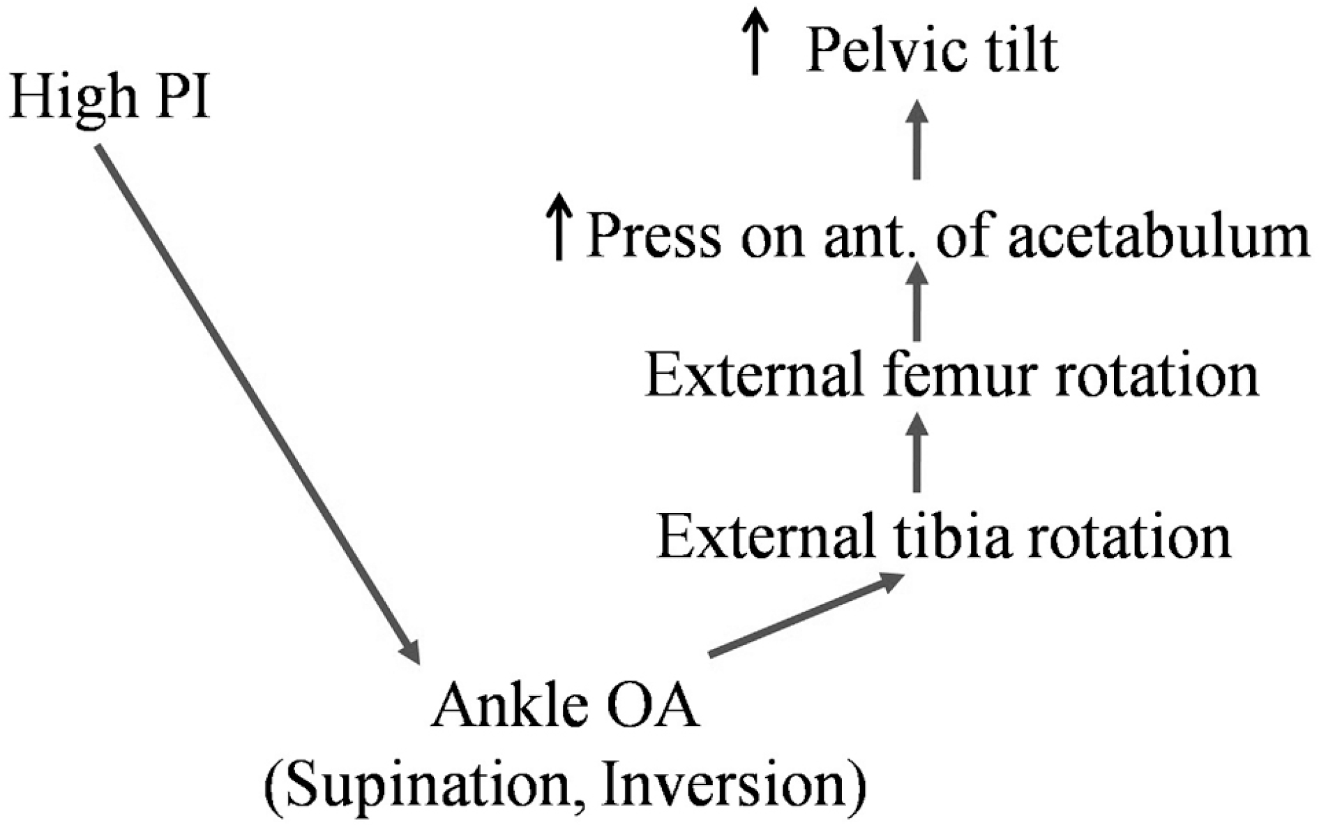
13. Duval K, Lam T, Sanderson D. The mechanical relationship between the rearfoot, pelvis and low-back. Gait Posture. 2010; 32:637–40.
14. Yoshimoto H, Sato S. Spinopelvic alignment in patients with osteoarthrosis of the hip: a radiographic comparison to patients with low back pain. Spine(Phila Pa 1976). 2005; 30:1650–7.
15. Lee CS, Park SJ, Chung SS, et al. The effect of simulated knee flexion on sagittal spinal alignment: novel interpretation of spinopelvic alignment. Eur Spine J. 2013; 22:1059–65.
16. Betsch M, Schneppendahl J, Dor L, et al. Influence of foot positions on the spine and pelvis. Arthritis Care Res. 2011; 63:1758–65.
17. Pinto R, Souza T, Trede R, et al. Bilateral and unilateral increases in calcaneal eversion affect pelvic alignment in standing position. Man Ther. 2008; 13:513–9.
18. Kumar MN, Baklanov A, Chopin D. Correlation between sagittal plane changes and adjacent segment degeneration following lumbar spine fusion. Eur Spine J. 2001; 10:314–9.
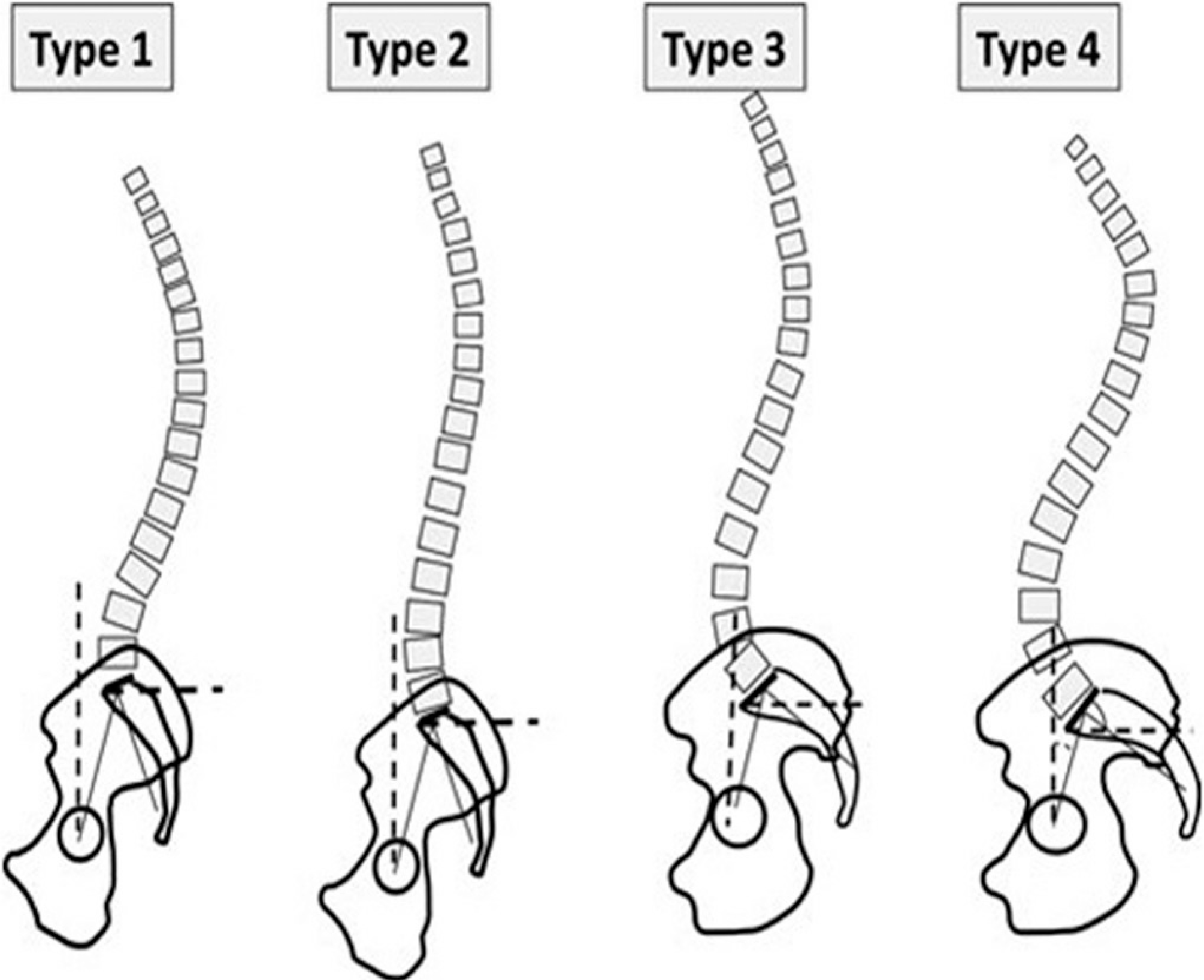
19. Roussouly P, Gollogly S, Berthonnaud E, et al. Classification of the normal variation in the sagittal alignment of the human lumbar spine and pelvis in the standing position. Spine (Phila Pa 1976). 2005; 30:346–53.
20. Schwab F, �ngar B, Blondel B, et al. Scoliosis research society-Schwab adult spinal deformity classification: a validation study. Spine(Phila Pa 1976). 2012; 37:1077–82.
21. Boissiere L, Vital JM, Aunoble S, et al. Lumbo-pelvic related indexes: impact on adult spinal deformity surgery. Eur Spine J. 2015; 24:1212–8.
22. Lafage V, Schwab F. Pelvic tilt and truncal inclination: two key radiographic parameters in the setting of adult with spinal deformity. Spine(Phila Pa 1976). 2009; 34:599–606.
23. Boissiere L, Bourghli A, Vita JMl, et al. The lumbar lordosis index: a new ratio to detect spinal malalignmnet with a therapeutic impact for sagittal balance correction decisions in adult scoliosis surgery. Eur Spine J. 2013; 22:1339–45.
24. During J, Goudfrooii H. Toward standards for posture. Postural characteristics of the lower back system in normal and parhologic conditions. Spine (Phila Pa 1976). 1985; 10:83–7.
25. Jackson RP, Hales C. Congruent spinopelvic alignment on standing lateral radiographs of adult volunteers. Spine (Phila Pa 1976). 2000; 25:2808–15.
26. Gnat R, Saulicz E. Induced static asymmetry of the pelvis is associated with functional asymmetry of the Lumbo-Pelvic-Hip complex. J Manipulative Physiol Ther. 2008; 31:204–11.
27. James CR, Lee T. Atkins. Kinematic and ground reaction force accommodation during weighted walking. Hum Mov Sci. 2015; 44:327–37.
 XML Download