Abstract
Summary of Literature Review
IDH is rare but there is a higher incidence of neurologic deficit in IDH. Therefore, it should be treated immediately.
REFERENCES
1. Epstein NE, Syrquin MS, Epstein JA, et al. Intradural disc herniations in the cervical, thoracic and lumbar spine: report of three cases and review of the literature. J Spinal Disord. 1990; 3:396–403.
2. D'Andrea G, Trillo G, Roperto R, et al. Intradural lumbar disc herniations: the role of MRI in preoperative diagnosis and review of the literature. Neurosurg Rev. 2004; 27:75–80.
3. Yildizhan A, Pasaoglu A, Okten T, et al. Intradural disc herniations: pathogenesis, clinical picture, diagnosis and treatment. Acta Neurochir. 1991; 110:160–5.

4. Ducati LG, Silva MV, Brandã o MM, et al. Intradural disc herniation: report of five cases with literature review. Eur Spine J. 2013; 22(Suppl):404–8.
5. Kataoka O, Nishibayashi Y, Sho T. Intradural lumbar disc herniation: report of three cases with a review of the literature. Spine. 1989; 14:529–33.
6. Kim KC, Choi JY, Kim JS, et al. Intradural lumbar disc herniation – A case report -. J Korean Soc Spine Surg. 1996; 3:274–9.
7. Borm W, Bohnstedt T. Intradural cervical disc herniation: case report and review of the literature. J Neurosurg Spine. 2000; 92:221–4.
8. Hida K, Iwasaki Y, Abe H, Shimazaki M, et al. Magnetic resonance imaging of intradural lumbar disc herniation. J Clin Neurosci. 1999; 6:345–7.

9. Aydin MV, Ozel S, Sen O, et al. Intradural disc mimicking a spinal tumor lesion. Spinal Cord. 2004; 42:52–4.

10. Agarwal N, Shah J, Hansberry DR, et al. Mammis A, Sharer LR, Goldstein IM. Presentation of cauda equina syndrome due to an intradural extramedullary abscess: a case report. Spine J. 2014; 14:E1–6.
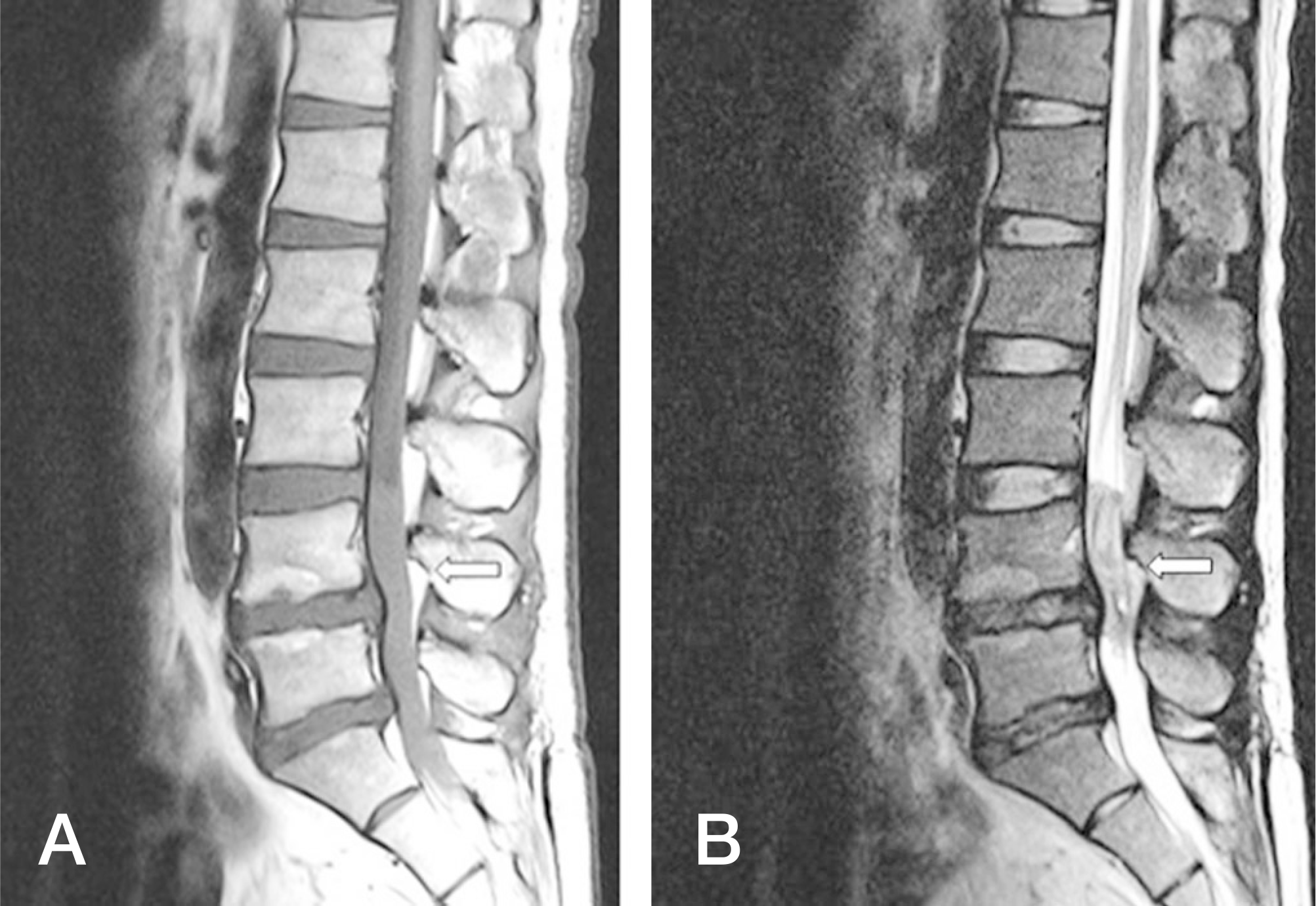
Fig. 1.
(A) The arrow indicates a huge isointense mass-like lesion located in the intradural space from the L4 to L5 level on a T1-weighted sagittal magnetic resonance (MR) image. (B) The arrow indicates a heteroge-neous isointense lesion in a T2-weighted sagittal MR image.


 PDF
PDF ePub
ePub Citation
 Print
Print


 XML Download