

 PDF
PDF ePub
ePub Citation
 Print
Print


Abstract
Summary of Literature Review
The intradural epidermoid cyst with extensive involvement is rare, and previous reports have reported only extensive intramedullary epidermoid cysts.
Materials and Methods
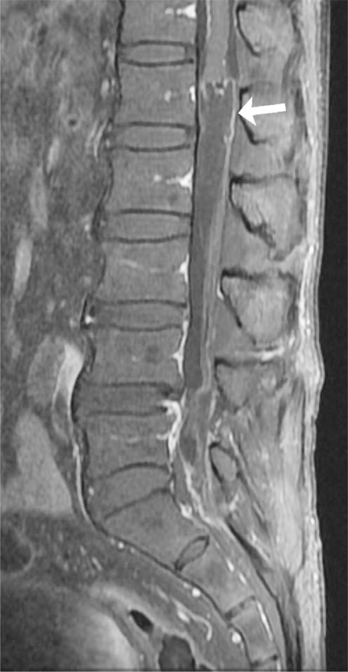
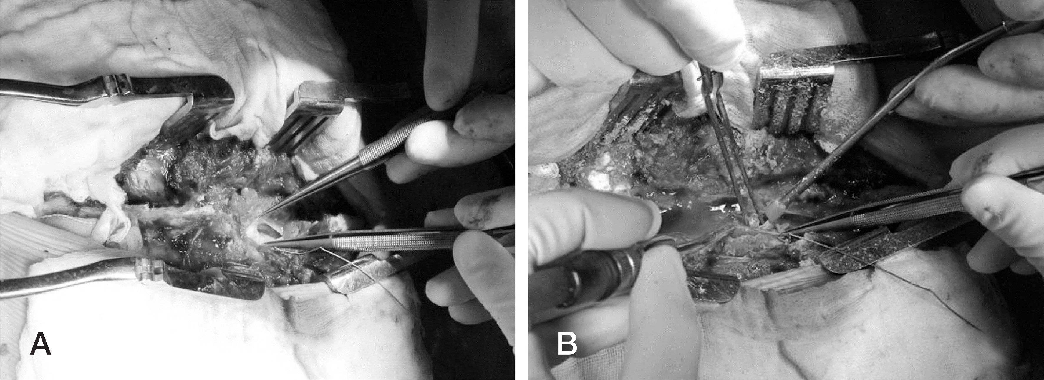
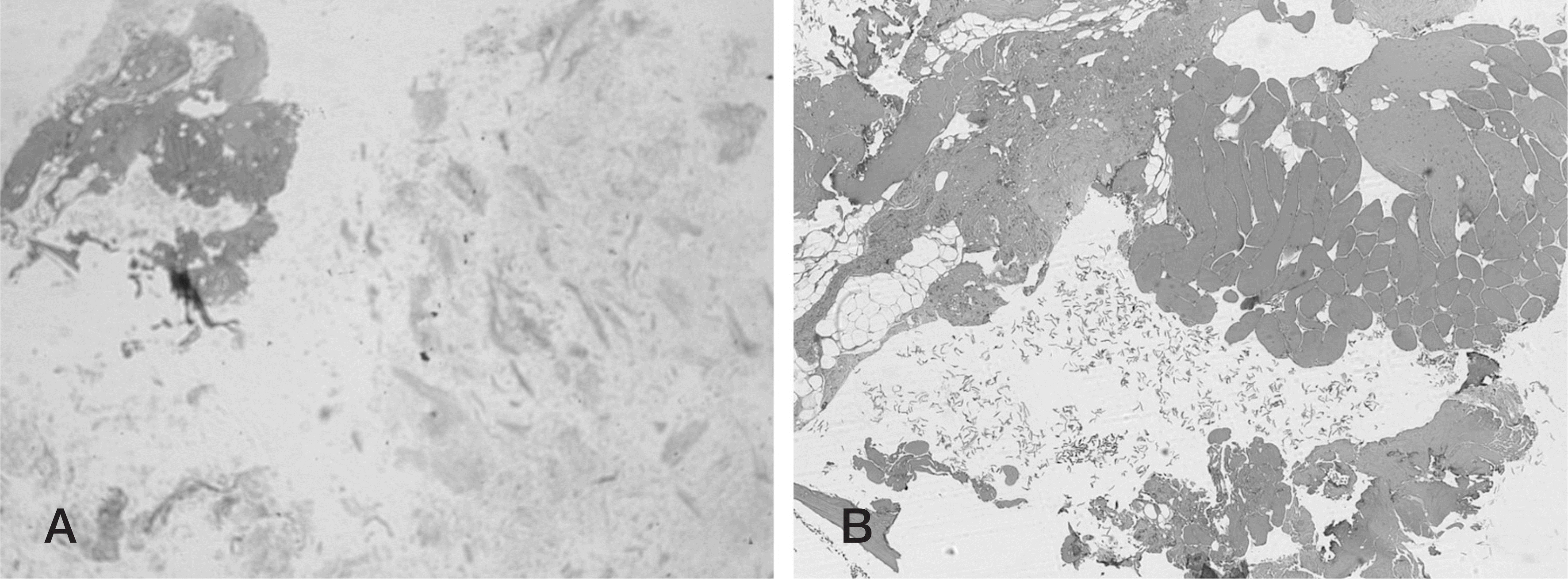
A 75-year-old male presented with progressive motor weakness of both extremities beginning 3 days prior. MRI showed extensive intradural extramedullary epidermoid cysts in the lumbosacral region. We performed total laminectomy from the L1 to the L5 level, and the cystic mass was removed.
REFERENCES
1. Teksam M, Casey SO, Michel E, et al. Intraspinalepi-dermoid cyst: diffusion weighted MRI. Neuroradiology. 2001; 43:572–4.
2. Gardner DJ, O'Gorman AM, Blundell JE. Intraspinal epi-dermoid tumour: Late complication of lumbar puncture. CMAJ. 1989; 141:223–5.
3. Cruveilhier J. Anatomic Pathologique du Corps Humain: Vol 1. Paris: Bailli6re, 1829, Book 2, Plate 6.
4. Gonzalvo A, Hall N, McMahon JH, et al. Intramedullary spinal epidermoid cyst of the upper thoracic region. J Clin Neurosci. 2009; 16:142–4.

5. Ozaras N, Sariyildiz M, Demir S, et al. A neglected case admitted with paraplegia: An intradural extramedullary epidermoid cyst. J ClinExp Invest. 2012; 3:270.
6. Min Ho P, Tack Geun C, Jae Gon M, et al. Iatrogenic Intraspinal Epidermoid Cyst. Korean J Spine. 2014; 11:195–7.
7. Sarang G, Deepak R, Shrikant S, et al. Giantintradural in-tramedullary epidermoid cyst Report of two cases with var-ied presentations. Asian J Neurosurg. 2014; 9:244.
8. Roux A, Mercier C, Larbrisseau A, et al. Intramedullary epidermoid cysts of the spinal cord: case report. J Neurosurg. 1992; 76:528–33.
9. Alves AM, Norrell H. Intramedullary epidermoid tumors of the spinal cord. Report of a case and review of the literature. Int Surg. 1970; 54:239–43.
10. Jee-Soo J, Sang-Ho L. Multiple Intramedullary and In-tradural Epidermoid Cystsin the Conus Medullaris and the Lumbar Spine. J Korean Neurosurg Soc. 2003; 33:512–3.
11. Haber MD, Nguyen DD, Li S. Differentiation of Id-iopathic Spinal Cord Herniation from CSF-isointense IntraspinalExtramedullary Lesions Displacing the Cord. RadioGraphics. 2014; 34:313–9.


 XML Download