

 PDF
PDF ePub
ePub Citation
 Print
Print


Abstract
Objectives
The purpose of this study was to evaluate risk factors for subsidence after posterior lumbar interbody fusion (PLIF).
Summary of Literature Review
Body mass index (BMI), bone mineral density (BMD), cage characteristics (titanium or poly-ether-ether-ketone (PEEK)) and degree of disc distraction are risk factors for cage subsidence after PLIF.
Materials and Methods
From January 2010 to January 2015, a total of 69 patients (93 segments) who were diagnosed with degenerative lumbar spine disease at the current authors’ institution and underwent follow-up for at least 1 year were included in this retrospective study. Data on all factors related to cage subsidence were taken into consideration. The degree of association for each of the factors was determined through the calculation of odds ratios (ORs) with a 95% confidence interval. Logistic regression analyses were performed. The P-value that represented statistical significance was set below 0.05.
Results
There were no significant associations between fused segment level and cage subsidence (p=0.588), nor were there any significant associations between the kind of cage (titanium or PEEK) and cage subsidence (p=0.371). Using logistic regression, the factors with a P-value below the 0.20 level in univariate analyses were included in the multivariate analyses. In multivariate analyses, diabetes mellitus (DM) (p=0.029, OR, 4.524), osteoporosis (p=0.026, OR, 6.046), and degree of disc distraction (p=0.010, OR, 1.446) had significant associations with cage subsidence. In addition, there were significant associations between cage subsidence and instrument failure (p=0.008, OR, 8.235).
Go to : 
REFERENCES
1. Oh KW, Lee JH, Lee JH, et al. The Correlation Between Cage Subsidence, Bone Mineral Density, and Clinical Results in Posterior Lumbar Interbody Fusion. J Spinal Disord Tech. 2015 Aug 18. [Epub ahead of print].

2. Park Y, Ha JW. Comparison of one-level posterior lumbar interbody fusion performed with a minimally invasive approach or a traditional open approach. Spine (Phila Pa 1976). 2007; 32:537–43.
3. Steffee AD, Sitkowski DJ. Posterior lumbar interbody fusion and plates. Clin Orthop Relat Res. 1988; 227:99–102.
4. McKenna PJ, Freeman BJ, Mulholland RC, et al. A prospective, randomised controlled trial of femoral ring allograft versus a titanium cage in circumferential lumbar spinal fusion with minimum 2-year clinical results. Eur Spine J. 2005; 14:727–37.
5. Schmieder K, Wolzik-Grossmann M, Pechlivanis I, et al. Subsidence of the wing titanium cage after anterior cervical interbody fusion: 2-year follow-up study. J Neurosurg Spine. 2006; 4:447–53.
6. Cabraja M, Abbushi A, Kroppenstedt S, et al. Cages with fixation wings versus cages plus plating for cervical reconstruction after corpectomy - is there any difference? Cent Eur Neurosurg. 2010; 71:59–63.
7. Cutler AR, Siddiqui S, Mohan AL, et al. Comparison of polyetheretherketone cages with femoral cortical bone allograft as a single-piece interbody spacer in transforaminal lumbar interbody fusion. J Neurosurg Spine. 2006; 5:534–9.
8. Chen Y, Wang X, Lu X, et al. Comparison of titanium and polyetheretherketone (PEEK) cages in the surgical treatment of multilevel cervical spondylotic myelopathy: a prospective, randomized, control study with over 7-year follow-up. Eur Spine J. 2013; 22:1539–46.
9. Barsa P, Suchomel P. Factors affecting sagittal malalignment due to cage subsidence in standalone cage assisted anterior cervical fusion. Eur Spine J. 2007; 16:1395–400.
10. Meier U, Kemmesies D. [Experiences with six different intervertebral disc spacers for spondylodesis of the cervical spine]. Orthopade. 2004; 33:1290–9.
11. Pechlivanis I, Thuring T, Brenke C, et al. Non-fusion rates in anterior cervical discectomy and implantation of empty polyetheretherketone cages. Spine (Phila Pa 1976). 2011; 36:15–20.
12. Steffen T, Tsantrizos A, Fruth I, et al. Cages: designs and concepts. Eur Spine J. 2000; 9(Suppl):89–94.
13. Kim MC, Chung HT, Cho JL, et al. Subsidence of poly-etheretherketone cage after minimally invasive transforaminal lumbar interbody fusion. J Spinal Disord Tech. 2013; 26:87–92.
14. Sasso RC, Kitchel SH, Dawson EG. A prospective, ran-domized controlled clinical trial of anterior lumbar interbody fusion using a titanium cylindrical threaded fusion device. Spine (Phila Pa 1976). 2004; 29:113–22.
15. Schiffman M, Brau SA, Henderson R, et al. Bilateral implantation of low-profile interbody fusion cages: subsidence, lordosis, and fusion analysis. Spine J. 2003; 3:377–87.
16. Kao TH, Wu CH, Chou YC, et al. Risk factors for subsidence in anterior cervical fusion with standalone poly-etheretherketone (PEEK) cages: a review of 82 cases and 182 levels. Arch Orthop Trauma Surg. 2014; 134:1343–51.
17. Borm W, Seitz K. Use of cervical standalone cages. Eur Spine J. 2004; 13:474–5.
18. Wang HR, Li XL, Dong J, et al. Skip-level anterior cervical discectomy and fusion with self-locking standalone PEEK cages for the treatment of 2 noncontiguous levels of cervical spondylosis. J Spinal Disord Tech. 2013; 26:286–92.
19. Shanbhogue VV, Mitchell DM, Rosen CJ, et al. Type 2 diabetes and the skeleton: new insights into sweet bones. Lancet Diabetes Endocrinol. 2016; 4:159–73.
20. Leslie WD, Rubin MR, Schwartz AV, et al. Type 2 diabetes and bone. J Bone Miner Res. 2012; 27:2231–7.
21. Schwartz AV, Sellmeyer DE, Ensrud KE, et al. Older women with diabetes have an increased risk of fracture: a prospective study. J Clin Endocrinol Metab. 2001; 86:32–8.
22. Halvorson TL, Kelley LA, Thomas KA, et al. Effects of bone mineral density on pedicle screw fixation. Spine (Phila Pa 1976). 1994; 19:2415–20.
23. Jost B, Cripton PA, Lund T, et al. Compressive strength of interbody cages in the lumbar spine: the effect of cage shape, posterior instrumentation and bone density. Eur Spine J. 1998; 7:132–41.
24. Polikeit A, Ferguson SJ, Nolte LP, et al. Factors influencing stresses in the lumbar spine after the insertion of intervertebral cages: finite element analysis. Eur Spine J. 2003; 12:413–20.
25. Tokuhashi Y, Ajiro Y, Umezawa N. Subsidence of metal interbody cage after posterior lumbar interbody fusion with pedicle screw fixation. Orthopedics. 2010; 34:226–7.
26. Kaito T, Hosono N, Fuji T, et al. Disc space distraction is a potent risk factor for adjacent disc disease after PLIF. Arch Orthop Trauma Surg. 2011; 131:1499–507.
27. Yang JJ, Yu CH, Chang BS, et al. Subsidence and non-union after anterior cervical interbody fusion using a standalone polyetheretherketone (PEEK) cage. Clin Orthop Surg. 2011; 3:16–23.
28. Francke EI, Demetropoulos CK, Agabegi SS, et al. Distractive force relative to initial graft compression in an in vivo anterior cervical discectomy and fusion model. Spine (Phila Pa 1976). 2010; 35:526–30.
29. Cabraja M, Oezdemir S, Koeppen D, et al. Anterior cervical discectomy and fusion: comparison of titanium and polyetheretherketone cages. BMC Musculoskelet Disord. 2012; 13:172.
Go to :
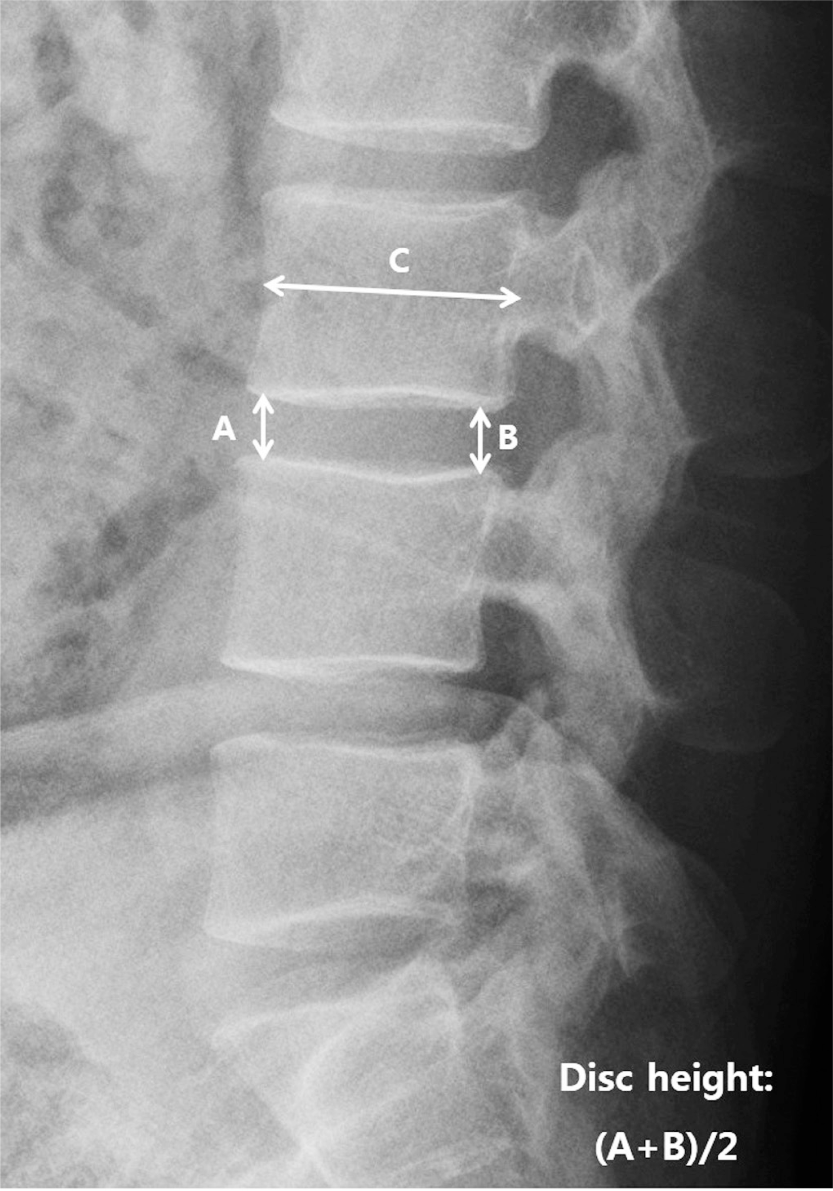 | Fig. 1.Measurement of preoperative and postoperative disc height in the lumbar lateral view (A: anterior disc height, B: posterior disc height, C: sagittal diameter of the vertebral body measured between the midpoints of the anterior and posterior surfaces). |
Table 1.
Demographic and clinical data of the patients (N=93 segments)
Table 2.
Ca ge subsidence followin ng the inserted level
| Level | No subsidence | Subsidence | p-value |
|---|---|---|---|
| L2-3 | 1 (1.1%) | 2 (2.2%) | 0.091 |
| L3-4 | 16 (17.2%) | 3 (3.2%) | 0.384 |
| L4-5 | 44 (47.3%) | 9 (9.7%) | 0.124 |
| L5-S1 | 11 (11.8%) | 7 (7.5%) | 0.061 |
Table 3.
Subsidence and clinical outcomes
| No subsidence | Subsidence | p-value | |
|---|---|---|---|
| VAS | 2.9±1.1 | 2.7±1.1 | 0.641 |
| ODI | 14.4±4.7 | 13.7±3.9 | 0.520 |
Table 4.
Strengths of associations between cage subsidence and various factors in the univariate and multivariate analyses
 XML Download