

 PDF
PDF ePub
ePub Citation
 Print
Print


Abstract
Objectives
To investiggaate radiologic aannd clinicaall outcomes of teripa.ratide in women with osteoporosis afafer instrume.nted lu.mbambar posterolateral fusion (PLF) or posterior lumbar interbody fusion (PLIF).
Summary of Literature Review
Summary of Literature Review: Teriparatide accelerated lumbar posterolateral fusion in women with postmenopausal osteoporosis. Materials and Methods: Eigh ty-six women older than 65 years old with osteoporosis underwent PLF or PLIF with bone graf t between Februar, 2011 ato May, 2012 pPatients were divided into four group: teriparatide group with local bone (A-1: 13 patients;, teriparatide group with composite bone (A-2: 27 patients; non-teriparatide group with local bone (B-1: 14 patients; and non-teriparatide group with composite bone (B-2: 32 patients). At 3, 6, and 12 months postoperatively, the Oswestry Disability Index (ODI), visual analog scale (VAS), fusion rate, and period of bone union were evaluated.
Results
VAS and ODI improved after surgery in all groups, but no significant differences were notell among the groupses Further, there was no sign ifican t diff erence am ong the groups for age fusion level, and fusiops(p>0.05). Fusion rate was 94.44% in the A-1 group, 92.59% in the A-2 group, 79.17% in the B-1 group, and 76.92% in the B-2 group. Average period of bone union was 3.25 months, 3.65 months, 5.67 months, and 5.65 months respectively. Fusion rate and averag e bone union time ma de no sign ifican t diff ereneen am ong the groups divided by graft materials (p>0.05). However, those in the teriparatide group were significantly superior to those in the nonteriparatide group (p<0.05).
REFERENCES
1. Cohen DB, Chotivichit A, Fujita T, et al. Pseudoarthrosis repair. autogenous iliac crest versus femoral ring allograft. Clin Orthop Relat Res. 2000; 371:46–55.
2. Fischgrund JS, Mackay M, Herkowitz HN, et al. 1997 Volvo Award winner in clinical studies. Degenerative lumbar spondylolisthesis with spinal stenosis: a prospective, randomized study comparing decompressive laminectomy and arthrodesis with and without spinal instrumentation. Spine (Phila Pa 1976). 1997; 22:2807–12.
3. Weinstein MA, McCabe JP, Cammisa FP Jr. Postoperative spinal wound infection: a review of 2,391 consecutive index procedures. J Spinal Disord. 2000; 13:422–6.

4. Rawlins BA, Michesen CB. Failed lumbosacral fusions: state of the art review. Spine (Phila Pa 1976). 1994; 8:563–71.
5. Miyazaki M, Tsumura H, Wang JC, et al. An update on bone substitutes for spinal fusion. Eur Spine J. 2009; 18:783–99.
6. Aspenberg P, Genant HK, Johansson T, et al. Teriparatide for acceleration of fracture repair in humans: a prospective, randomized, double-blind study of 102 postmenopausal women with distal radial fractures. J Bone Miner Res. 2010; 25:404–14.
7. Lindsay R, Nieves J, Formica C, et al. Randomized controlled study of effect of parathyroid hormone on vertebral bone mass and fracture incidence among postmenopausal women on estrogen with osteoporosis. Lancet. 1997; 350:550–5.
8. Alkhiary YM, Gerstenfeld LC, Krall E, et al. Enhancement of experimental fracture-healing by systemic administration of recombinant human parathyroid hormone (PTH 1-34). J Bone Joint Surg. Am. 2005; 87:731–41.
9. Aspenberg P, Johansson T. Teriparatide improves early callus formation in distal radial fractures. Acta Orthop. 2000; 81:234–6.
10. Seiji Ohtori, Gen Inoue, Sumihisa Orita, et al. Teriparatide accelerates lumbar posterolateral fusion in women with postmenopausal osteoporosis. Spine (Phila Pa 1976). 2012; 37:E1464–8.
11. Tokuhashi Y, Matsuzaki H, Oda H, et al. Clinical course and significance of the clear zone around the pedicle screws in the lumbar degenerative disease. Spine (Phila Pa 1976). 2008; 8:903–8.
12. Tokuhashi Y, Nishimura T, Matsuzaki Y. Clinical results of more than 10 years after posterolateral fusion with pedicle screw fixation for degenerative lumbar spondylolisthesis. Spine (Phila Pa 1976). 2004; 17:185–92.
13. Tan JS, Singh S, Zhu QA, et al. The effect of cement aug-mentation and extension of posterior instrumentation on stabilization and adjacent level effects in the elderly spine. Spine (Phila Pa 1976). 2008; 33:2728–40.
14. Cheong US, Kim DY, Cho JL, et al. Comparison of the effect of hydroxyapatite and allogenous bone as an adjunct to autogenous iliac bone grafting in posterolateral spinal fusion. J Korean Orthop Assoc. 2008; 43:347–52.
15. Park IH, Lee KB, Song KW, et al. Donor site pain from the ilium in spinal fusion. J Korean Soc Spine Surg. 1995; 2:90–7.
16. Hu SS. Internal fixation in the osteoporotic spine. Spine (Phila Pa 1976). 1997; 22:43–8.
17. Skripitz R, Aspenberg P. Implant fixation enhanced by intermittent treatment with parathyroid hormone. J Bone Joint Surg Br. 2001; 83:437–40.
18. Neer RM, Arnaud CD, Zanchetta JR, et al. Effect of parathyroid hormone (1-34) on fractures and bone mineral density in postmenopausal women with osteoporosis. N Engl J Med. 2001; 344:1434–41.
19. Lehma RA Jr, Dmitriev AE, Cardoso MJ, et al. Effect of teriparatide [rhPTH(1,34)] and calcitonin on intertransverse process fusion in a rabbit model. Spine (Phila Pa 1976). 2010; 35:146–52.
20. O'Loughlin PF, Cunningham ME, Bukata SV, et al. Paramass volume, and fusion mass quality in a rabbit spinal fusion model. Spine (Phila Pa 1976). 2009; 34:121–30.
21. Abe Y, Takahata M, Ito M, et al. Enhancement of graft bone healing by intermittent administration of human parathyroid hormone (1–34) in a rat spinal arthrodesis model. Bone. 2007; 41:775–85.
22. Brodsky AE, Kovalsky ES, Khalil MA. Correlation of radiologic assessment of lumbar spine fusions with surgical exploration. Spine (Phila Pa 1976). 1991; 16:261–5.
23. Kant AP, Daum WJ, Dean SM, et al. Evaluation of lumbar spine fusion. Plain radiographs versus direct surgical exploration and observation. Spine (Phila Pa 1976). 1995; 21:2313–7.
24. Fischgrund JS, Mackay M, Herkowitz HN, et al. Degenerative lumbar spondylolisthesis with spinal stenosis: a prospective, randomized study comparing decompressive laminectomy and arthrodesis with and without spinal instrumentation. Spine (Phila Pa 1976). 1997; 22:2807–12.
25. Herkowitz HN, Kurz LT. Degenerative lumbar spondylolisthesis with spinal stenosis. J Bone Joint Surg Am. 1991; 73:802–7.
26. Neer RM, Arnaud CD, Zanchetta JR, et al. Effect of parathyroid hormone (1-34) on fractures and bone mineral density in postmenopausal women with osteoporosis. N Engl J Med. 2001; 344:1434–41.
27. Bransford R, Goergens E, Briody J, et al. Effect of zoledronic acid in an L6-L7 rabbit spine fusion model. Eur Spine J. 2007; 16:557–62.
28. Nagahama K, Kanayama M, Togawa D, et al. Does alendronate disturb the healing process of posterior lumbar interbody fusion? A prospective randomized trial. J Neurosurg Spine. 2011; 14:500–7.
29. Etah S. Kurland, Samantha L. Heller, Beverly Diamond, et al. The importance of bisphosphonate therapy in maintaining bone mass in men after therapy with teriparatide. Osteoporosis Int. 2004; 15:992–7.
Figures and Tables%
Fig. 1.
At the six month foll ow- up of the 73-year-old woman with posterior lumba r fusion for spinal stenosis L4-L5, L5-S1, dynamic flexion (A) and extension (B) radiographs show less than 2˚ angular motion on L4-L5 after instrumentation withoutscrew loosening.

Fig. 2.
CT scans 6 months after surgery from a 74-year-old woman who underwent posterolateral lumbar fusion of L3-L4, L4-L5with composite bone graft of local bone and allograft. (A) An axial CT image shows big solid trabeculated bilateral fusion massformation bilaterally on L4-L5. (B) A sagittal CT image of the same patient shows continuously sufficient trabecular bone formationin the right transverse processes of L3 to L5.
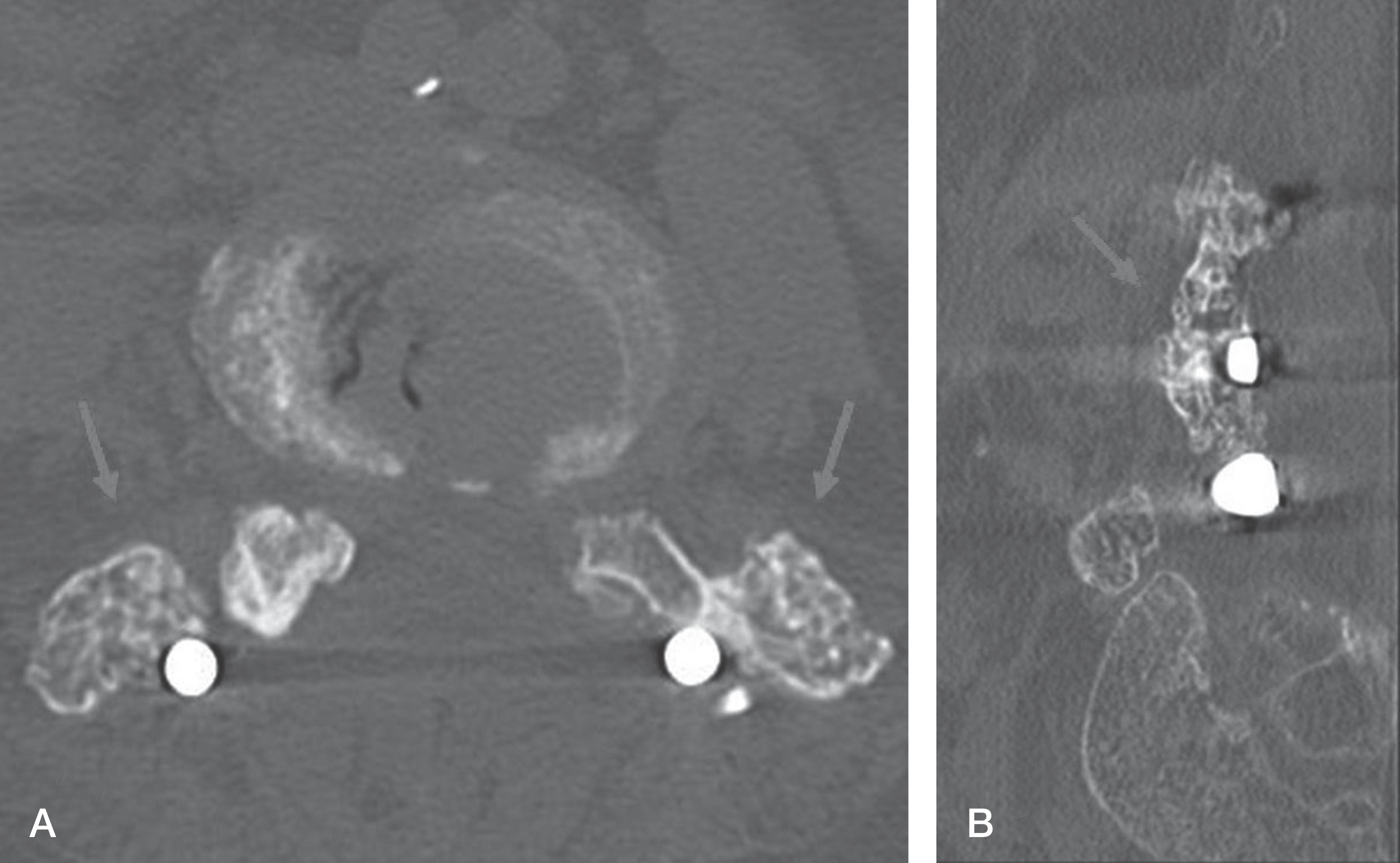
Table 1.
Demographic Characteristics of the Patients
Table 2.
Clinical Pain Scores of Patients
Table 3.
Multivariate Analysis
Table 4.
Tests of Between-Subjects Effect
Table 5.
Evaluation of Bone Union
 XML Download