

 PDF
PDF ePub
ePub Citation
 Print
Print


Abstract
Objectives
Using ultrasound to evaluate association of minor cutaneous stigmas with occult spinal dysraphism (OSD) according to the presence of co-morbidities.
Summary of Literature Review
OSD can be associated with various cutaneous markers. Ultrasound of the spine is an effective, non-invasive screening method.
Materials and Methods
Over a 5-year period (2009-2013), a total of 180 infants with various skin stigmas were evaluated. Ninety- seven patients were normal infants, eighty-three had other co-morbidities. The type of skin stigmata and/or co-morbidities as well as lumbar ultrasound results were reviewed for all patients.
Results
Three of the 97 normal infants had abnormalities. One of the three had OSD. Eighteen of the 83 infants with congenital anomalies had abnormalities, and eleven of the 18 had OSD. Infants with congenital anomalies were 6 times more likely to have OSD than normal infants (OR 5.98, 95% CI 1.927 to 18.612, p=0.001) and there was no significant correlation between the presence of minor skin lesions and the presence of dysraphism.
Go to : 
REFERENCES
1. Byrd SE, Darling CF, McLone DG. Developmental dis-orders of the pediatric spine. Radio Clin North Am. 1991; 29:11–52.
2. Kriss VM, Desai NS. Occult spinal dysraphism in neonates: assessment of high-risk cutaneous stigmata on sonography. AJR. 1998; 178:1687–92.

3. Saitsu H, Yamada S, Uwabe C, et al. Development of the posterior neural tube in human embryos. Anat Embryol (Berl). 2004; 209:107–17.
4. Northrup H, Volcik KA. Spina bifida and other neural tube defects. Curr Probl Pediatr. 2000; 30:313–32.
5. Venkataramana NK. Spinal dysraphism. J Pediatr Neurosci. 2011; 6:31–40.
6. Soonawala N, Overweg-Plandsoen WC, Brouwer OF. Early clinical signs and symptoms in occult spinal dysraphism: a retrospective case study of patients. Clin Neurol Neurosurg. 1999; 101:11–4.
7. Colak A, Tahta K, Ozcan OE, Eryilmaz M. Congenital lumbosacral lipomas presenting as a form of occult spinal dysraphism. A report of surgically treated cases. Zentralbl Neurochir. 1992; 53:15–9.
8. Peter JC, Sinclair-Smith C, de Villiers JC. Midline dermal sinuses and cysts and their relationship to the central nervous system. Eur J Pediatr Surg. 1991; 1:73–9.
9. Kriss VM, Kriss TC, Desai NS, Warf BC. Occult spinal dysraphism in the infant. Clin Pediatr. 1995; 34:650–4.
10. Chapman PH. Congenital spinal Lipomas: anatomic considerations and surgical treatment. Child Brain. 1982; 9:37–47.
11. Lew SM, Kothbauer KF. Tethered cord syndrome: an up-dated review. Pediatr Neurosurg. 2007; 43:236–48.
12. Tubbs RS, Wellons JC III, Iskandar BJ, Oakes WJ. Iso-lated flat capillary midline lumbosacral hemangiomas as indicators of occult spinal dysraphism. J Neurosurg. 2004; 100:86–9.
13. Powell KR, Cherry JD, Hougen TJ, Blinderman EE, Dunn MC. A prospective search for congenital dermal abnormalities of the craniospinal axis. J Pediatr. 1975; 87:744–50.
14. Robinson AJ, Russell S, Rimmer S. The value of ultrasonic examination of the lumbar spine in infants with specific reference to cutaneous markers of occult spinal dysraphism. Clin Radiol. 2005; 60:72–7.
15. Sardana K, Gupta R, Garg VK, et al. A prospective study of cutaneous manifestations of spinal dysraphism from India. Pediatr Dermatol. 2009; 26:688–95.
16. Scheible W, James HE, Leopold GR, Hilton SV. Occult spinal dysraphism in infants: screening with high resolution realtime ultrasound. Radiology. 1983; 146:743–6.
Go to :
Figures and Tables%
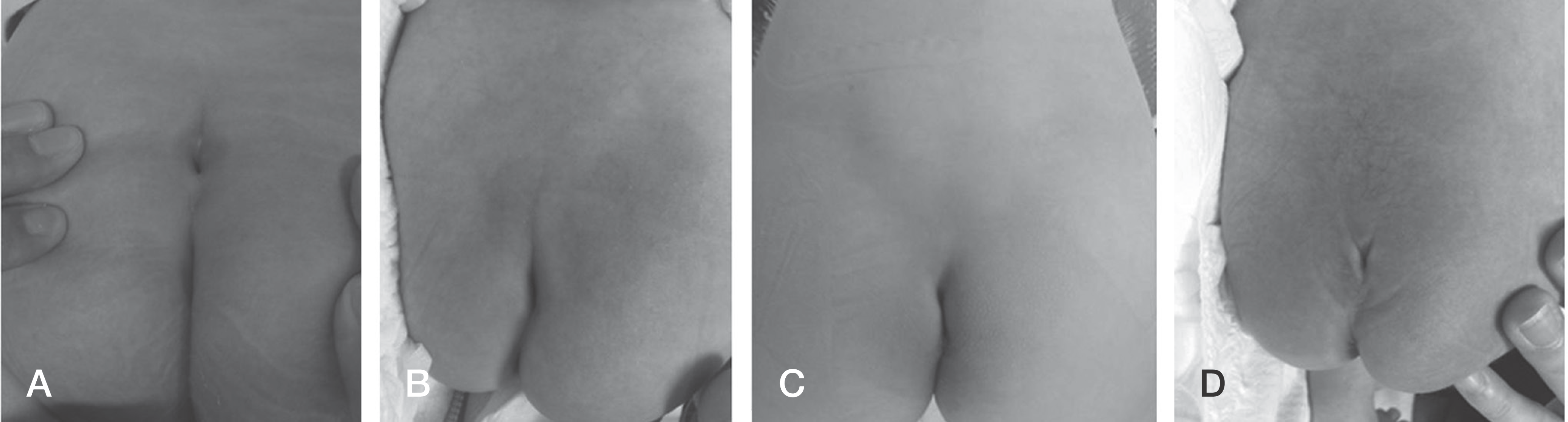 | Fig. 1.Clinical features of lumbosacral skin stigmas. (A) Dimple; (B) Midline deviation, (C) Mass, (D) Dimple + hair. |
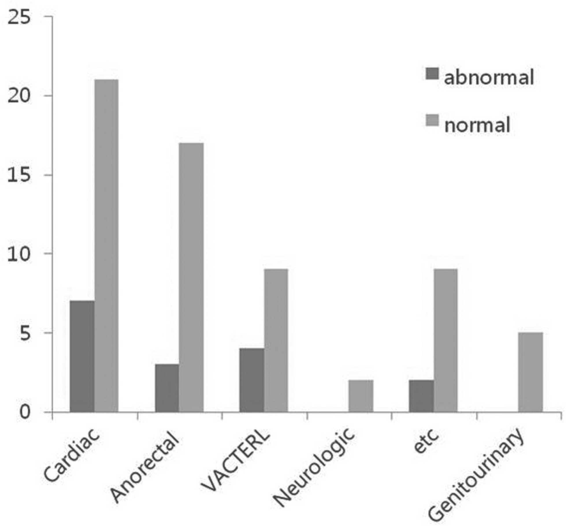 | Fig. 2.Summary of patients referred for lumbar ultrasonography because of the presence of congenital abnormalities. |
 | Fig. 3.Newborns with combined sacral cutaneous lesions. (A) Clinical characteristics of a sacral dimple with hair. (B) A sonogram showing a longitudinal view of a lipoma. (C) A sonogram showing a longitudinal view of a tethered cord. |
Table 1.
Summary of patients referred for lumbar ultrasonography because of the presence of cutaneous markers
 XML Download