

 PDF
PDF ePub
ePub Citation
 Print
Print


Abstract
Summary of Literature Review
Epidermoid cyst in the spinal canal is rare. Idiopathic epidermoid cyst in the spinal canal not associated with a trauma or infection is even rarer.
Material and Methods
A 73 year-old female presented with a 1 year history of progressive paresthesia and motor weakness of both lower extremeties. MRI showed a cystic mass on the 7th thoracic canal. We performed total laminectomy at the T6 –T8 level. The cystic mass was excised after durotomy using a posterior approach.
REFERENCES
1. Alvisi C, Cerisoli M, Giulioni M, Guerra L. Longterm results of surgically treated congenital intradural spinal arachnoid cysts. J Neurosurg. 1987; 67:333–5.

2. Manno NJ, Uihlein A, Kernohan JW. Intraspinal epider-moids. J Neurosurg. 1962; 19:754–65.
3. Alves AM, Norrell H. Intramedullary epidermoid tumors of the spinal cord. Report of a case and review of the literature. Int Surg. 1970; 54:239–43.
4. Roux A, Mercier C, Larbrisseau A, Dube LJ, Dupuis C, Del Carpio R. Intramedullary epidermoid cysts of the spinal cord. Case report. J Neurosurg. 1992; 76:528–33.
5. Hatfield MK, Udesky RH, Strimling AM, Kim BH, Sibergleit R. MR imaging of a spinal epidermoid tumor. AJNR Am J Neuroradiol. 1989; 10:S95–6.
6. Lai SW, Chan WP, Chen CY, Chien JC, Chu JS, Chiu WT. MRI of epidermoid cyst of the conus medullaris. Spinal Cord. 2005; 43:320–3.
7. Horowitz BL, Chari MV, James R, Bryan RN. MR of intracranial epidermoid tumors: correlation of in vivo imaging with in vitro 13C spectroscopy. AJNR Am J Neuroradiol. 1990; 11:299–302.
8. Dunn RC Jr., Archer CA, Rapport RL 2nd, Looi LM. Unusual CT-dense posterior fossa epidermoid cyst: case report. J Neurosurg. 1981; 55:654–6.
9. Debray MP, Ricolfi F, Brugieres P, Khalil A, Adle-Biassette H, Gaston A. Epidermoid cyst of the conus medullaris: atypical MRI and angiographic features. Neuroradiology. 1996; 38:526–8.
10. Cervoni L, Celli P, Cantore G, Fortuna A. Intradural tumors of the cauda equina: a single institution review of clinical characteristics. Clin Neurol Neurosurg. 1995; 97:8–12.
Fig. 1.
MRI finding of lumbar spine (A) Cystic mass with hyper-intense signal area in T2 weighted image at the T7. (B) Tumor mass occupies the almost entire canal in axial views.

Fig. 2.
Operative Findings : Huge irregularly shaped nodular tumor mass wall is seen, when the dura was open.
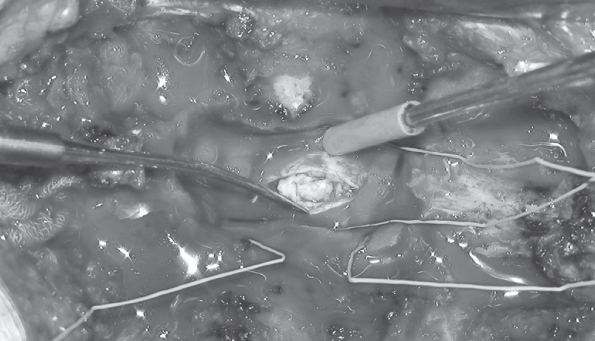
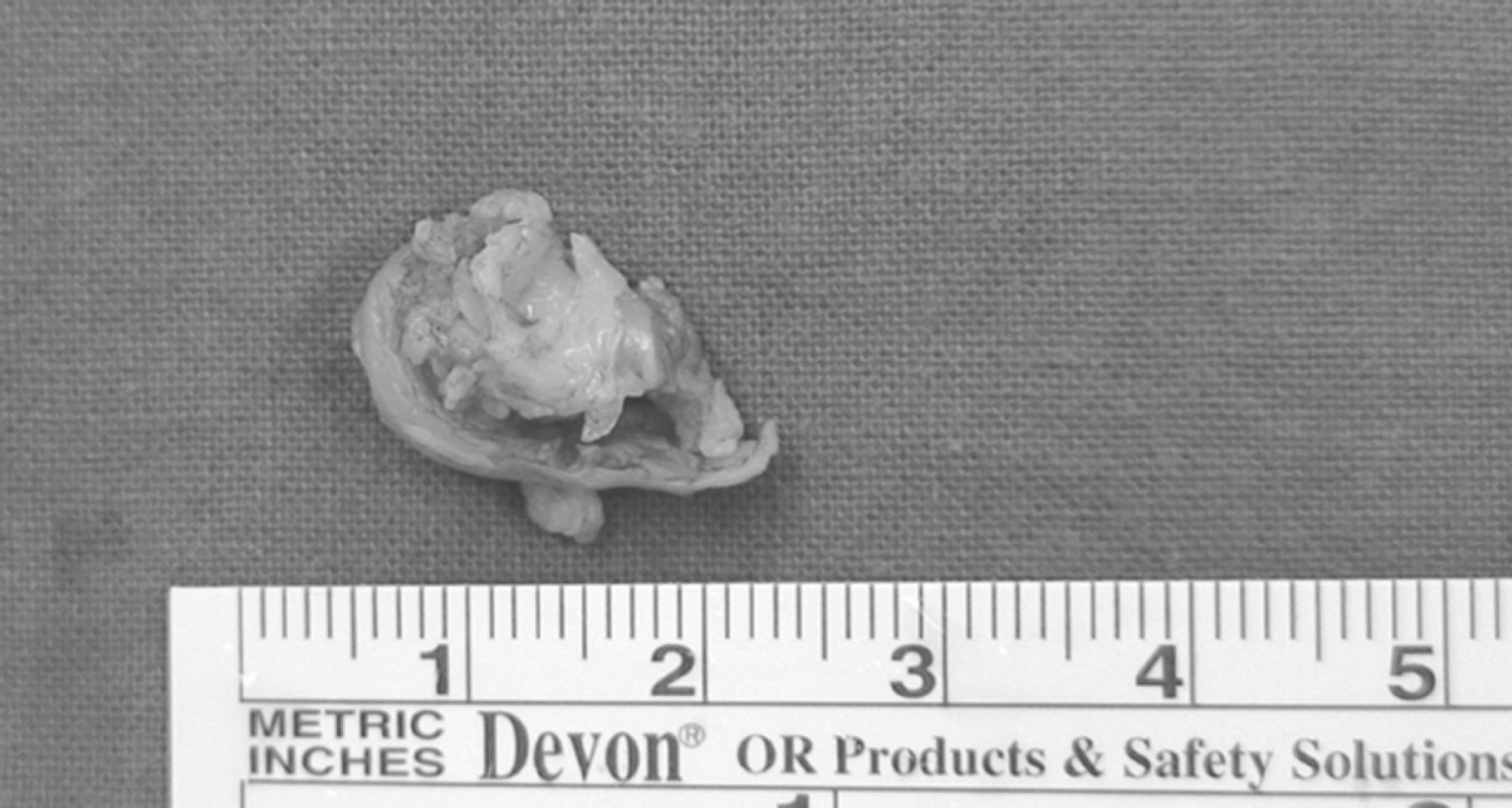
Fig. 4.
Sagittal and axial magnetic resonance images 1 weeks after surgery reveal normalization of the previously compressed spinal cord.

Fig. 5.
(A) The microscopic finding shows keratin laid down by the well-developed squamous epithelium. The stroma reveals abun-dant skin appendages. But no sweat gland and hair follicles are seen. (B) The well-developed squamous epithelia are shown. The underlying stroma demonstrates loose mesenchymal tissue(x100).

 XML Download