Abstract
Summary of Literature Review
Various surgical techniques have been attempted to decrease postoperative axial neck pain.
Material and Methods
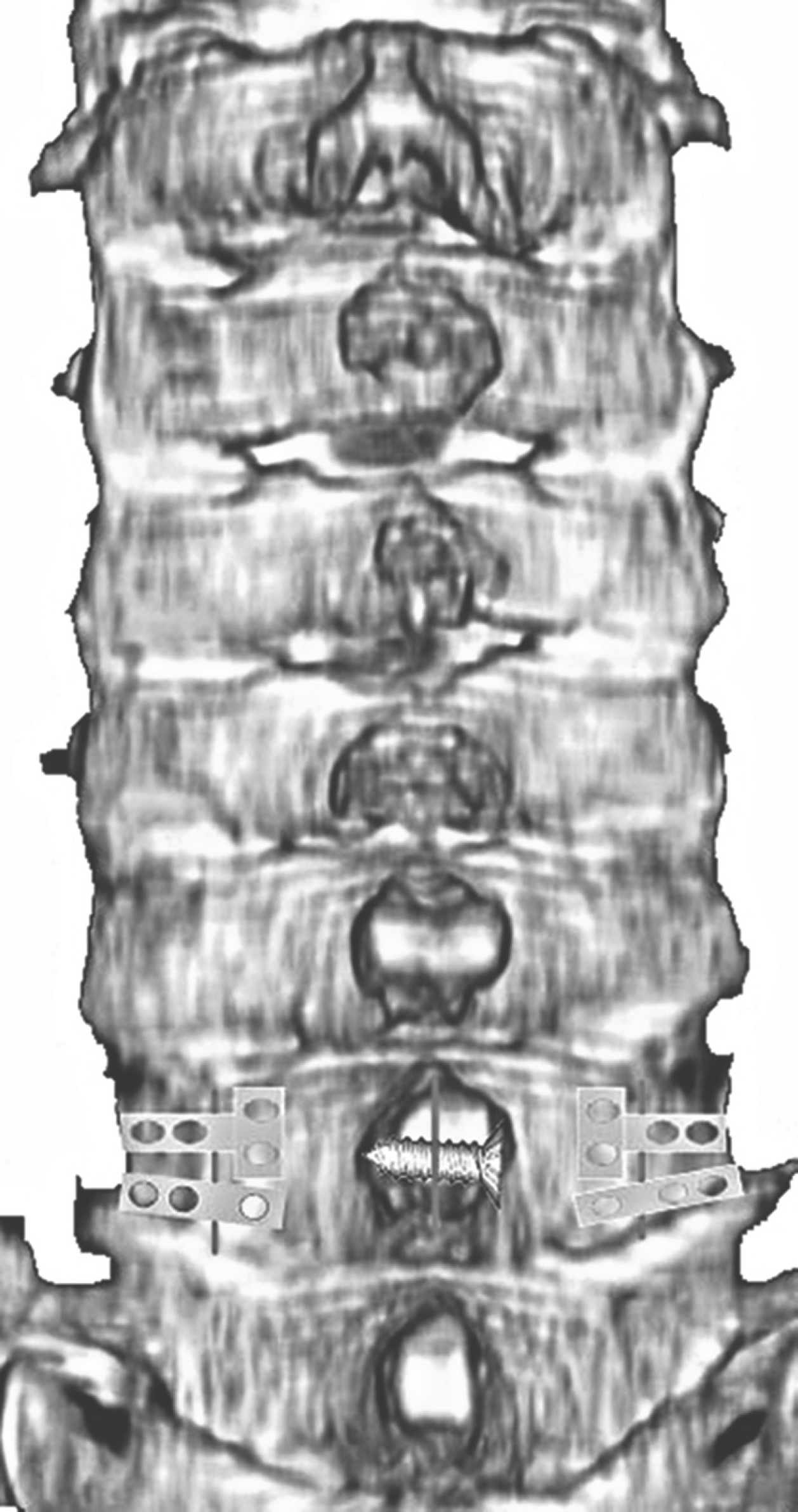
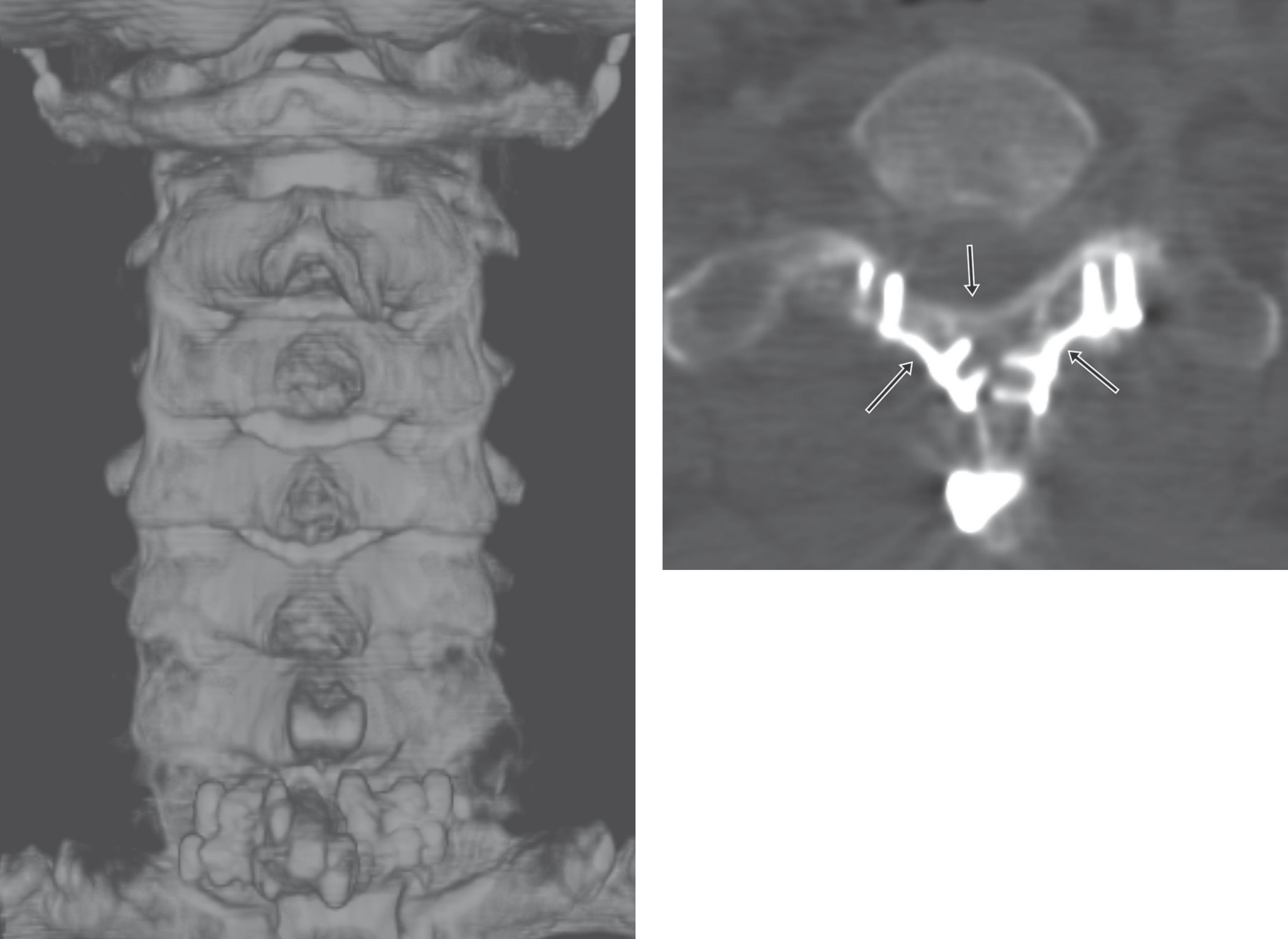
Kurokawa laminoplasty of C7 was performed. Autogenous bone graft material was harvested from partial T1 laminectomy. Intradural tumor was removed without any complications. Four mini plates were applied at hinge sites of laminoplasty and one lag screw was fixed at the longitudinally splitted lamina of C7.
Results
Early range of motion without braces was possible following laminoplasty and recapping procedure. Solid union was achieved at the hinge sites of laminoplasty at the 3-month postoperative followup. No instability was observed at the 2-year postoperative followup. The visual analog scale of axial neck pain at the 2-year postoperative followup was 2.
REFERENCES
1. Wiemels J, Wrensch M, Claus EB. Epidemiology and etiol-ogy of meningioma. J Neurooncol. 2010; 99:307–14.

2. Lee GW, Kang SS, Padua MRA, et al. C2 en bloc hemilaminectomy and recapping using laminar screws: A new approach to preserve the C2 extensor muscle during intradural tumor resection at the C2 level. J Korean Orthop Assoc. 2012; 47:452–6.
3. Xie T, Qian J, Wu X, et al. Unilateral, multilevel, interlami-nar fenestration in the removal of a multisegment cervical intramedullary ependymoma. Spine J. 2013; 13:747–53.

4. Sivaraman A, Bhadra AK, Altaf F, et al. Skip laminectomy and laminoplasty for cervical spondylotic myelopathy. J Spinal Disord Tech. 2010; 23:96–100.
5. Dehcordi SR, Marzi S, Ricci A, et al. Less invasive approaches for the treatment of cervical schwannomas: our experience. Eur Spine J. 2012; 21:887–96.

6. Shiraishi T, Kato M, Yato Y, et al. New Techniques for Exposure of Posterior Cervical Spine Through Intermus-cular Planes and Their Surgical Application. Spine (Phila Pa 1976). 2012; 37:E286–96.

7. Miyakoshi N, Hongo M, Kasukawa Y, et al. Huge thoracolumbar extradural arachnoid cyst excised by recapping T-saw laminoplasty. Spine J. 2010; 10:E14–8.

8. Hida S, Naito M, Arimizu J, et al. The transverse placement laminoplasty using titanium miniplates for the reconstruction of the laminae in the thoracic and lumbar lesion. Eur Spine J. 2006; 15:1292–7.
9. Vasavada AN, Li S, Delp SL. Influence of muscle mor-phometry and moment arms on the moment-generating capacity of human neck muscles. Spine (Phila Pa 1976). 1998; 23:412–22.

10. Iplikcioqlu AC, Hatiboqlu MA, Ozek E, et al. Surgial removal of spinal mass with open door laminoplsty. 2010; 71:213–8.
Fig. 1.
Gadolinium-enhanced magnetic resonance images show an intradural extramedullary tumor at C7-T1 level.

Fig. 3.
Intraoperative photographs are shown. (A) Lamina was longitudinally splitted with T-saw TM. (B) Intradural extramedullary tumor was dissected. (C) Intradural extramedullary tumor was successfully removed. (D) The longitudinally splitted lamina was reattached with a cortical lag screw and each hinge site of Kurokawa laminoplasty was fixed with Hinge plate TM, and mini plate.


 PDF
PDF ePub
ePub Citation
 Print
Print


 XML Download