Abstract
Objectives
To investigate the potential risk factors for subsequent vertebral fractures according to the treatment of primary vertebral fractures.
Summary of Literature Review
Many previous studies have been reported on bone mineral density, bone loss, and mechanical properties as risk factors for osteoporotic vertebral fractures. However, few studies have investigated subsequent osteoporotic vertebral fractures.
Materials and Methods
57 patients who had undergone followup magnetic resonance imaging (MRI) of the spine were divided into two groups depending on the development of subsequent vertebral fractures: the fracture group with 40 cases and the non-fracture group with 17 cases. The patients’ clinical and radiographic data including bone mineral density, medication for osteoporosis, body mass index, vertebroplasty of primary vertebral fractures, thoracic kyphotic angle and lumbar lordotic angle, fat infiltration of the back extensor muscle, and primary multiple fractures were examined.
Results
The subsequent new vertebral fractures occurred at a mean of 24 ± 19 months after primary osteoporotic vertebral fractures. Vertebroplasty for primary fractures was associated with a higher incidence of subsequent new vertebral fractures (p=0.001). There was a significant increase in fat infiltration of the back extensor muscle after the primary vertebral fractures in the fracture group (p=0.001). A multiple logistic regression analysis showed the significance of vertebroplasty (odds’ ratio: 4.623, 95% confidence interval: 1.145–18.699, p=0.031).
Go to : 
REFERENCES
1. Johnell O, Kanis JA. An estimate of the worldwide prevalence and disability associated with osteoporotic fractures. Osteoporos Int. 2006; 17:1726–33.

3. Lindsay R, Silverman SL, Cooper C, et al. Risk of new vertebral fracture in the year following a fracture. JAMA. 2001; 285:320–3.

4. Syed MI, Patel NA, Jan S, et al. New symptomatic vertebral compression fractures within a year following vertebroplasty in osteoporotic women. AJNR Am J Neuroradiol. 2005; 26:1601–4.
5. Klotzbuecher CM, Ross PD, Landsman PB, et al. Patients with prior fractures have an increased risk of future fractures: a summary of the literature and statistical synthesis. J Bone Miner Res. 2000; 15:721–39.

6. Hayes WC, Piazza SJ, Zysset PK. Biomechanics of fracture risk prediction of the hip and spine by quantitative computed tomography. Radiol Clin North Am. 1991; 29:1–18.
7. Hayes WC, Myers ER. Biomechanical considerations of hip and spine fractures in osteoporotic bone. Instr Course Lect. 1997; 46:431–8.
8. Myers ER, Wilson SE. Biomechanics of osteoporosis and vertebral fracture. Spine (Phila Pa 1976). 1997; 22(Suppl):25S–31S.

9. Lee JC, Cha JG, Kim Y, et al. Quantitative analysis of back muscle degeneration in the patients with the degenerative lumbar flat back using a digital image analysis: comparison with the normal controls. Spine (Phila Pa 1976). 2008; 33:318–25.
10. Yuan HA, Brown CW, Phillips FM. Osteoporotic spinal deformity: a biomechanical rationale for the clinical conse-quences and treatment of vertebral body compression fractures. J Spinal Disord Tech. 2004; 17:236–42.
11. Zhang Z, Fan J, Ding Q, et al. Risk factors for new osteoporotic vertebral compression fractures after vertebroplasty: a systematic review and meta-analysis. J Spinal Disord Tech. 2013; 26:E150–7.
12. Lin WC, Cheng TT, Lee YC, et al. New vertebral osteoporotic compression fractures after percutaneous vertebroplasty: retrospective analysis of risk factors. J Vasc Interv Radiol. 2008; 19:225–31.

13. Lee WS, Sung KH, Jeong HT, et al. Risk factors of developing new symptomatic vertebral compression fractures after percutaneous vertebroplasty in osteoporotic patients. Eur Spine J. 2006; 15:1777–83.

14. Kim SH, Kang HS, Choi JA, et al. Risk factors of new compression fractures in adjacent vertebrae after percutaneous vertebroplasty. Acta Radiol. 2004; 45:440–5.

15. Moon ES, Kim HS, Park JO, et al. The incidence of new vertebral compression fractures in women after kyphoplasty and factors involved. Yonsei Med J. 2007; 48:645–52.

16. Berlemann U, Ferguson SJ, Nolte LP, et al. Adjacent vertebral failure after vertebroplasty. A biomechanical investigation. J Bone Joint Surg Br. 2002; 84:748–52.
17. Ma X, Xing D, Ma J, et al. Risk factors for new vertebral compression fractures after percutaneous vertebroplasty. Spine (Phila Pa 1976). 2013; 38:E713–22.

18. Sanfelix-Genoves J, Reig-Molla B, Sanfelix-Gimeno G, et al. The population-based prevalence of osteoporotic vertebral fracture and densitometric osteoporosis in postmenopausal women over 50 in Valencia, Spain (the FRAVO study). Bone. 2010; 47:610–6.

19. Briggs AM, Greig AM, Wark JD, et al. A review of ana-tomical and mechanical factors affecting vertebral body integrity. Int J Med Sci. 2004; 1:170–80.

20. Cunha-Henriques S, Costa-Paiva L, Pinto-Neto AM, et al. Postmenopausal women with osteoporosis and muscu-loskeletal status: a comparative crosssectional study. J Clin Med Res. 2011; 3:168–76.

21. So KY, Kim DH, Choi DH, et al. The influence of fat infiltration of back extensor muscles on osteoporotic vertebral fractures. Asian Spine J. 2013; 7:308–13.

22. Sinaki M, Wollan PC, Scott RW, et al. Can strong back extensors prevent vertebral fractures in women with osteoporosis? Mayo Clin Proc. 1996; 71:951–6.

23. Sinaki M, Itoi E, Wahner HW, et al. Stronger back muscles reduce the incidence of vertebral fractures: a prospective 10 year followup of postmenopausal women. Bone. 2002; 30:836–41.

24. Nevitt MC, Thompson DE, Black DM, et al. Effect of alendronate on limited-activity days and bed-disability days caused by back pain in postmenopausal women with existing vertebral fractures. Fracture Intervention Trial Research Group. Arch Intern Med. 2000; 160:77–85.
Go to : 
Figures and Tables%
 | Fig. 1.
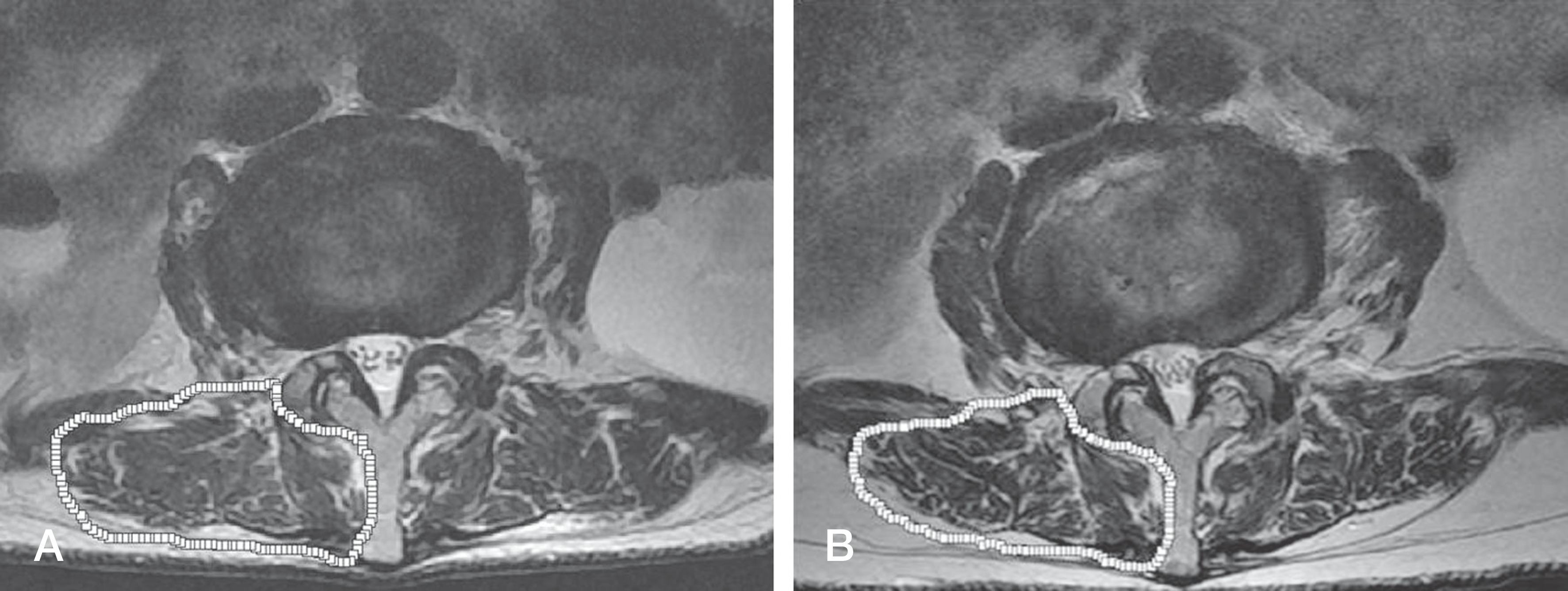
(A) An 80- years -old female patient with a primary L2 osteoporoti vertebral fracture., The paravertebral muscle sity was measured by using a pseudocoloring tool on the L3 axial image. Muscle densty: 1111.87 (74.6%)., In this case, vertebroplas-trformed. (B) After 18Eighteen months after the primary fracture, a subsequent L4 vertebral frcture occurred. The Pparavertebral muscle density was measured by ug the same method on atthe same level. The Mmuscle denity had decreased to: 924.01 (64.0%). |
Table 1.
Demographic Characteristics of the Study Subjects
| Study Group (n=40) | Control Group (n=17) | p-value | |
|---|---|---|---|
| Median age (yr) | 71±8.4 | 70.3±7.7 | 0.474 |
| Sex | 0.031 | ||
| Male | 6 (15.0%) | 7 (41.2%) | |
| Female | 34 (85.0%) | 10 (58.8%) | |
| Body mass index (Kg/m2) | 23.1±3.4 | 23.1±2.53 | 0.935 |
Table 2.
Baseline Characteristics of the Study Subjects
Table 3.
Changes from Baseline to Final Follow up of Both Groups
| Variables | Study Group | Control Group | ||||
|---|---|---|---|---|---|---|
| 1st | 2nd | P | 1st | 2nd | P | |
| Extensor muscle volume of L3 (mm2) | 1980.8 | 1871.3 | 0.006* | 1788.5 | 1605.1 | <0.001* |
| Muscle-fat infiltration ratio of L3 (%) | 61.6 | 57.72 | 0.001* | 59.5 | 57.4 | 0.394 |
| BMD (T-score) | -2.57 | -2.81 | 0.089 | -2.41 | -2.53 | 0.600 |
| BMI (Kg/m2) | 23.1 | 22.9 | 0.331 | 23.1 | 22.9 | 0.612 |
| T-Kyphotic angle(°) | 19.5 | 21.2 | 0.310 | 21.5 | 22.7 | 0.715 |
| L-Lordortic angle (°) | 40.7 | 39.6 | 0.401 | 42.7 | 41.5 | 0.381 |
Table 4.
Univariate Logistic Regression Analysis for New Vertebral Fracture
| Variables | B | OR | 95% Cl | p-value |
|---|---|---|---|---|
| Primary factured age (yr) | 0.023 | 1.024 | 0.955-1.096 | 0.509 |
| Sex | 1.378 | 4.325 | 1.082-14.533 | 0.038* |
| Primary multiple fracture | 0.511 | 1.667 | 0.454-6.112 | 0.441 |
| Vertebroplasty | 2.148 | 10.201 | 2.293-32.015 | 0.001* |
| ΔBMD | -0.900 | 0.995 | 0.069-2.381 | 0.318* |
| ΔBMI (Kg/m2) | 0.002 | 0.995 | 0.723-1.389 | 0.986 |
| ΔThoracic kyphosis (°) | 0.017 | 1.018 | 0.946-1.095 | 0.641 |
| ΔLumbar lordorsis (°) | 0.002 | 1.003 | 0.934-1.076 | 0.940 |
| ΔExtensor muscle of L3 (mm2) | 0.001 | 1.054 | 0.998-1.004 | 0.304* |
| ΔExtensor muscle of L3 (%) | -0.044 | 0.911 | 0.867-1.055 | 0.378 |


 PDF
PDF ePub
ePub Citation
 Print
Print


 XML Download