

 PDF
PDF ePub
ePub Citation
 Print
Print


Abstract
Summary of Literature Review
Few reports of tuberculous spondylitis have discussed atypical cases, which resulted in a poor prognosis due to the delay in early diagnosis and proper treatment.
Materials and Methods
A 74-year-old female underwent an incision and drainage, and posterior decompression and fusion (PDF) due to tuberculous epidural abscess after vertebroplasty of a compression fracture at T12. A 52-year-old female underwent interbody fusion and posterior lateral fusion (PLF) because of aggravation of an abscess and neurologic symptoms following non-invasive intervention to treat atypical tuberculous spondylitis.
Results
Clinical symptoms and serological tests of the patients were improved at postoperative 6 months.
Conclusions
When a patient presents with focal bony or soft tissue abnormality on an image study, the possibility of non-typical tuberculous spondylitis has to be considered when infective spondylitis or a tumor is detected. Moreover, an invasive diagnosis tool such as biopsy will be needed for proper management.
REFERENCES
1. An HS, Seldomridge JA. Spinal infections: diagnostic tests and imaging studies. Clin Orthop Relat Res. 2006; 444:27–33.
2. De Backer AI, Mortele KJ, Vanschoubroeck IJ, et al. Tuberculosis of the spine: CT and MR imaging features. JBR-BTR. 2005; 88(2):92–7.
3. Pande KC, Pande SK, Babhulkar SS. An atypical pre-sentation of tuberculosis of the spine. Spinal Cord. 1996; 34(12):716–9.

4. Ha K-Y, Na K-T, Kee S-R, Kim Y-H. Tuberculosis of the Spine: A new Understanding of an Old Disease. J Korean Soc Spine Surg. 2014; 21(1):41–7.
5. Batson OV. The Function of the Vertebral Veins and Their Role in the Spread of Metastases. Ann Surg. 1940; 112(1):138–49.
6. Laloum E, Zeller V, Graff W, et al. Salmonella typhi osteitis can mimic tuberculosis. A report of three cases. Joint Bone Spine. 2005; 72(2):171–4.
7. Torii H, Takahashi T, Shimizu H, Watanabe M, Tominaga T. Intramedullary spinal tuberculoma–case report. Neurol Med Chir (Tokyo). 2004; 44(5):266–8.
8. Garcia-Monco JC. Central nervous system tuberculosis. Neurol Clin. 1999; 17(4):737–59.
9. Mak KC, Cheung KM. Surgical treatment of acute TB spondylitis: indications and outcomes. Eur Spine J. 2013; 22(Suppl 4):603–11.
10. Alg VS, Demetriades AK, Naik S, Gunasekera L. Isolated subacute tuberculous spinal epidural abscess of the cervical spine: a brief report of a special case. Acta Neurochir (Wien). 2009; 151(6):695–6.
Fig. 1.
(A) T1-weighted sagittal image shows low signal intensity in the T12 vertebral body found by usual acute compression fracture. Lateral radiographs show the site after vertebroplasty T12 and kyphotic deformity. (B) MR images show T11 superior end plate destruction and epidural abscess at the T10-12 level after 8 weeks of vertebroplasty. (C) MR images show a small amount of epidural abscess at postoperative 6 months. (D) The histologic findings show caseous necrosis with chronic granulated inflammation (H-E staining, 200× magnification).

Fig. 2.
(A) The evidence of brain magnetic resonance (MR) images that show tiny disseminated enhancing nodules with mild edema in the whole brain results in brain infection from Tb spondylitis. (B) Brain MR images show that the previously disseminated small enhancing nodules have nearly completed disappeared at postoperative 4 months.
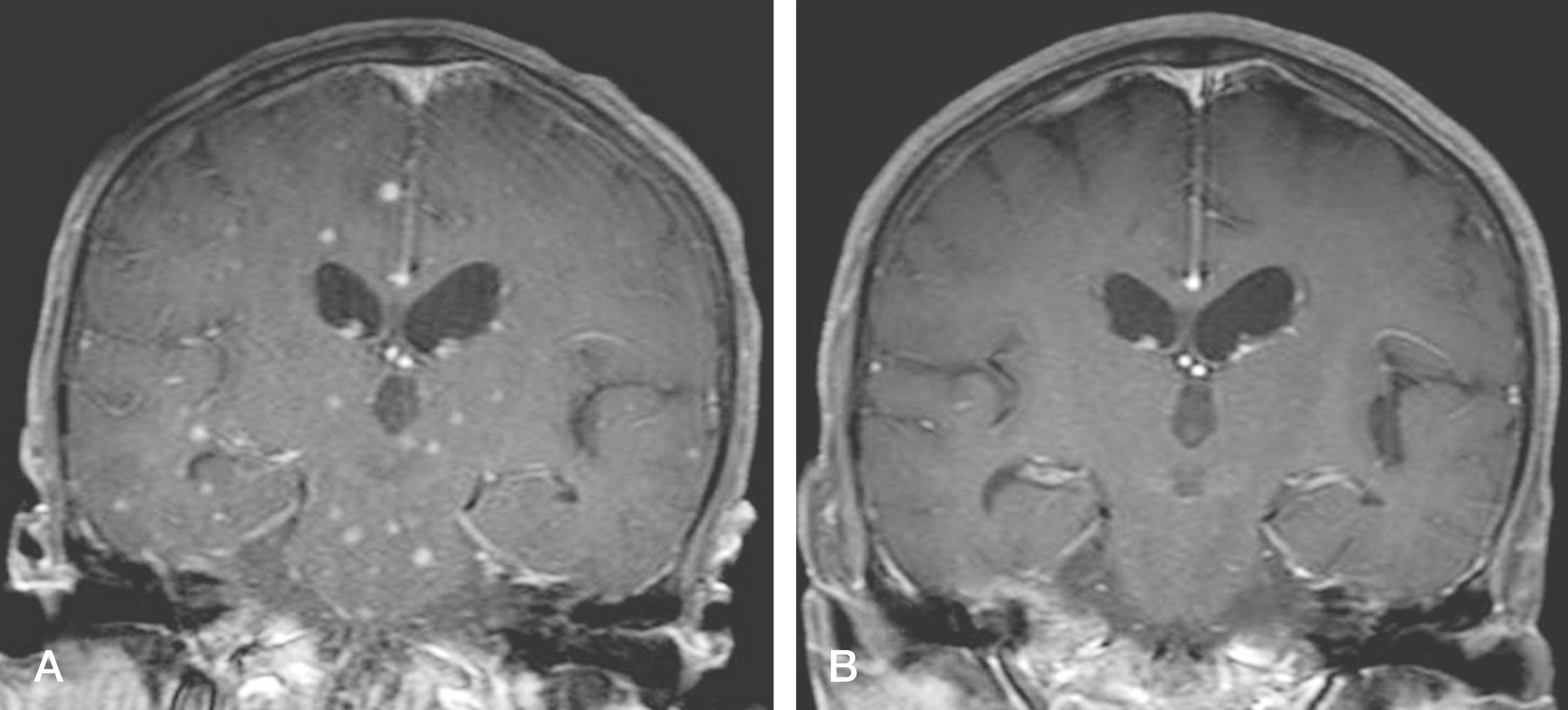
Fig. 3.
(A) Preoperative magnetic resonance (MR) images show the signal change of bone in an epidural abscess and infective spondylitis at the L5-S1 level. (B) MR images show progression of infective spondylitis as well as a larger abscess than in previous study 8 weeksafter discectomy and laminotomy. (C) AP and lateral radiographs taken at the 6-month followup after interbody fusion show solid bony fusion.

 XML Download