

 PDF
PDF ePub
ePub Citation
 Print
Print


Abstract
Summary of Literature Review
Extradural arachonid cysts of the spine are a rare cause of spinal cord and nerve root compression. There are few reports about it, and the etiology remains unclear.
Results
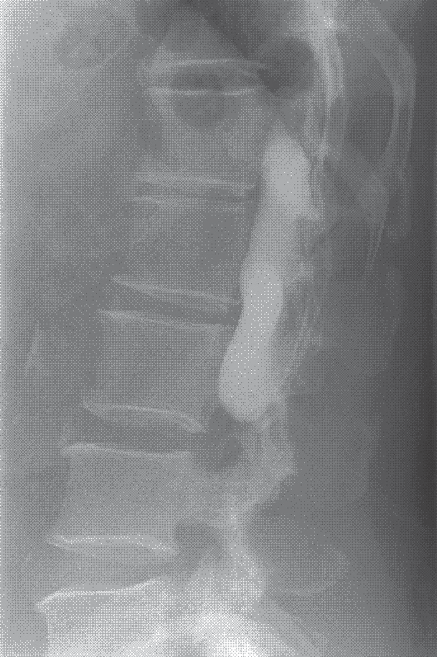
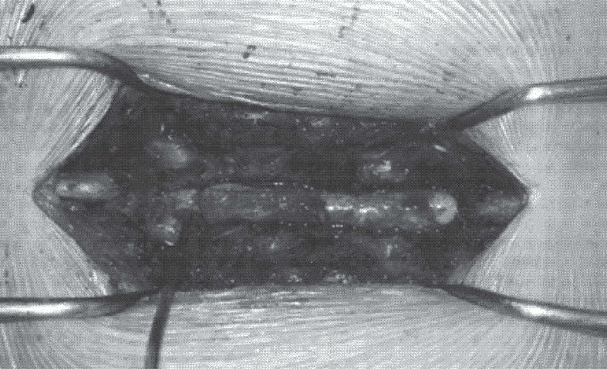
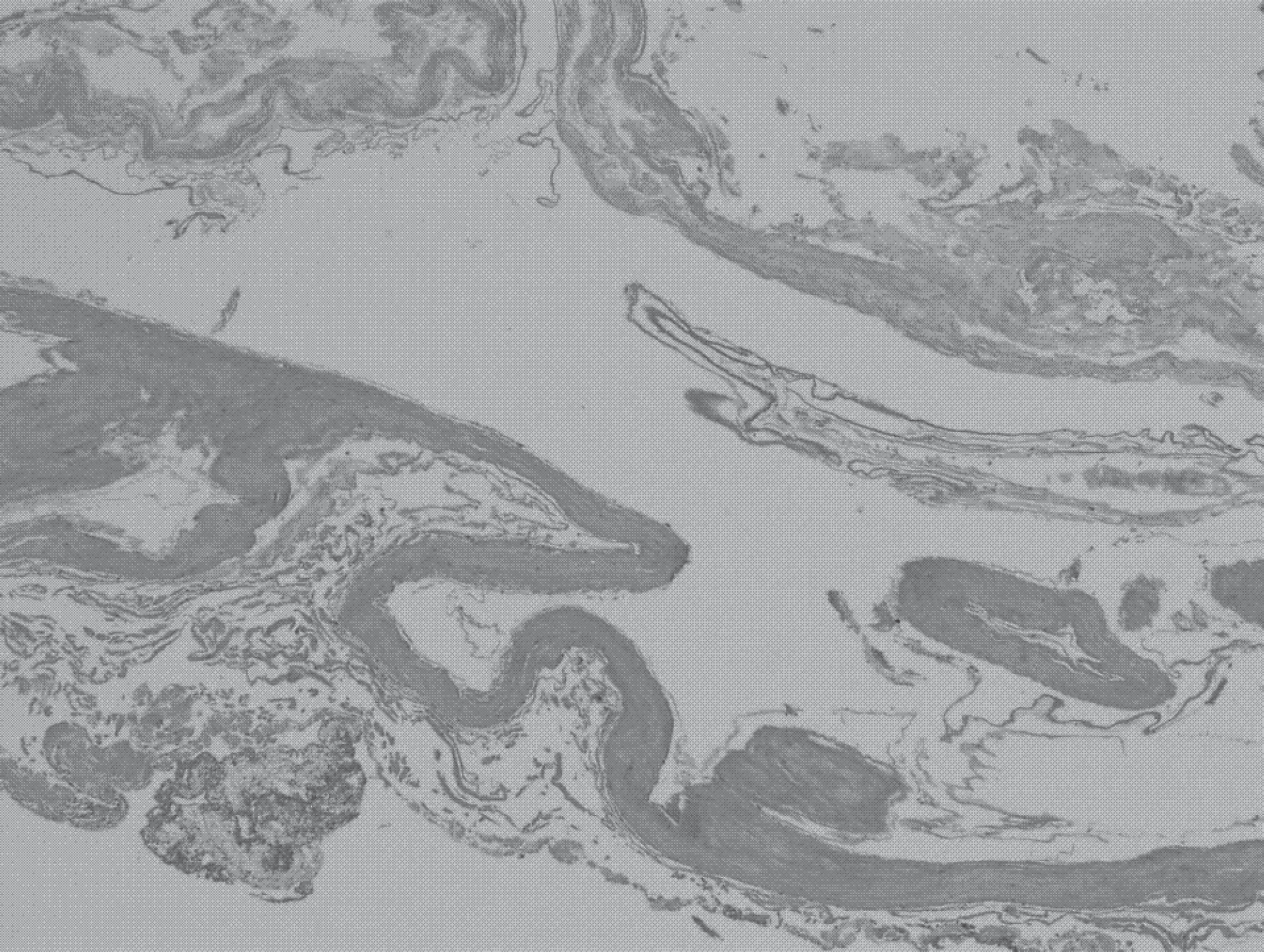
A 56-year-old male patient presented with both lower extremity radiating pain and tingling sensation in both feet for four years. His MRI revealed a large, well-demarcated extradural lesion, isointense to cerebrospinal fluid from L1 to L3. We performed dural repair and laminectomy for partial resection of the cyst. The outcome was good in the immediate postoperative period, and the patient made a full recovery without complications.
REFERENCES
1. Fortuna A, La Torre E, Ciappetta P. Arachnoid diverticula: a unitary approach to spinal cysts communicating with the subarachnoid space. Acta Neurochir. 1977; 39:259–68.

2. Cilluffo JM, Gomez MR, Reese DF, et al. �diopathic (�congenital”) spinal arachnoid diverticula. Clinical diagnosis and surgical results. Myo Clin Proc. 1998; 56:93–101.
3. Fortuna A, La Torre E, Ciappetta P. Arachnoidal diverticula: a unitary approach to spinal cysts communicating with the subarachnoid space. Acta Neurochir. 1977; 39:259–68.
4. Nabors MW, Pait TG, Byrd EB, et al. Updated assessment and current classification of spinal meningeal cysts. J Neurosurg. 1988; 68:366–77.
5. Bergland RM. Congenital intraspinal extradural cyst. Report of three cases in one family. J Neurosug. 1968; 28:495–9.
6. Liu JK, Cole CD, Kan P, et al. Spinal extradural arachnoid cysts: clinical, radiological, and surgical features. Neurosurg Focus. 2007; 22:E6.
7. Ersahin Y, Yildizhan A, Seber N. Spinal extradural arachnoid cyst. Childs Nerv Syst. 1993; 9:250–2.
8. Lee HJ, Cho WH, Han � H, et al. Large thoracolumbar extradural arachnoid cyst excised by minimal skipped hemilaminectomy: A case report. Korean J spine. 2013; 10:28–31.
9. Neo M, Koyoma T, Sakamoto T, et al. Detection of a dural defect by cinematic magnetic resonance imaging and its se-lective closure as a treatment for a spinal extradural arachnoid cyst. Spine (Phila Pa 1976). 2004; 29:E426–30.
10. Stechison MT, Hendrick EB, Cohen E. Spinal extradural arachnoid cyst. Pedatr Neurosci. 1989; 15:36–8.

 XML Download