Abstract
Objectives
We report a case of posterior ring apophysis fracture (PRAF) with lumbar disc herniation treated by immobile bony fragment excision.
REFERENCES
1. Akhaddar A, Belfquih H, Oukabli M, Boucetta M. Posterior ring apophysis separation combined with lumbar disc herniation in adults: a 10-year experience in the surgical management of 87 cases. J Neurosurg Spine. 2011; 14:475–83.

3. Yang IK, Bahk YW, Choi KH, Paik MW, Shinn KS. Posterior lumbar apophyseal ring fractures: a report of 20 cases. Neuroradiology. 1994; 36:453–5.

4. Savini R, Di Silvestre M, Gargiulo G, Picci P. Posterior lumbar apophyseal fractures. Spine (Phila Pa 1976). 1991; 16:1118–23.

5. Epstein NE. Lumbar surgery for 56 limbus fractures em-phasizing noncalcified type III lesions. Spine (Phila Pa 1976). 1992; 17:1489–96.

6. Chang CH, Lee ZL, Chen WJ, Tan CF, Chen LH. Clinical significance of ring apophysis fracture in adolescent lumbar disc herniation. Spine (Phila Pa 1976). 2008; 33:1750–4.

7. Asazuma T, Nobuta M, Sato M, Yamagishi M, Fujikawa K. Lumbar disc herniation associated with separation of the posterior ring apophysis: analysis of five surgical cases and review of the literature. Acta Neurochir (Wien). 2003; 145:461–6.

8. Shirado O, Yamazaki Y, Takeda N, Minami A. Lumbar disc herniation associated with separation of the ring apophysis: is removal of the detached apophyses manda-tory to achieve satisfactory results? Clin Orthop Relat Res. 2005; 431:120–8.
Fig. 1.
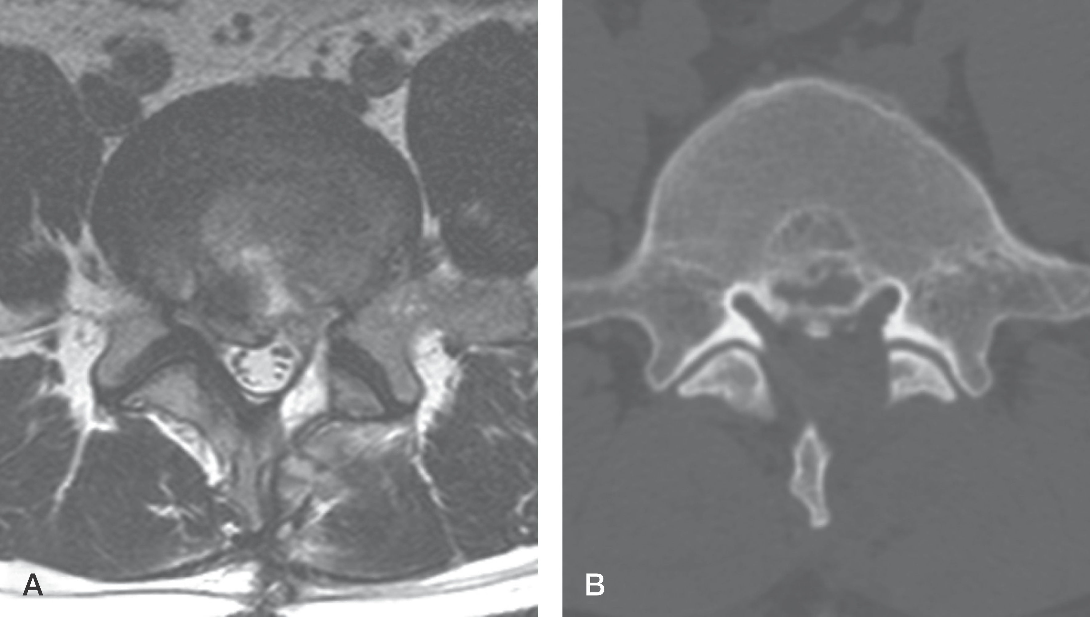
(A-B) A sagittal and axial T2 weighted MRI of L4-5 level shows herniated disc material and bony fragment (white arrow) located centrally and markedly occupying the spinal canal space. Bony defect (asterisk) was noted at the posterior endplate of L5.

Fig. 2.
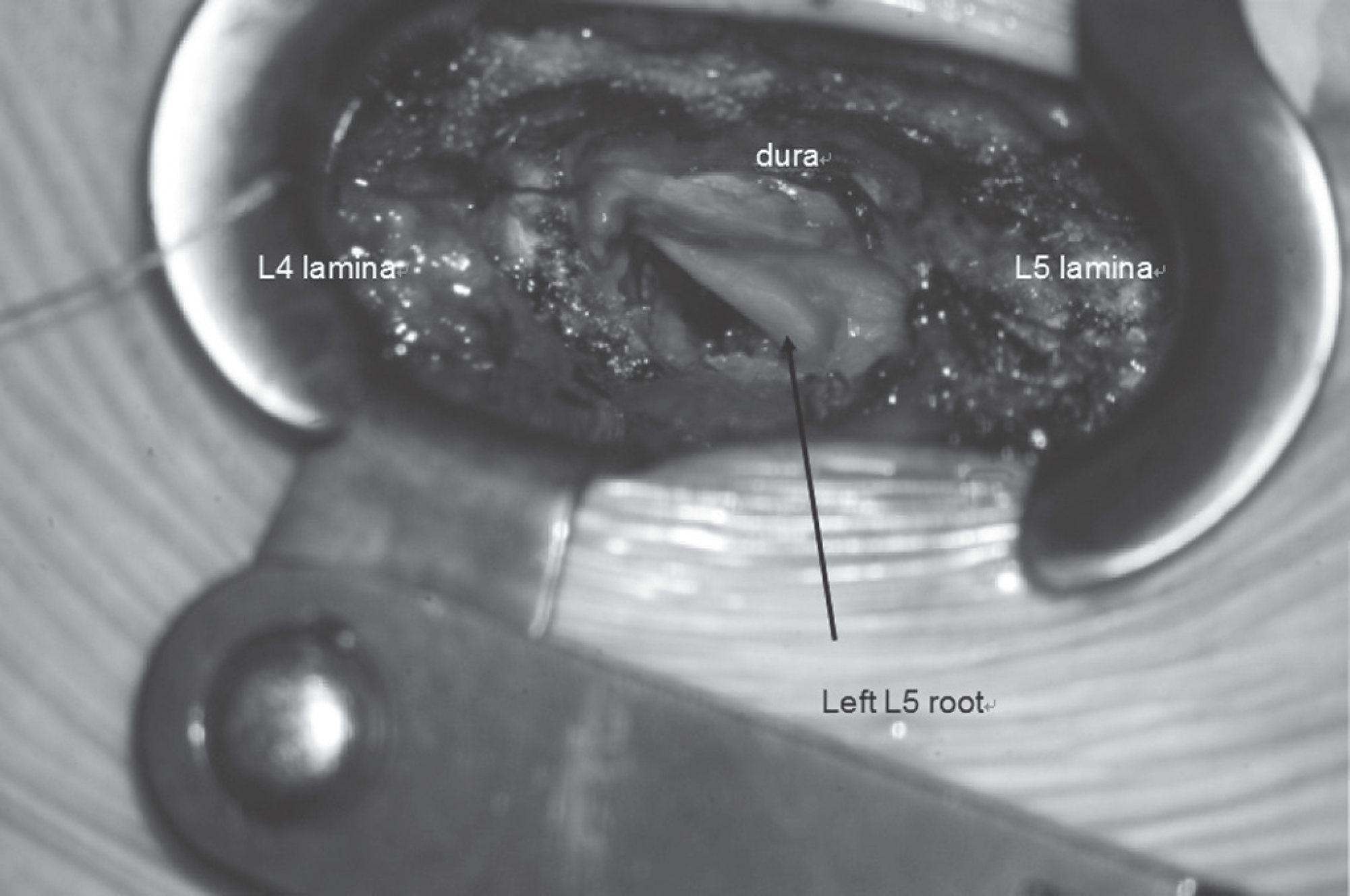
An intraoperative microscopic photo shows adequate decompression at the lateral recess to the foramen and mobilization of the left L5 root.


 PDF
PDF ePub
ePub Citation
 Print
Print


 XML Download