

 PDF
PDF ePub
ePub Citation
 Print
Print


Abstract
Summary of Literature Review
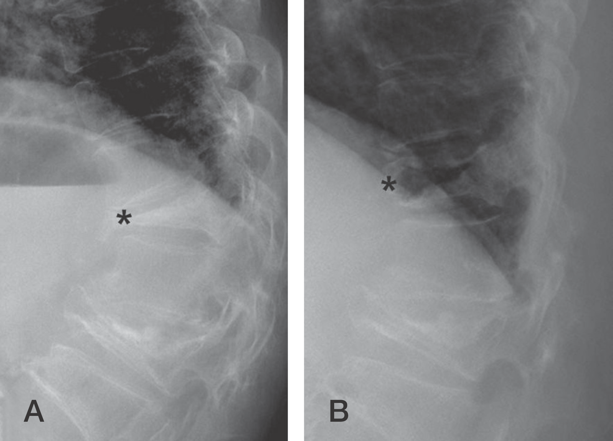
Conventional modalities including simple radiographs, bone mineral density (BMD) tests, and bone scans are sufficient for diagnosis of OSFs. However, other clinical and radiographic clues should be considered for prediction of the prognosis and differential diagnosis.
Results
Clinical clues including morphometric changes in the vertebral body are sufficient for diagnosis of OSFs. BMD testing is helpful for diagnosis of osteoporosis. However, simple radiographs and BMD tests do not present sufficient information on the prognosis of OSFs. The location of the involved segments, morphological characteristics, and other co-morbidities should be taken into consideration in the initial management of OSFs. Moreover, pathologic conditions leading to spinal fractures should be taken into account in some clinical situations.
REFERENCES
1. Green AD, Colon-Emeric CS, Bastian L, et al. Does this woman have osteoporosis? JAMA. 2004; 292:2890–900.

2. Genant HK, Jergas M, Palermo L, et al. Comparison of semiquantitative visual and quantitative morphometric assessment of prevalent and incident vertebral fractures in osteoporosis The Study of Osteoporotic Fractures Research Group. J Bone Miner Res. 1996; 11:984–96.
3. Kuet KP, Charlesworth D, Peel NF. Vertebral fracture assessment scans enhance targeting of investigations and treatment within a fracture risk assessment pathway. Osteoporos Int. 2013; 24:1007–14.
4. Vokes T, Bachman D, Baim S, et al. Vertebral fracture assessment: the 2005 ISCD Official Positions. J Clin Densitom. 2006; 9:37–46.
5. Lewiecki EM, Laster AJ. Clinical review: Clinical applications of vertebral fracture assessment by dual-energy x-ray absorptiometry. J Clin Endocrinol Metab. 2006; 91:4215–22.
6. Sugita M, Watanabe N, Mikami Y, et al. Classification of vertebral compression fractures in the osteoporotic spine. J Spinal Disord Tech. 2005; 18:376–81.
7. Ha KY, Kim YH. Risk factors affecting progressive collapse of acute osteoporotic spinal fractures. Osteoporos Int. 2013; 24:1207–13.
8. Tsujio T, Nakamura H, Terai H, et al. Characteristic radiographic or magnetic resonance images of fresh osteoporotic vertebral fractures predicting potential risk for nonunion: a prospective multicenter study. Spine (Phila Pa 1976). 2011; 36:1229–35.
9. Kanis JA, McCloskey EV, Johansson H, et al. European guidance for the diagnosis and management of osteoporosis in postmenopausal women. Osteoporos Int. 2013; 24:23–57.
10. Kanis JA, McCloskey EV, Johansson H, et al. Case finding for the management of osteoporosis with FRA�–assess-ment and intervention thresholds for the UK. Osteoporos Int. 2008; 19:1395–408.
11. Marshall D, Johnell O, Wedel H. Meta-analysis of how well measures of bone mineral density predict occurrence of osteoporotic fractures. BMJ. 1996; 312:1254–9.
12. Watts NB. Fundamentals and pitfalls of bone densitometry using dual-energy �-ray absorptiometry (D�A). Osteoporos Int. 2004; 15:847–54.
13. Muijs SP, Akkermans PA, van Erkel AR, et al. The value of routinely performing a bone biopsy during percutaneous vertebroplasty in treatment of osteoporotic vertebral compression fractures. Spine (Phila Pa 1976). 2009; 34:2395–9.
14. Schoenfeld AJ, Dinicola NJ, Ehrler DM, et al. Retrospective review of biopsy results following percutaneous fixation of vertebral compression fractures. Injury. 2008; 39:327–33.
15. Sung JK, Jee WH, Jung JY, et al. Differentiation of acute osteoporotic and malignant compression fractures of the spine: use of additive qualitative and quantitative axial dif-fusion-weighted MR imaging to conventional MR imaging at 3.0 T. Radiology. 2014; 271:488–98.
16. Geith T, Schmidt G, Biffar A, et al. Quantitative evaluation of benign and malignant vertebral fractures with diffusion-weighted MRI: what is the optimum combination of b values for ADC-based lesion differentiation with the single-shot turbo spin-echo sequence? AJR Am J Roentgenol. 2014; 203:582–8.
17. Murakami H, Kawahara N, Gabata T, et al. Vertebral body osteonecrosis without vertebral collapse. Spine (Philadelphia, Pa 1976). 2003; 28:E323–8.
18. Kumpan W, Salomonowitz E, Seidl G, et al. The intravertebral vacuum phenomenon. Skeletal radiology. 1986; 15:444–7.
19. Kim YC, Kim YH, Ha KY. Pathomechanism of intravertebral clefts in osteoporotic compression fractures of the spine. Spine J. 2014; 14:659–66.
20. Ryan PJ, Fogelman I. Osteoporotic vertebral fractures: diagnosis with radiography and bone scintigraphy. Radiology. 1994; 190:669–72.
21. Handel SF, Lee YY. Computed tomography of spinal fractures. Radiol Clin North Am. 1981; 19:69–89.
Fig. 1.
Radiographic characteristics for differential diagnosis of osteoporotic spinal fractures with pathologic fractures. A 52-year-old man presenting multiple compression fractures. T2 weighted (A), T1 weighted (B) and T2 fat suppression (C) images show multiple spinal fractures with posterior bulging of the vertebral body, involvement of the posterior column, and decreased signal intensity of the marrow compared to the intervertebral disc (*) in the T1 weighted image.

 XML Download