

 PDF
PDF ePub
ePub Citation
 Print
Print


Abstract
Objectives
We present two cases of quadriplegia after posterior decompression with fusion caused by a suspicious reperfusion injury of spinal cord without remarkable surgical insult.
Summary of Literature Review
Posterior decompression and posterolateral fusion have been reported as effective procedures in patients with multilevel myelopathy. However, postoperative spinal cord injury without remarkable intraoperative technical damage has been reported in a few articles. Reperfusion mechanism was suggested as one of the leading causes and reported in some animal models.
Materials and Methods
There was one case of ossification of the posterior longitudinal ligament and one developmental multilevel stenosis that underwent laminectomy with lateral mass instrumentation. After surgery, the patients presented with quadriplegia; MRI demonstrated swelling of the spinal cord and intramedullary lesion in two cases.
REFERENCES
1. Hukuda S, Mochizuki T, Ogata M, Schichikwa K, Shimo-mura Y. Operations for cervical spondylotic myelopathy. A comparison of the results of anterior posterior procedures. J Bone Joint Surg. 1985; 67:609–15.
2. Lee CW, Kang JH, Lee KY, Sung HW. Laminoplasty versus laminectomy and fusion for multilevel cervical spondylosis. J Korean Spine Surg. 2010; 17:147–53.

3. Chiba K, Toyama Y, Matsumoto M, Maruiwa H, Wata-nabe M, Hirabayashi K. Segmental motor paralysis after expansive open-door laminoplasty. Spine (Phila Pa 1976). 2002; 27:2108–15.
4. Shimizu T, Shimada H, Edakuni H. Postlaminoplasty palsy of upper extremities, with special reference to the spinal cord factors in Japanese. Bessatsu Seikeigeka. 1996; 29:188–93.
5. Okada M, Minamide A, Endo T, et al. A prospective randomized study of clinical outcomes in patients with cervical compressive myelopathy treated with open-door or French-door laminoplasty. Spine (Phila Pa 1976). 2009; 34:1119–26.
6. Gebarski SS, Maynard FW, Gabrielsen TO, Knake JE, Latack JT, Hoff JT. Posttraumatic progressive myelopathy: Clinical and radiologic correlation employing MR imaging, delayed CT metrizamide myelopathy, and intraoperative sonography. Radiology. 1985; 157:379–85.
7. Schwartz ED, Falcone SF, Quencer RM, Green BA. Post-traumatic syringomyelia: pathogenesis, imaging, and treatment. AJR Am J Roentgenol. 1999; 173:487–92.
8. Dumont RJ, Okonkwo DO, Verma S, et al. Acute spinal cord injury, part I: pathophysiologic mechanisms. Clin Neuropharmacol. 2001; 24:254–64.
9. Kim-Lee MH, Stokes BT, Yates AJ. Reperfusion paradox: A novel mode of glial cell injury. Glia. 1992; 5:56–64.
10. Oyinbo CA. Secondary injury mechanisms in traumatic spinal cord injury: a nugget of this multiply cascade. Acta Neurobiol Exp (Wars). 2011; 71:281–99.
Fig. 1.
49-year-old man with OPLL. (A, B) Preoperative lateral radiogram and sagittal reconstruction CT scan show a posterior ossification to the vertebral bodies and mixed configuration at C3-5 (White box). (C) Postoperative lateral radiogram after posterior decompression with posterior fusion.
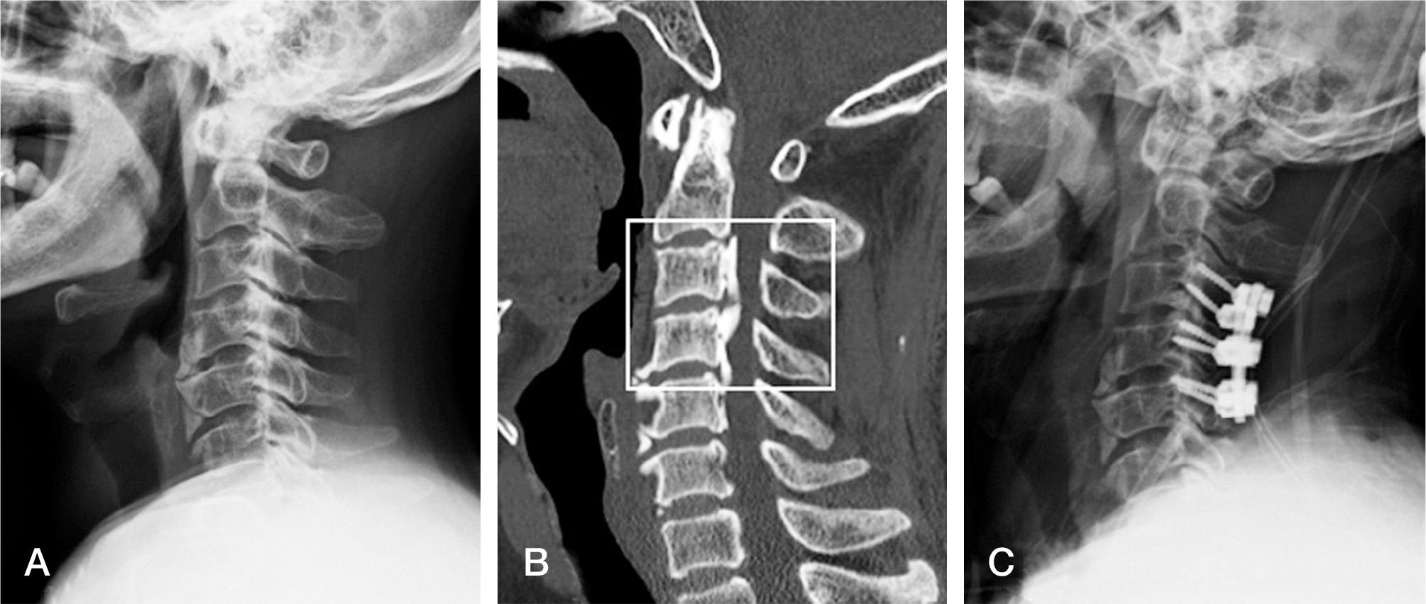
Fig. 2.
Preoperative and postoperative MR images of a 49-year-old man with OPLL who developed distal sensory decrease of both arms. (A) Pre-operative T2-MRI showed the presence of a high intensity area at C3-C4. (B) Postoperative T-2 MRI 2 hours after surgery revealed swelling of the spinal cord with wide spreading of the high-intensity area between C3-C4. (C) Postoperative T-2 MRI 1 week after surgery showed some degree of decrease of the swelling of the spinal cord and residual high-intensity area.
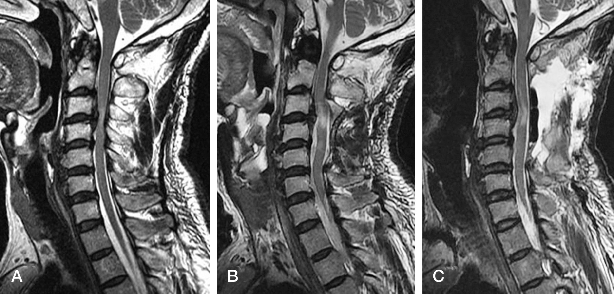
Fig. 3.
71-year-old man with multilevel spondylosis. (A, B) Preoperative lateral radiogram and sagittal reconstruction CT scan show some loss of lordosis and a posterior continuous configuration at the vertebral bodies of C3-5. (C) Postoperative lateral radiogram after posterior decompression with posterior fusion.
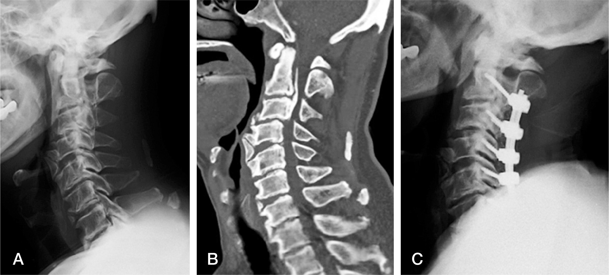
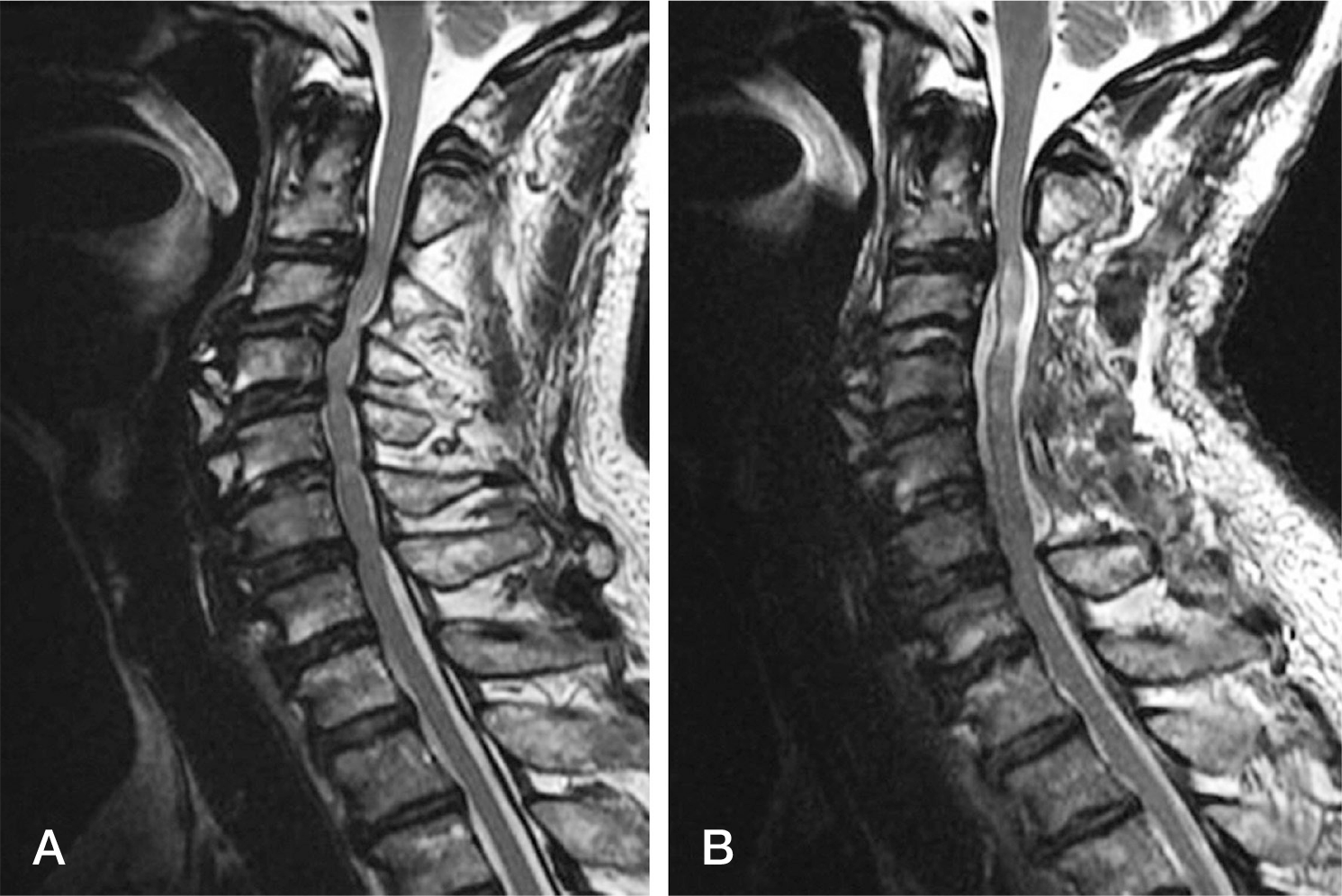
Fig. 4.
Preoperative and postoperative MR images of a 71-year-old-man. (A) Preoperative MRI showing severe cord compression caused by multilevel spondylosis. (B) Postoperative MRI showing decompression and presence of T2 high-signal intensity zones at C2-3 and C3-4, C4-5, high-signal intensity zones are present in the
 XML Download