Abstract
Objectives
Sagittal imbalance cannot be predicted depending on the degree of lumbar lordosis. Thus, we tried to evaluate the necessity of whole spine standing lateral radiograph through comparison of the spinal and pelvic parameter between supine lumbar lateral radiograph and whole spine standing lateral radiograph.
Summary of the Literature Review
No studies in the literature compare supine lumbar lateral radiograph and whole spine standing lateral radiograph.
Materials and Methods
We randomly selected 50 males and 50 females among the patients over the age of 50 who visited our hospital for outpatient due to degenerative lumbar disease. Lumbar lordosis (sLL/wLL), sacral slope (sSS/wSS), and pelvic tilt (sPT/ wPT) were measured and compared respectively by supine lumbar lateral radiograph and whole spine standing lateral radiograph. We categorized as group AI (sLL<30˚) and group AII (sLL≥30˚) by supine lumbar lateral radiograph and analyzed them. We also categorized as group BI (SVA≤5 cm) and group BII (SVA>5 cm) by whole spine standing lateral radiograph and analyzed them.
Results
There were no statistical difference in lumbar lordosis (sLL/wLL: 35.1˚/37.7˚) and pelvic parameter (sSS/wSS: 32˚/31.7˚, sPT/ wPT: 24.3˚/24.2˚. sPI/wPI: 56.3˚/58.2˚) between supine lumbar lateral radiograph and whole spine standing lateral radiograph, and there were also no statistical difference between two groups (group AI & AII) in SVA, lumbar lordosis and pelvic parameter. Pelvic parameter compared by supine lumbar lateral radiograph and whole spine standing lateral radiograph based on sagittal balance was no significant difference, but lumbar lordosis appeared statistical difference.
REFERENCES
1. Benz RJ, Ibrahim ZG, Afshar P, Garfin SR. Predicting complications in elderly patients undergoing lumbar decompression. Clin Orthop Relat Res. 2001. 116–21.

2. Deyo RA, Ciol MA, Cherkin DC, Loeser JD, Bigos SJ. Lumbar spine fusion. A cohort study of complications, reoperations, and resource use in the Medicare population. Spine (Phila Pa 1976). 1993; 18:1463–70.
3. Gelb DE, Lenke LG, Bridwell KH, Blanke K, McEnery KW. An analysis of sagittal spinal alignment in 100 asymptomatic middle and older aged volunteers. Spine (Phila Pa 1976). 1995; 20:1351–8.

4. Vedantam R, Lenke LG, Keeney JA, Bridwell KH. Comparison of standing sagittal spinal alignment in asymptomatic adolescents and adults. Spine (Phila Pa 1976). 1998; 23:211–5.

5. Propst-Proctor SL, Bleck EE. Radiographic determination of lordosis and kyphosis in normal and scoliotic children. J Pediatr Orthop. 1983; 3:344–6.

6. Lenke LG, Bridwell KH, Blanke K, Baldus C, Weston J. Radiographic results of arthrodesis with Cotrel-Dubousset instrumentation for the treatment of adolescent idiopathic scoliosis. A five to ten-year followup study. J Bone Joint Surg Am. 1998; 80:807–14.

7. Stagnara P, De Mauroy JC, Dran G, et al. Reciprocal an-gulation of vertebral bodies in a sagittal plane: approach to references for the evaluation of kyphosis and lordosis. Spine (Phila Pa 1976). 1982; 7:335–42.

9. Bernhardt M, Bridwell KH. Segmental analysis of the sagittal plane alignment of the normal thoracic and lumbar spines and thoracolumbar junction. Spine (Phila Pa 1976). 1989; 14:717–21.

10. Barrey C, Jund J, Noseda O, Roussouly P. Sagittal balance of the pelvis-spine complex and lumbar degenerative diseases. A comparative study about 85 cases. Eur Spine J. 2007; 16:1459–67.

11. Kawakami M, Tamaki T, Ando M, Yamada H, Hashizume H, Yoshida M. Lumbar sagittal balance influences the clinical outcome after decompression and posterolateral spinal fusion for degenerative lumbar spondylolisthesis. Spine (Phila Pa 1976). 2002; 27:59–64.

12. Korovessis P, Dimas A, Iliopoulos P, Lambiris E. Correlative analysis of lateral vertebral radiographic variables and medical outcomes study short-form health survey: a comparative study in asymptomatic volunteers versus patients with low back pain. J Spinal Disord Tech. 2002; 15:384–90.
13. Kumar MN, Baklanov A, Chopin D. Correlation between sagittal plane changes and adjacent segment degeneration following lumbar spine fusion. Eur Spine J. 2001; 10:314–9.

14. Videbaek TS, Bunger CE, Henriksen M, Neils E, Chris-tensen FB. Sagittal spinal balance after lumbar spinal fusion: the impact of anterior column support results from a randomized clinical trial with an eight- to thirteen-year radiographic followup. Spine (Phila Pa 1976). 2011; 36:183–91.
15. Roussouly P, Nnadi C. Sagittal plane deformity: an overview of interpretation and management. Eur Spine J. 2010; 19:1824–36.

16. Roussouly P, Nnadi C. Sagittal plane deformity: an overview of interpretation and management. European Spine Journal. 2010; 19:1824–36.

17. Barrey C, Jund J, Noseda O, Roussouly P. Sagittal balance of the pelvis-spine complex and lumbar degenerative diseases. A comparative study about 85 cases. European Spine Journal. 2007; 16:1459–67.

18. Kim YJ, Bridwell KH, Lenke LG, Glattes CR, Rhim S, Cheh G. Proximal junctional kyphosis in adult spinal deformity after segmental posterior spinal instrumentation and fusion: minimum five-year followup. Spine (Phila Pa 1976). 2008; 33:2179–84.
19. Faro FD, Marks MC, Pawelek J, Newton PO. Evaluation of a functional position for lateral radiograph acquisition in adolescent idiopathic scoliosis. Spine (Phila Pa 1976). 2004; 29:2284–9.

20. Kim MS, Chung SW, Hwang CJ, Lee CK, Chang BS. A radiographic analysis of sagittal spinal alignment for the standardization of standing lateral position. J Korean Orthop Assoc. 2005; 40:861–8.

22. Moon MS, Lee KS, Lim CI. A clinical study of degenerative lumbar scoliosis. Proc 5th Conf Lumbar Fusion and Stabilization. Tokyo, Springer-Verlag. 1993. 98–112.
23. Glassman SD, Berven S, Bridwell K, Horton W, Dimar JR. Correlation of radiographic parameters and clinical symptoms in adult scoliosis. Spine (Phila Pa 1976). 2005; 30:682–8.

24. Pritchett JW, Bortel DT. Degenerative symptomatic lumbar scoliosis. Spine (Phila Pa 1976). 1993; 18:700–3.

25. Kim JH, Suk SS, Chung ER, et al. Epidemiologic study of lumbar scoliosis with plain abdominal x-ray. J Kor Soc Spine Surg. 2004; 11:246–52.

26. Bridwell KH, Dewald RL. The textbook of spinal surgery. 3rd ed.Philadelphia: Wolters Kluwer/Lippincott Williams & Wilkins Health;2011.
27. Schwab F, Patel A, Ungar B, Farcy JP, Lafage V. Adult spinal deformity-postoperative standing imbalance: how much can you tolerate? An overview of key parameters in assessing alignment and planning corrective surgery. Spine (Phila Pa 1976). 2010; 35:2224–31.
28. Prieto L, Lamarca R, Casado A. [Assessment of the reliabil-ity of clinical findings: the intraclass correlation coefficient]. Med Clin (Barc). 1998; 110:142–5.
29. Philippot R, Wegrzyn J, Farizon F, Fessy MH. Pelvic balance in sagittal and Lewinnek reference planes in the standing, supine and sitting positions. Orthop Traumatol Surg Res. 2009; 95:70–6.

30. Suzuki H, Endo K, Mizuochi J, Kobayashi H, Tanaka H, Yamamoto K. Clasped position for measurement of sagittal spinal alignment. European Spine Journal. 2010; 19:782–6.

31. Jackson RP, Kanemura T, Kawakami N, Hales C. Lum-bopelvic lordosis and pelvic balance on repeated standing lateral radiographs of adult volunteers and untreated patients with constant low back pain. Spine (Phila Pa 1976). 2000; 25:575–86.

32. Jackson RP, McManus AC. Radiographic analysis of sagittal plane alignment and balance in standing volunteers and patients with low back pain matched for age, sex, and size. A prospective controlled clinical study. Spine (Phila Pa 1976). 1994; 19:1611–8.
Figures and Tables%
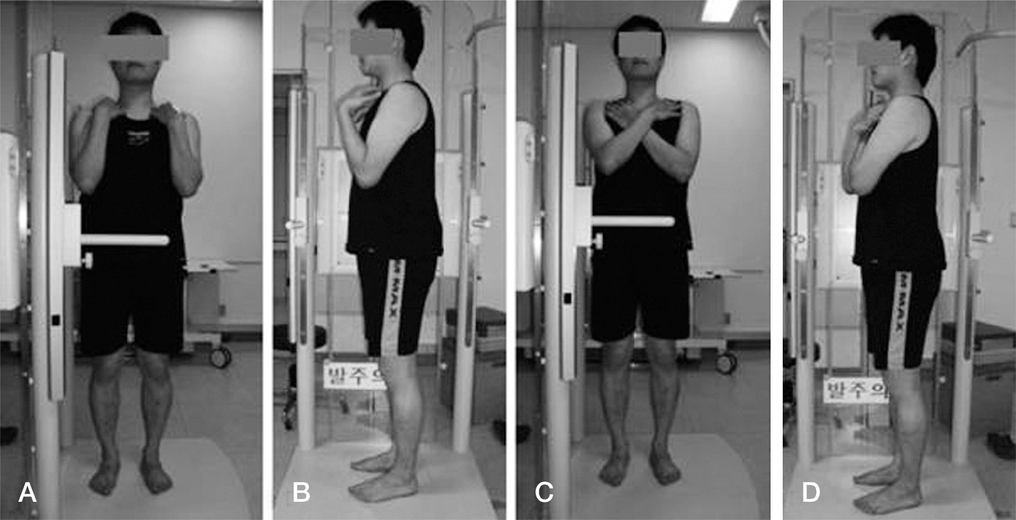
Fig. 1.
Fits-on-clavicle position (A, B) or cross-arm position (C, D) is recommended during taking radiographs.

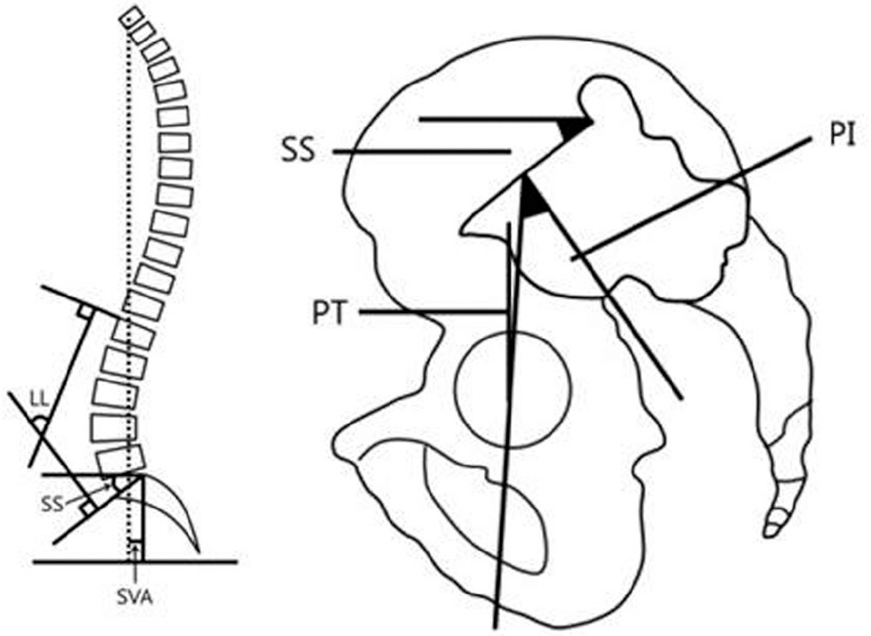
Fig. 2.
Spinal & Pelvic parameters. LL: lumbar lordosis, SS: Sacral slope, SVA: Sagittal vertical axis, PI: Pelvic incidence, PT: Pelvic tilt.

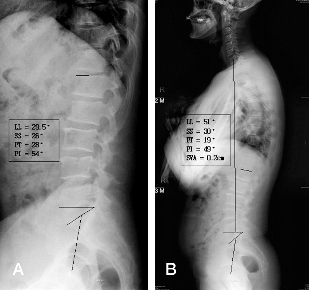
Fig. 3.
A 53-year-old man's supine lumbar lateral radiograph (A) and whole spine lateral radiograph with SVA ≤ 5 cm (B).

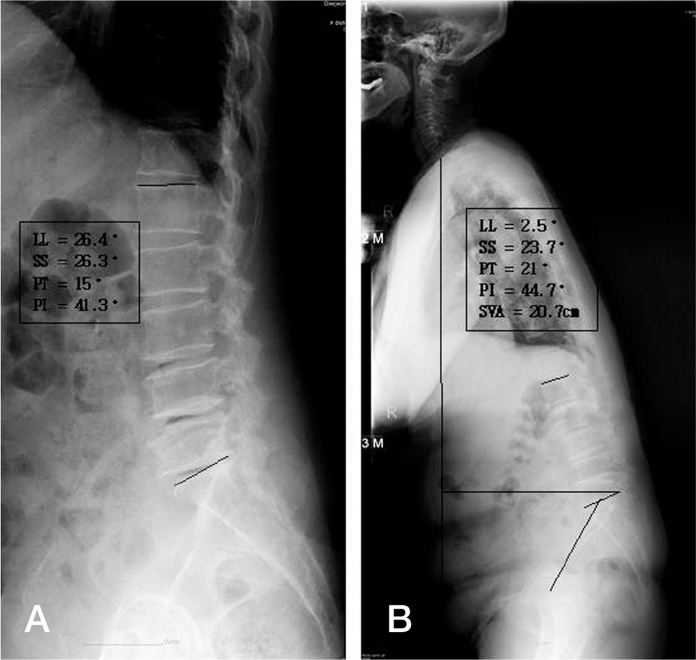
Fig. 4.
A 75-year-old woman's supine lumbar lateral radiograph (A) and whole spine lateral radiograph with SVA > 5 cm (B).

Table 1.
Supine lumbar lateral radiograph vs whole spine lateral radiograph
| Supine Lumbar Lateral Radiograph | Whole spine Lateral Radiograph | p-value | |
|---|---|---|---|
| LL | 35.1˚(±14.4˚) | 37.7˚(±16.8˚) | 0.170 |
| SS | 32˚(±9.6˚) | 31.7˚(±9.8˚) | 0.085 |
| PT | 24.3˚(±10.2˚) | 24.2˚(±11.1˚) | 0.132 |
Table 2.
Spine & pelvic parameter(˚) vs SVA(cm)
| p-value | |
|---|---|
| sLL* and SVA | 0.612 |
| sSS and SVA | 0.820 |
| sPT and SVA | 0.510 |
Table 3.
Lumbar lordosis(˚) & SVA(cm) in AI and AII
| sLL* | wLL | p-value | |
|---|---|---|---|
| AI (<30˚) | 20.0˚(±7.6˚) | 23.5˚(±13.5) | 0.520 |
| AII (≥30˚) | 43.5˚(±9.6˚) | 45.7˚(±12.7) | 0.174 |
Table 4.
BI vs BII


 PDF
PDF ePub
ePub Citation
 Print
Print


 XML Download