Abstract
Objectives
To evaluate the usefulness of iliac screws in the surgical correction of sagittal imbalance by changes of spinopelvic parameters.
Summary of Literature Review
Although reports exist regarding the fusion rates on lumbosacral fusion by iliac screws, no previous studies address the issue of changes of spinopelvic parameters on surgical correction of sagittal imbalance by iliac screws.
Materials and Methods
We analyzed a total of 23 patients who were operated on by pedicle subtraction osteotomy and posterior fusion on sagittal imbalance. Patients were divided into two groups: 1) non-iliac screw fixation and; 2) iliac screw fixation. The two groups were compared during the preoperative and postoperative stages, and the last followup spinopelvic parameters of two groups.
Results
Spinopelvic parameters, except for pelvic incidence, were corrected after surgery; some corrected values of spinopelvic parameters were lost during followup. There was a statistically significant difference in the last followup period between lumbar lordosis and pelvic tilt. Values of postoperative lumbar lordosis and pelvic tilt was similar to each other; however, during the followup period corrected values of spinopelvic parameters of non-iliac screw fixation group were more lost. There were no statistically significant changes in postoperative and last followup sacral slope and pelvic incidence.
Conclusions
Sagittal imbalance could be corrected by pedicle subtraction osteotomy, and corrected values of lumbar lordosis and pelvic tilt of iliac screw fixation group could be maintained well compared to non-iliac screw fixation. Iliac screw fixation could be useful for maintenance of corrected values of spinopelvic parameters in surgical correction of sagittal imbalance.
Go to : 
REFERENCES
1. Vialle R, Levassor N, Rillardon L, Templier A, Skalli W, Guiqui P. Radiographic analysis of the sagittal alignment and balance of the spine in asymptomatic subjects. J Bone Joint Surg Am. 2005; 87:260–7.

2. Jackson RP, Peter MD, McManus AC, Hales C. Compen-satory spinopelvic balance over the hip axis and better reli-ability in measuring lordosis to the pelvic radius on standing lateral radiographs of adult volunteers and patients. Spine (Phila Pa 1976). 1998; 23:1750–67.

3. Legaye J, Duval-Beaupere G, Hecquet J, Marty C. Pelvic incidence: a fundamental pelvic parameter for three-dimensional regulation of spinal sagittal curves. Eur Spine J. 1998; 7:99–103.

4. Vaz G, Roussouly P, Berthonnaud E, Dimnet J. Sagittal morphology and equilibrium of pelvis and spine. Eur Spine J. 2002; 11:80–7.

5. Schwab F, Lafage V, Patel A, Farcy JP. Sagittal plane considerations and the pelvis in the adult patient. Spine (Phila Pa 1976). 2009; 34:1828–33.

6. Duval-Beaupere G, Schmidt C, Cosson P. A barycentre-metric study of the sagittal shape of spine and pelvis: the conditions required for an economic standing position. Ann Biomed Eng. 1992; 20:451–62.

7. Vaz G, Roussouly P, Berthonnaud E, Dimnet J. Sagittal morphology and equilibrium of pelvis and spine. Eur Spine J. 2002; 11:80–7.

8. Mangione P, Gomez D, Senegas J. Study of the course of the incidence angle during growth. Eur Spine J. 1997; 6:163–7.

9. Hyun SJ, Rhim SC, Kim YJ, Kim YB. A midterm followup result of spinopelvic fixation using iliac screws for lumbosacral fusion. J Korean Neurosurg Soc. 2010; 48:347–53.

10. Moshirfar A, Rand FF, Sponseller PD, et al. Pelvic fixation in spine surgery. Historical overview, indications, biomechanical relevance, and current techniques. J Bone Joint Surg Am. 2005; 87:89–106.
11. Jackson RP, McManus AC. The iliac buttress. A computed tomographic study of sacral anatomy. Spine (phila Pa 1976). 1993; 18:1318–28.
12. Emami A, Deviren V, Berven S, Smith JA, Hu SS, Bradford DS. Outcome and complications of long fusion to the sacrum in adult spine deformity: luque-galvestone, combined iliac and sacral screw, and sacral fixation. Spine (phila Pa 1976). 2002; 27:776–86.
13. Kasten MD, Rao LA, Priest B. Longterm results of iliac wing fixation below extensive fusions in ambulatory adult patiens with spinal disorders. J Spinal Disord Tech. 2010; 23:E37–42.
14. Kuklo TR, Bridewell KH, Lewis SJ, et al. Minimum 2-year analysis of L5-S1 fusion using S1 and iliac screws. Spine (phila Pa 1976). 2001; 26:1976–83.
15. Tsuchiya K, Bridewell KH, Kuklo TR, Lenke LG, Baldus C. Minimum 5-year analysis of L5-S1 fusion using sacropelvic fixation(bilateral S1 and iliac screws) for spinal deformity. Spine (phila Pa 1976). 2006; 31:303–8.
16. Philips JH, Gutheil JP, Knapp DR. Iliac screw fixation in neuromuscular scoliosis. Spine (phila Pa 1976). 2007; 32:1566–70.
17. Zahi R, Vialle R, Abelin K, Mary P, Khouri N, Damsin J. Spinopelvic fixation with iliosacral screws in neuromuscular spinal deformities: results in a prospective cohort of 62 patients. Childs Nerv Syst. 2010; 26:81–6.

18. Kim WJ, Kang JW, Kim HY, et al. Change of Pelvic Tilt before and after Gait in Patients with Lumbar Degenerative Kyphosis. J Korean Soc Spine Surg. 2009; 16:95–103.

19. Kim WJ. Optimal Standing Radiographic Positioning in Patients with Sagittal Imbalance. J Korean Soc Spine Surg. 2010; 17:198–204.

20. Jackson RP, McManus AC. Radiographic analysis of sagittal plane alignment and balance in standing volunteers and patients with low back pain matched for age, sex, and size. A prospective controlled clinical study. Spine (Phila Pa 1976). 1994; 19:1611–8.
21. Barrey C, Jund J, Noseda O, Roussouly P. Sagittal balance of the pelvis-spine complex and lumbar degenerative diseases. A comparative study about 85 cases. Eur Spine J. 2007; 16:1459–67.

22. Gottfried ON, Daubs MD, Patel AA, Dailey AT, Brodke DS. Spinopelvic parameters in postfusion flatback deformity patients. Spine J. 2009; 9:639–47.

23. Tanguay F, Mac-Thiong JM, de Guise JA, Labelle H. Relation between the sagittal pelvic and lumbar spine geometries following surgical correction of adolescent idiopathic scoliosis. Eur Spine J. 2007; 16:531–6.

24. Schwab F, Patel A, Ungar B, Farcy JP, Lafage V. Adult spinal deformity-postoperative standing imbalance: how much can you tolerate? An overview of key parameters ini assessing alignment and planning corrective surgery. Spine (Phila Pa 1976). 2010; 35:2224–31.
25. Rose PS, Bridwell KH, Lenke LG, et al. Role of pelvic incidence, thoracic kyphosis, and patient factors on sagittal plane correction following pedicle subtraction osteotomy. Spine (Phila Pa 1976). 2009; 34:785–91.

26. Lafage V, Schwab F, Vira S, Patel A, Ungar B, Farcy JP. Spinopelvic parameters after surgery can be predicted: a preliminary formula and validation of standing alignment. Spine (Phila Pa 1976). 2011; 36:1037–45.
27. Edwards CC 2nd, Bridwell KH, Patel A, Rinella AS, Berra A, Lenke LG. Long adult deformity fusions to L5 and the Sacrum. A matched cohort analysis. Spine (Phila Pa 1976). 2004; 29:1996–2005.

28. Kim WJ, Kang JW, Yang DS, et al. Radiologic Analysis of Postoperative Sagittal Plane Correction in Lumbar Degenerative Kyphosis (LDK). J Korean Soc Spine Surg. 2009; 16:177–85.

29. Kim WJ, Kang JW, Kim KH, et al. Analysis of Correction Loss after Pedicle Subtraction Osteotomy in Patients with Sagittal Imbalance: Radiologic Aspects. J Korean Orthop Assoc. 2004; 39:629–35.

30. Kim YJ, Bridewell KH, Lenke LG, Rhim S, Cheh G. Pseudoarthrosis in Long Adult Spinal Deformity Instrumentation and Fusion to the sacrum: Prevalence and Risk Factor Analysis of 144 Cases. Spine (Phila Pa 1976). 2006; 31:2329–36.
Go to : 
Figures and Tables%
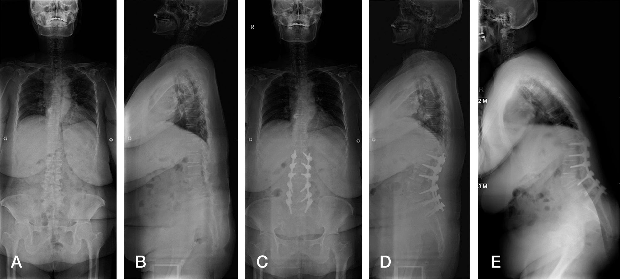
 | Fig. 1.Serial five whole spine x-ray shows initial, postoperative & last followup changes of spinopelvic parameters on non-iliac screw group. (A) Initial whole spine AP x-ray, (B) initial whole spine lateral x-ray, (C) postoperative whole spine AP x-ray, (D) postoperative whole spine lateral x-ray, (E) last followup whole spine lateral x-ray. |
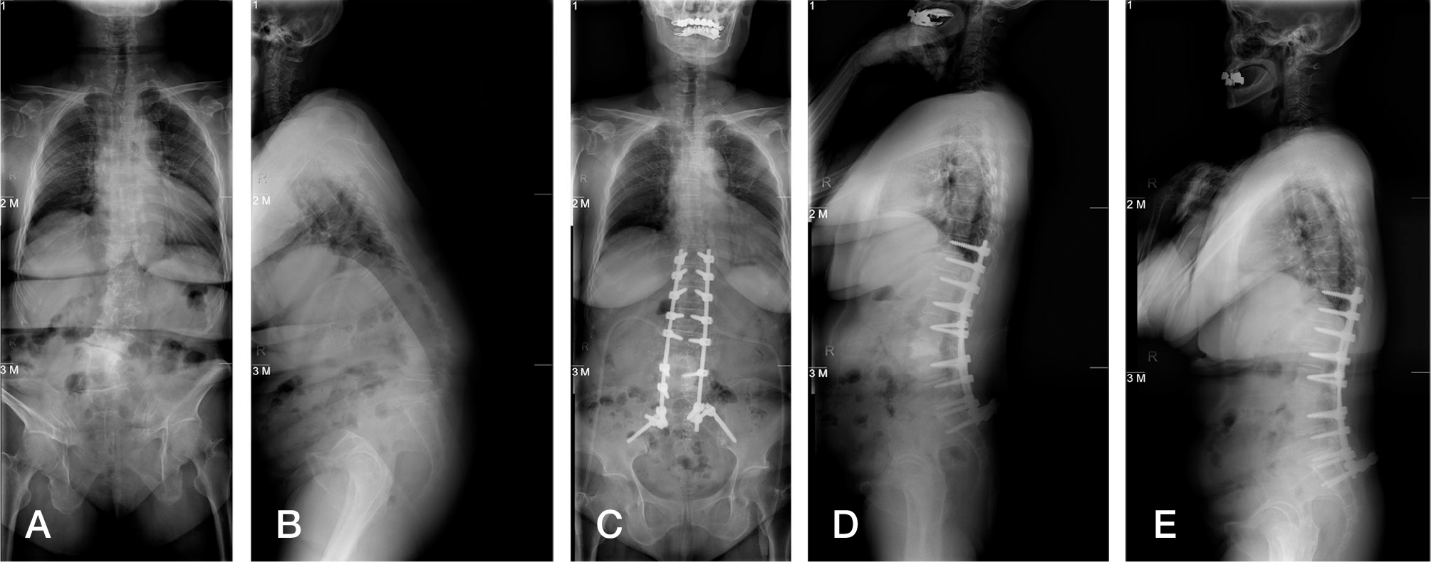
 | Fig. 2.Serial five whole spine x-ray shows initial, postoperative & last followup changes of spinopelvic parameters on iliac screw group. (A) Initial whole spine AP x-ray, (B) initial whole spine lateral x-ray, (C) postoperative whole spine AP x-ray, (D) postoperative whole spine lateral x-ray, (E) last followup whole spine lateral x-ray. |
Table 1.
Demographics of the patients.
Table 2.
The initial Spinopelvic parameters.
| Parameter | Group A | Group B | P-value |
|---|---|---|---|
| SVA | 12.8←5.3 | 13.5←7.1 | 0.880 |
| TK | 13.1←9.1 | 12.8←8.1 | 0.880 |
| LL | 21.8←12.0 | 21.3←6.8 | 0.976 |
| SS | 23.3←4.8 | 20.7←6.4 | 0.379 |
| PT | 36.6←6.5 | 37.3←7.0 | 0.928 |
| PI | 59.0←7.4 | 58.9←3.6 | 0.316 |
Table 3.
The postoperative changes of Spinopelvic parameters.


 PDF
PDF ePub
ePub Citation
 Print
Print


 XML Download