

 PDF
PDF ePub
ePub Citation
 Print
Print


Abstract
Objectives
We studied the clinical results and prognostic factors for the postoperative caudaequinasyndrome (CES). Summary of Literature Review: The CES is a rare complication, but its aftereffects are serious. And no satisfactory discussion about its accurate treatment guidelines and prognosis has been provided yet.
Materials and Methods
10 patients who were diagnosed with a postoperative CES were enrolled from June 2004 to February 2011. Patients were classified into group I with a favorable neurologic prognosis and groupII without neurologic improvement. The medical history, diagnosis, involved segmentand duration till CES was obtained, the duration was performed till second decompression and the clinical symptoms and the outcome of surgical treatment were investigated.
Results
Group I contained of 6cases and group 4 of cases. On average were 1.25(0.5-3) hours required for group I and 22(8-38) hours for group II until CES was diagnosed. The time span for the second operation was 7(3-12) hours for group I and 12.25(5-24) hours for group II. Of 6 cases showing motor losswere 4 cases classified as group II at the last followup. Of 10 cases with voiding difficulties belonged 4 cases to the group II. Voiding difficulty was continued as clinical symptom in 4 patients of group II after the secondary decompression.
Conclusion
The less the motor loss and voiding difficulty before the secondary decompression and the faster diagnosis and surgical decompression, the better the prognosis. In particular, as voiding difficulty showed the lowest recovery rate, it is considered to affect prognosis and satisfaction most seriously.
REFERENCES
1. Mixter WJ, Barr JS. Rupture of the intervertebral disc with involvement of the spinal cord. N Engl J Med. 1934; 211:210–4.
2. Kim HT, Hong SM, Lee KI, Jung JW, Park YM. Cauda Equina Syndrome in the Lumbar disc Herniation. J Korean Soc Spine Surg. 1998; 5:116–21.
3. Cho YH, Chang SA, Park JY, Han JH, Shin JH. Posterior Epidural Migration of a Sequestrated Intervertebral lumbar disc with Cauda Equina Syndrome. J Korean Soc Spine Surg. 2008; 15:277–80.
4. Eyre Brook AL. A study of lateresults from disc operations. Present employment and residual complaints. Br J Surg. 1952; 39:289–96.
5. Jennett WB. A study of 25 cases of compression of the cauda equina by prolapsed intervertebral discs. J Neurol Neurosurg Psychiatry. 1956; 19:109–16.

6. O'Connell JEA. Protrusion of the lumbar intervertebral disc. A clinical review based on five hundred cases treated by ex-cision of the protrusion. J Bone Joint Surg Br. 1951; 33:8–30.
7. Shapiro S. Cauda Equina syndrome secondary to lumbar disc herniation. Neurosurgery. 1993; 32:743–7.
8. Lawton MT, Porter RW, Heiserman JE, Jacobowitz R, Sonntag VK, Dickman CA. Surgical management of spinal epidural hematoma: Relationship between surgical timing and neurological outcome. J Neurosurg. 1995; 83:1–7.
9. Sokolowski MJ, Garvey TA, Perl J 2nd, et al. Postoperative lumbar epidural hematoma: does size really matter? Spine (Phila Pa 1976). 2008; 33:114–9.
10. Uribe J, Moza K, Jimenez O, Green B, Levi AD. De-layed postoperative spinal epidural hematomas. Spine J. 2003; 3:125–9.
11. Park BM, Won YY. Clinical observation on 8 cases of cauda equina syndrome. J Korean Orthop Assoc. 1988; 23:184–92.
12. McCarthy MJ, Aylott CE, Grevitt MP, Hegarty J. Cauda equina syndrome: factors affecting longterm functional and sphincteric outcome. Spine (Phila Pa 1976). 2007; 32:207–16.
13. Gleave JR, Macfarlane R. Cauda equina syndrome: what is the relationship between timing of surgery and outcome? Br J Neurosurg. 2002; 16:325–8.
14. Scavarda D, Peruzzi P, Bazin A, et al. Postoperative spinal extradural hematomas. 14 cases. Neurochirurgie. 1997; 43:220–7.
15. O'Laoire SA, Corckard HA, Thomas DG. Prognosis for sphincteric recovery after operation for cauda equine compression owing to lumbar disc prolapse. BMJ. 1981; 282:1852–4.
16. Hussain SA, Gullan RW, Chitnavis BP. Cauda equina syndrome: outcome and implications for management. Br J Neurosurg. 2003; 17:164–7.
17. Gleave JRW, McFarlane R. Prognosis for recovery of bladder function following lumbar central disc prolapse. Br J Neurosurg. 1990; 76:205–10.
18. Kohles SS, Kohles DA, Karp AP, Erlich VM, Polissar NL. Time-dependent surgical outcomes following cauda equina syndrome diagnosis: comments on a meta-analysis. Spine (Phila Pa 1976). 2004; 29:1281–7.
19. Ahn UM, Ahn NU, Buchowski JM, Garrett ES, Sieber AN, Kostuik JP. Cauda equina syndrome secondary to lumbar disc herniation: a meta analysis of surgical outcomes. Spine (Phila Pa 1976). 2000; 25:1515–22.
Figures and Tables%
Fig. 1.
Post primary operative MRI of 70 year-old female with spinal stenosis L3-4, 4-5 c degenerative spondylolisthesis L4 on L5. Dorsally located epidural hematoma from T12 to L5 with compressed spinal cord, L4-5 level. (A) T2 weighted sagittal image demonstrates large epidural hematoma showing heterogenous signal intensity. (B) T1 weighted sagittal image demonstrate isointense or increased signal intensity. (C) T2-weighted axial image shows that cauda equine is compressed by hematoma at L4-5 level.
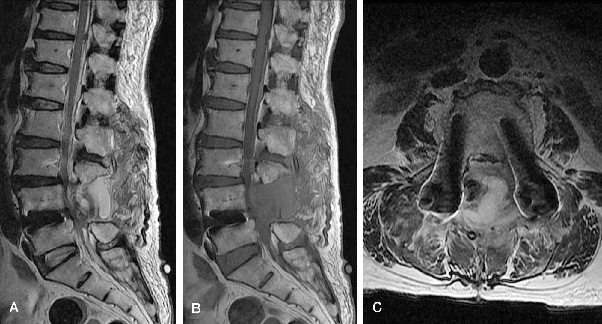
Table 1.
Patient data profile
| Group | Case | Sex | Age (year) | Etiology | Level | Duration of symptom* (hours) | Time to decompression† (hours) |
|---|---|---|---|---|---|---|---|
| I | 1 | M | 36 | Stenosis | L4-S1 | 0.5 | 3 |
| 2 | M | 56 | Stenosis | L4-S1 | 3 | 10 | |
| 3 | F | 79 | Stenosis | L3-S1 | 1.25 | 12 | |
| 4 | M | 78 | HNP | L3-L4 | 0.5 | 4 | |
| 5 | F | 70 | Stenosis | L4-S1 | 0.75 | 3 | |
| 6 | F | 80 | HNP | L5-S1 | 1.5 | 10 | |
| II | 7 | M | 40 | Stenosis | L4-S1 | 8 | 5 |
| 8 | M | 62 | Stenosis | L4-S1 | 18 | 8 | |
| 9 | F | 66 | Stenosis | L3-L5 | 24 | 12 | |
| 10 | M | 62 | Stenosis | L3-S1 | 38 | 24 |
Table 2.
The effect of early diagnosis and decompression on outcome in cauda equina syndrome
| Outcome | Duration from primary operation to 2nd operation* | |
|---|---|---|
| <24 hours (N) | 24-62 hours (N) | |
| Group I (n=6) | 6 | 0 |
| Group II (n=4) | 1 | 3 |
Table 3.
Clinical manifestations of postoperative cauda equina syndrome
Table 4.
Residual symptoms of postoperative cauda equina syndrome after 2nd surgery in group II at longterm follow up
| Group II (n=4)* | |
|---|---|
| Lower back pain | 0 |
| Sciatic neuropathy | 0 |
| Saddle anesthesia | 1 |
| Motor loss of the lower extremities | 1 |
| Sensory loss of the lower extremities | 0 |
| Voiding difficulty | 4 |
| Anal sphincter tone loss | 1 |
 XML Download