

 PDF
PDF ePub
ePub Citation
 Print
Print


Abstract
Summary of Literature Review
Epidural air pseudocyst at the lumbar spine that provokes radiating pain and neurologic symptoms can be misdiagnosed as an epidural tumor or HIVD. Consequently, proper diagnosis and treatment of the epidural air pseudocyst at the lumbar spine is necessary.
Materials and Methods
We report on two patients with radiculopathy and neurologic symptoms resulting from epidural air pseudocysts. In one patient, the epidural air pseudocyst was found within the epidural ligament flavum area on an MRI, and fluoroscopic-guided FNA (fine needle aspiration) was performed. In the other, the epidural air pseudocyst was found behind the posterior longitudinal ligament and was accompanied by spinal stenosis. In this patient, we conducted open cystectomy and posterior decompression surgery.
REFERENCES
1. Lee DY, Lee S. L2 radicular compression caused by a foraminal extradural gas pseudocyst. J Korean Neurosurg Soc. 2010; 47:232–4.

2. Kakitsubata Y, Theodorou SJ, Theodorou DJ, et al. Symptomatic epidural gas cyst associated with discal vacuum phenomenon. Spine(Phila Pa 1976). 2009; 34:E784–9.
3. Qasho R, Santoro A, Vangelista T, et al. Nerve root compression by a gascontaining cyst associated with stenotic lateral recess. Case report and reviewof the literature. J Neurosurg Sci. 2001; 45:181–4.
4. Sei A, Mizutamari M, Fujimoto T, et al. Gas-filled intradural cysts of the lumbar spine and the possible pathogenesis. Spine J. 2009; 9:E6–8.
5. Giraud F, Fontana A, Mallet J, et al. Sciatica caused by epidural gas. Four case reports. Joint Bone Spine. 2001; 68:434–7.
6. Bosser V, Dietemann JL, Warter JM, et al. L5 radicular pain related to lumbar extradural gascontaining pseudocyst. Role of CT-guided aspiration. Neuroradiology. 1990; 31:552–3.
7. Kaymaz M, Oztanir N, Emmez H, et al. Epidural air en-trapment after spinal surgery. Clin Neurol Neurosurg. 2005; 107:421–4.
8. Kuh SU, Heo DH, Kim KS, Cho YJ. Lumar epidural gas-contining pseudocysts as a cause of severe radicular pain. Joint Bone Spine. 2011; 78:398–401.
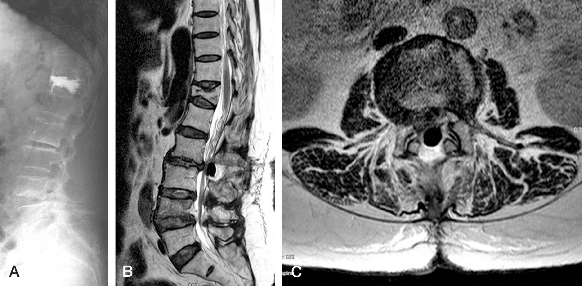
Fig. 1.
Multiple level degenerative disease (narrowing of the intervertabral) space and central vacuum phenomenon at lateral radiograph of the lumbar spine.
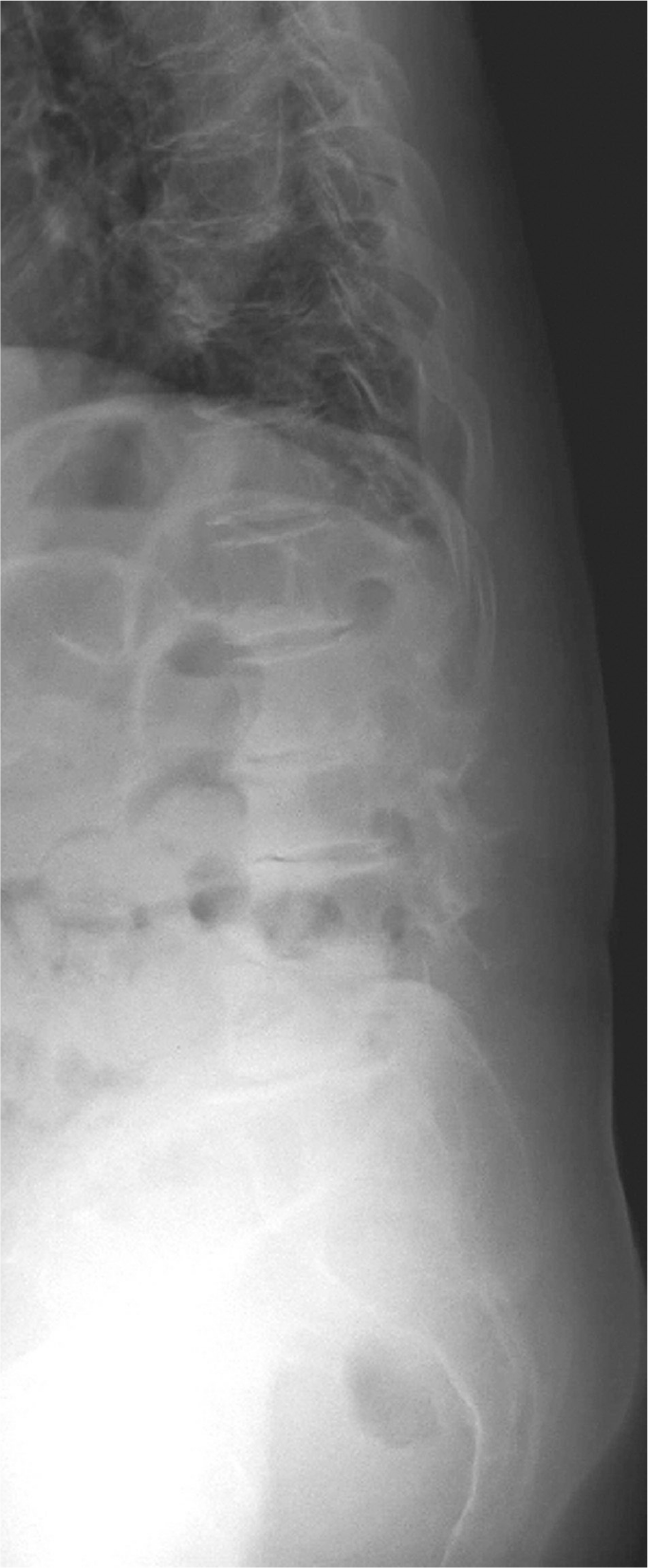
Fig. 2.
T2-weighted MRI sagittal (A) and T1-weighted MRI sagittal (B) of the lumbar spine. A round space occupying the spinal shows a very low signal, indicating the presence of gas (arrow).
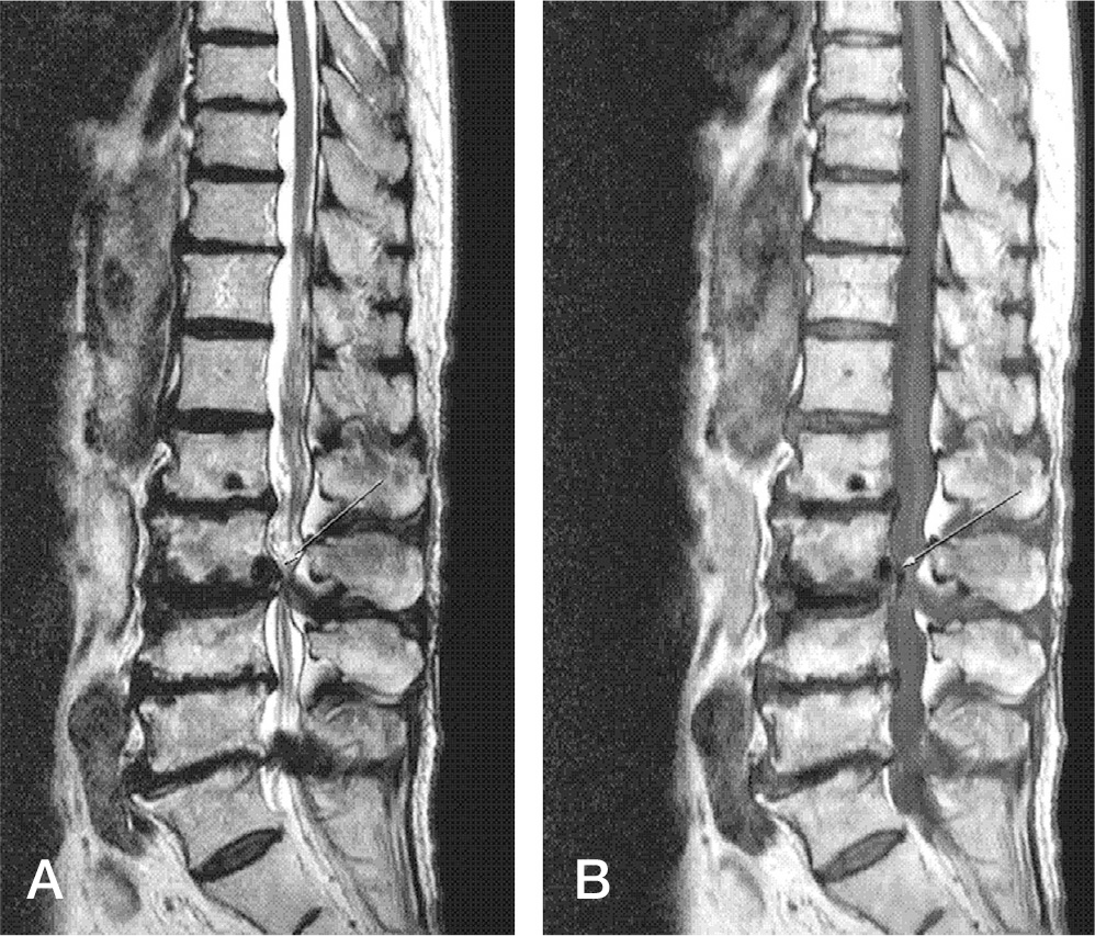
Fig. 3.
T2-weighted MRI axial image at L3-4 (A), L4-5 (B) and L5-S1 (C) of the lumbar spine. Air bubble (arrow) from intervertebral vacuum at L3-4. Right lateral recess and right foraminal stenosis at L4-5. Both lateral recess and left neural foraminal stenosis at L5-S1.
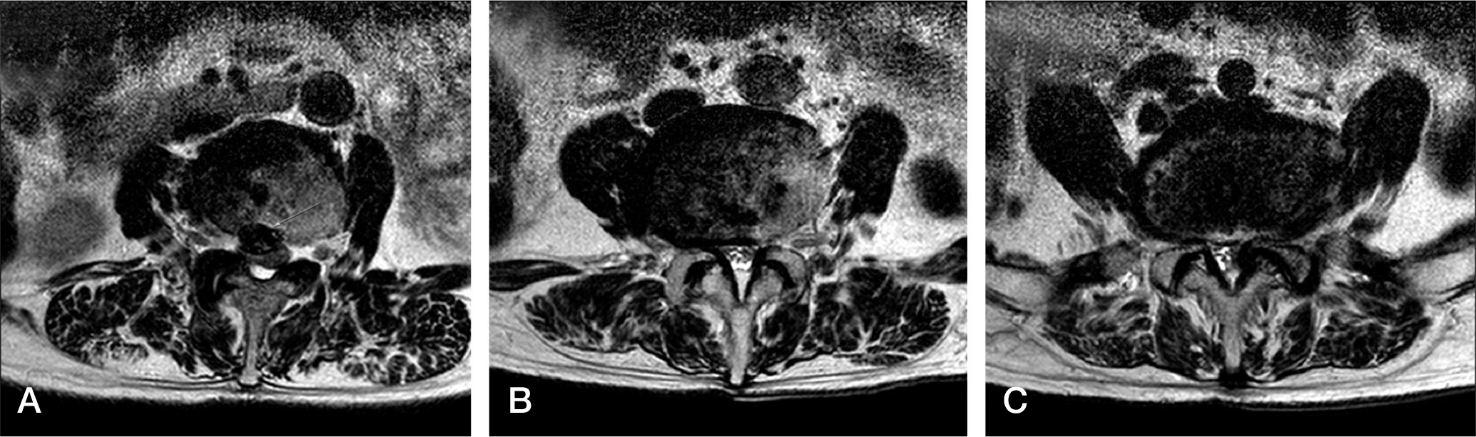
Fig. 4.
L3-4 Computed tomography demonstrate an air bubble(arrow) within the anterior part of the epidural space (A). Sagittal Computed tomography image demonstrate an air bubble(arrow) within the posteroinferior area of the L3 body (B).
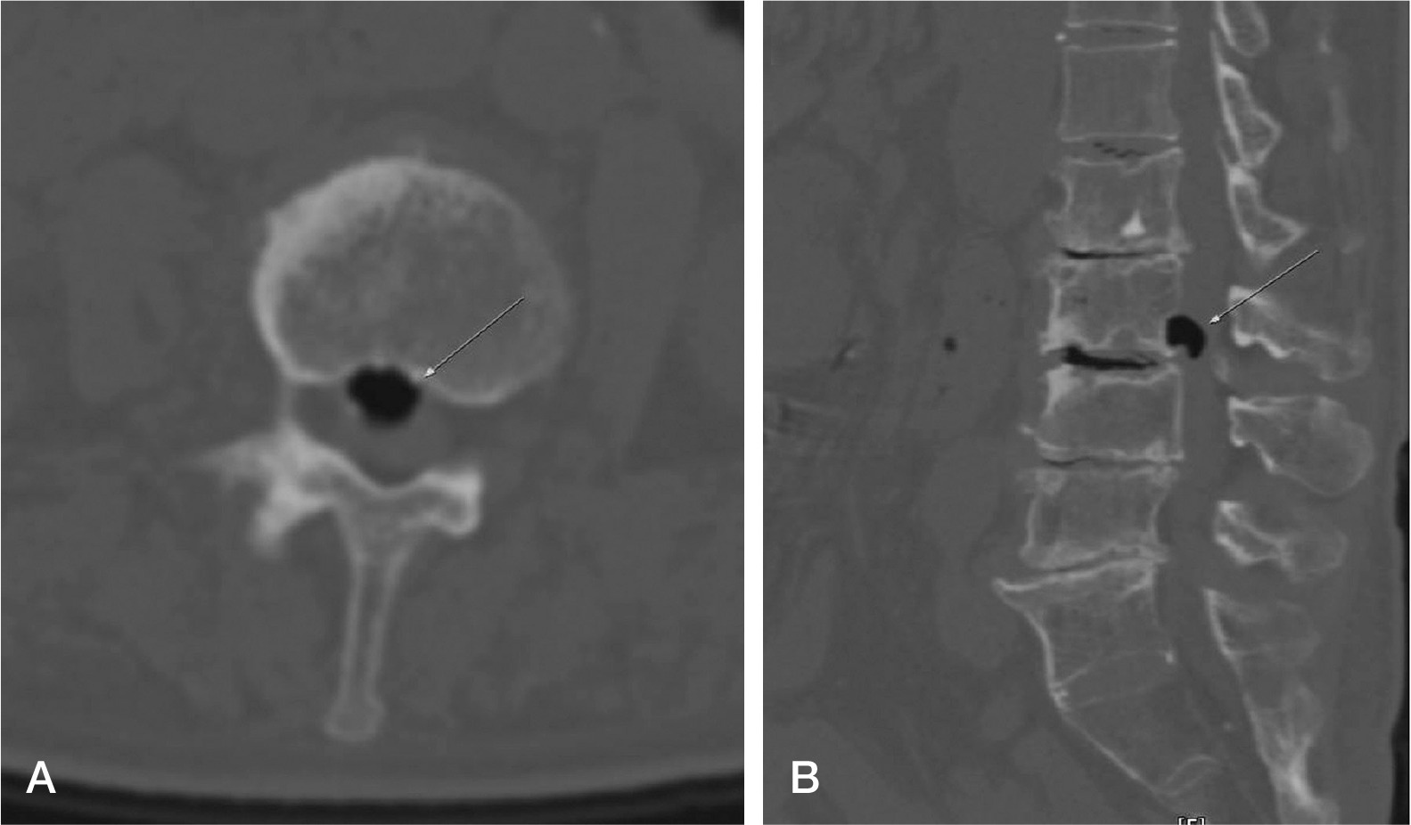
Fig. 5.
L3-4 Immediate post operative computed tomography demonstrate an air bubble within the anterior parth of the epidural space (A). Sagittal Computed tomography image demonstrate an air bubble within the posteroinferior area of the L3 body (B). both image demonstrate decreased air bubble.
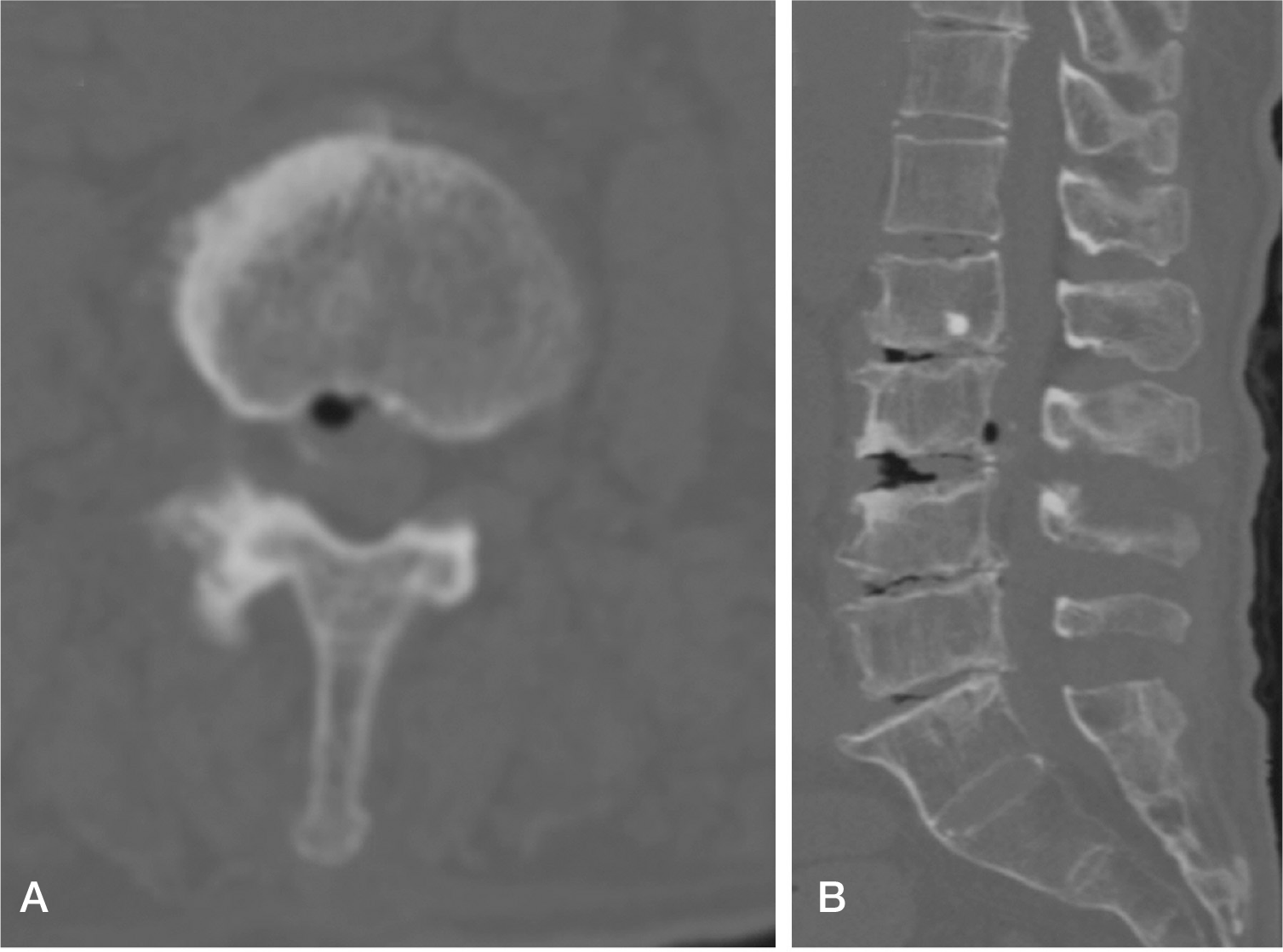
Fig. 6.
Lateral radiograph of the lumbar spine between L3-4 degenerative disease (narrowing of the intervertabral) space and central vacuum phenomenon (A). T2-weighted MRI sagittal (B) and axial (C) of the lumbar spine. A round space occupying the spinal shows a very low signal, indicating the presence of gas (S/p vertabroplasty in T12).
 XML Download