Abstract
Objectives
To observe the short term effect of selective nerve root block (sNRB) depending on the contrast pattern and spinal canal size.
Summary of Literature Review
A number of studies have demonstrated that sNRB is quite effective not only for patients with herniated intervertebral discs but also for those with spinal stenosis.
Materials and Methods
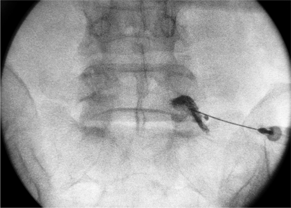
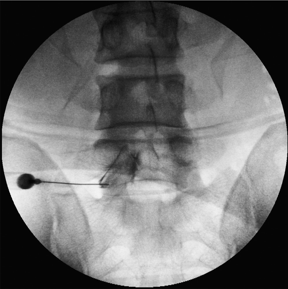
The Visual Analog Scale(VAS) score was collected before and after the procedure from 217 subjects with lumbar spinal stenosis and underwent sNRB. Two types were classified after observing the contrast's spreading pattern, Type I contrast reaching the spinal canal and Type II not reaching the spinal canal. Efficacy of the treatment for each type was also compared. In addition, the spinal canal size was classified into three categories. Treatment efficacy depending on the contrast pattern was also compared in each category.
Results
When divided into two types based on the contrast pattern, type I showed a more significant reduction in VAS score according to T-test although both types showed a decrease in VAS score after the procedure. In regards to spinal canal dimension, both types showed decreased VAS scores after the procedure in patients with spinal canal size larger than 172.2mm2; however, there were no changes in VAS score before and after the procedure for those with spinal canal size smaller than 73mm2.
Conclusions
There was a short term effect of selective nerve root block (sNRB) in patients with spinal stenosis regardless of their contrast pattern, type I group showing a stronger correlation. In regards to spinal canal dimension, patients with larger spinal canal sizes not only showed a significant decrease in VAS score after selective nerve root block (sNRB) but also showed differences depending on the contrast pattern. On the contrary, there was no significant difference in VAS score before and after selective nerve root block (sNRB) in patients with small spinal canal sizes, and there was also no difference in the outcome depending on the contrast pattern in patients with small spinal canal sizes. Therefore, when performing selective root nerve block (sNRB), the operator should remember to manipulate the angle and position of the spinal needle when injecting the appropriate drug after confirming that the contrast material reached the spinal canal. The operator should also consider surgical management when performing selective nerve root block (sNRB) in patients with severe central spinal stenosis.
Go to : 
REFERENCES
1. Bokduk N. Epidural steroids. Spine (Phila Pa 1976). 1995; 20:845–8.
2. Cuckler JM, Bernini PA, Wiesel SW, Booth RE, Rothman RH, Pickens GT. The use of epidural steroids in the treatment of lumbar radicular pain. A prospective, randomized, double-blind study. J Bone Joint Surg. 1985; 67:63–6.

3. Derby R, Kine G, Saal JA. Response to steroid and duration of raducular pain as predictors of surgical outcome. Spine (Phila Pa 1976). 1992; 17:176–83.
4. Van Tulder MW, Koes BW, Bouter LM. Conservative treatment of acute and chronic nonspecific low back pain. A systematic review of randomized controlled trials of the most common interventions. Spine (Phila Pa 1976). 1997; 22:2128–56.
5. Macnab I. Negative disc exploration. An analysis of the causes of nerve root involvement in sixty-eight patients. J Bone Joint Surg. 1971; 53:891–903.
6. Ng L, Chaudhary N, Sell P. The efficacy of corticosteroid in periradicular infiltration for chronic radicular pain. a randomized, double-blind, controlled trial. Spine (Phila Pa 1976). 2005; 30:857–62.
7. Narozny M, Zanetti M, Boos N. Therapeutic efficacy of selective nerve root blocks in the treatment of lumbar radicular leg pain. Swiss Med Wkly. 2001; 131:75–80.
8. Tajima T, Furukawa K, Kuramochi E. Selective lumbosacral radiculography and block. Spine (Phila Pa 1976). 1980; 5:68–77.

9. Hong YK, Sa SJ, Kim JD. Selective spinal nerve root block for the treatment of sciatica. J Korean Orthop Assoc. 1997; 32:1056–62.

10. Lee DH, Yang SH, Yang BR, Yi SR, Chung SW, Kim MS. The short term results of selective nerve root block in herniated lumbar disc patients. J Korean Soc Spine Surg. 2004; 11:216–22.

11. Shim DM, Kim TK, Song HH, You SS, Cho JD. The usefulness of selective spinal nerve root block. J Korean Soc Spine Surg. 2004; 11:48–54.

12. Kim SB, Jeon TS, Park WK, Jo SK, Kim YS, Hwang CM. Transforaminal selective nerve root blocks for treating single lumbosacral radiculopathy. J Korean Orthop Assoc. 2009; 44:619–26.
13. Lindahl D, Rexed B. Histologic changes in spinal nerve roots of operated cases of sciatica. Acta Orthop Scand. 1951; 20:215.

14. Kelman H. Epidural injection therapy of sciatic pain. Am J Surg. 1944; 64:183.
15. Kikuchi S, Hasue M, Nishyama K. Anatomic and clinical studies of radicular symptom. Spine (Phila Pa 1976). 1984; 9:23–30.
16. Derby R, Kine G, Saal JA. Response to steroid and duration of raducular pain as predictors of surgical outcome. Spine (Phila Pa 1976). 1992; 17:176–83.
17. Yokoyama M, Hanazaki M, Fujii H, Mizobuchi S, Nakat-suka H, Takahashi T. Correlation between the distribution of contrast medium and the extent of blockade during epidural anesthesia. Anesthesiology. 2004; 100:1504–10.

18. Amundsen T, Weber H, Nordal HJ, Magnaes B, Abdelnoor M, Lilleas F. Lumbar spinal stenosis: conservative or surgical management. A prospective 10-years study. Spine (Phila Pa 1976). 2000; 25:1424–36.
19. Atlas SJ, Keller RB, Robson D, Deyo RA, Singer DE. Surgial and nonsurgical management of lumbar spinal stenosis: four-year outcomes from the Maine lumbar spine study. Spine (Phila Pa 1976). 2000; 25:556–62.
Go to : 
Figures and Tables%
 | Fig. 2.Fluorography shows contrast extending to proximal and distal portion of L5 nerve root but not the spinal canal. |
 | Fig. 3.We measured the spinal canal dimension by using free line ROI calculator of Infinitt PACS system. |
Table 1.
Demographics of study participants
| (Age) | Male | Female | Total |
|---|---|---|---|
| 30-39 | 7 | 4 | 11 (5.0%) |
| 40-49 | 14 | 11 | 25 (11.5%) |
| 50-59 | 38 | 21 | 59 (27.1%) |
| 60-69 | 44 | 23 | 67 (30.8%) |
| 70-79 | 24 | 16 | 40 (18.4%) |
| 80- | 8 | 7 | 15 (6.9%) |
| Total | 135 (62.2%) | 82 (37.8%) | 217 (100%) |
Table 2.
Comparison of VAS score before and after 1 hour, 1month and 3months in conjunction with Type I and II in each time period
Table 3.
Comparison of VAS score for Type I and Type 1month and 3months depending on the canal size


 PDF
PDF ePub
ePub Citation
 Print
Print


 XML Download