Abstract
Summary of Literature Review
Little has been known about the sagittal curve patterns of cervical spine in AIS patients.
Materials and Methods
One-hundred-thirty-three AIS patients were checked by scanographs and followed up for more than 2 years were divided into cervical kyphosis (≥+5°), lordosis (≤-5°) and straight (-4°~+4°) groups according to the sagittal curves of cervical spine (C2~C7). Each group was evaluated for thoracic kyphosis, lumbar lordosis, sagittal balance and Cobb's angle on coronal plane. Of the patients, 49 were treated by braces, 84 were surgically corrected (rod derotation in 52, direct vertebral rotation (DVR) in 32).
Results
At the initial radiographs, cervical kyphosis was found in 97, lordosis in 23 and straight in 13 patients. In the kyphosis group, cervical kyphosis showed typical patterns of angular kyphosis. Thoracic and upper T-kyphosis (T1~T5) were lower than those in the cervical lordosis group (p=0.000, 0.001, respectively.) Other factors showed no significant differences between the groups. Patients treated by conservative management or by rod derotation had no significant differences in cervical kyphosis during the followup periods, though the thoracic hypokyphosis was surgically corrected. On the contrary, patients who were treated by DVR restored cervical lordosis (14/32=43.8%) from initial state showed significant differences in both conservative and rod derotation groups (p=0.008, 0.002, respectively)
Go to : 
REFERENCES
1. Hilibrand AS, Tannenbaum DA, Graziano GP, Loder RT, Hensinger RN. The sagittal alignment of the cervical spine in adolescent idiopathic scoliosis. J Pediatr Orthop. 1995; 15:627–32.

2. Canavese F, Turcot K, De Rosa V, de Coulon G, Kaelin A. Cervical spine sagittal alignment variations following posterior spinal fusion and instrumentation for adolescent idiopathic scoliosis. Eur Spine J. 2011; 20:1141–8.

3. Winter RB, Lovell WW, Moe JH. Excessive thoracic lordosis and loss of pulmonary function in patients with idiopathic scoliosis. J Bone Joint Surg Am. 1975; 57:972–6.

4. Berthonnaud E, Dimnet J, Roussouly P, Labelle H. Analysis of the sagittal balance of the spine and pelvis using shape and orientation parameters. J Spinal Disord Tech. 2005; 18:40–7.

5. Takeshima T, Omokawa S, Takaoka T, Araki M, Ueda Y, Takakura Y. Sagittal alignment of cervical flexion and extension: lateral radiographic analysis. Spine (Phila Pa 1976). 2002; 27:E348–55.
6. Penning L. Acceleration injury of the cervical spine by hy-pertranslation of the head: effect of normal translation of the head on cervical spine motion. Eur Spine J. 1992; 1:7–19.
7. Faro FD, Marks MC, Pawelek J, Newton PO. Evaluation of a functional position for lateral radiograph acquisition in adolescent idiopathic scoliosis. Spine (Phila Pa 1976). 2004; 29:2284–9.

8. Marks M, Stanford C, Newton P. Which lateral radiographic positioning technique provides the most reliable and functional representation of a patient's sagittal balance? Spine (Phila Pa 1976). 2009; 34:949–54.

9. Bernhardt M, Bridwell KH. Segmental analysis of the sagittal plane alignment of the normal thoracic and lumbar spines and thoracolumbar junction. Spine (Phila Pa 1976). 1989; 14:717–21.

10. Harrison DE, Harrison DD, Janik TJ, Holland B, Siskin LA. Slight head extension: does it change the sagittal cervical curve? Eur Spine J. 2001; 10:149–53.

11. Erkan S, Yercan HS, Okcu G, Ozalp RT. The influence of sagittal cervical profile, gender and age on the thoracic kyphosis. Acta Orthop Belg. 2010; 76:675–80.
Go to : 
Figures and Tables%
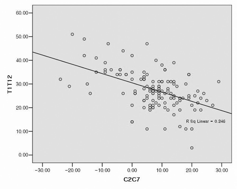
 | Fig. 1.Negative correlations of sagittal angles between Cervical and Thoracic curves (r=-.496, P <0.01, Pearson coefficients). |
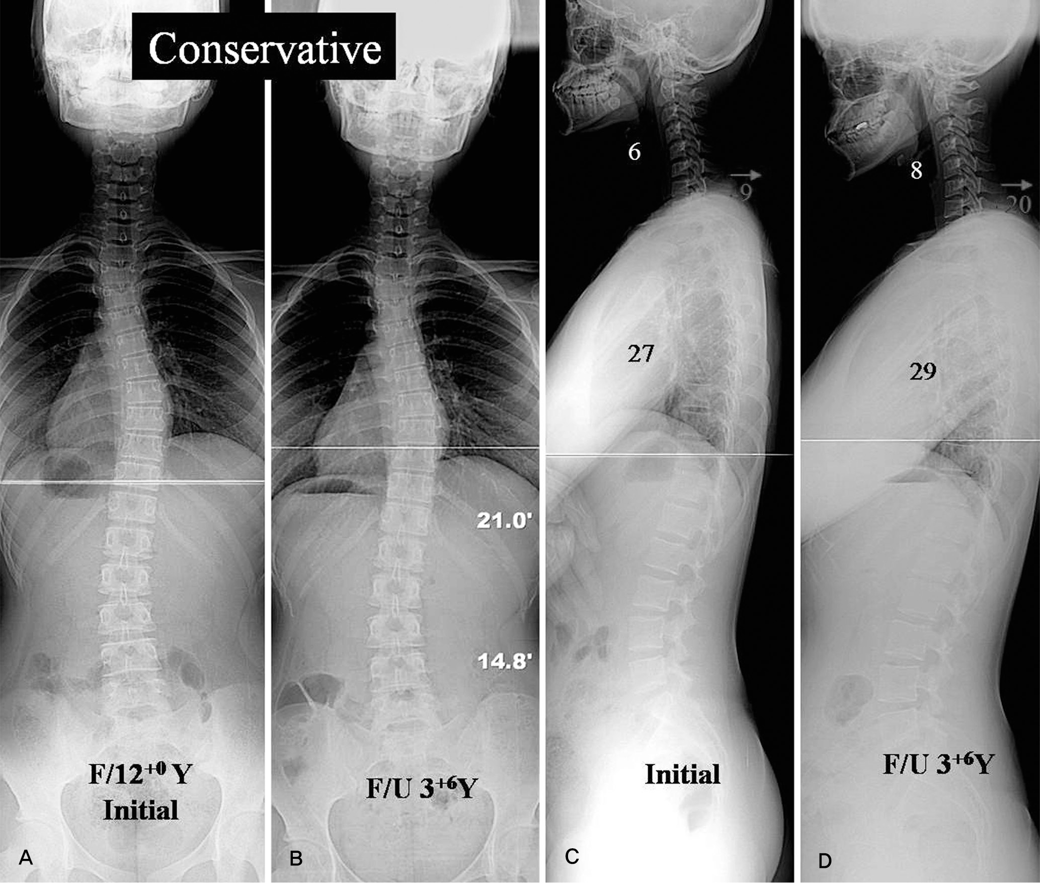
 | Fig. 2.(A, B) A twelve-year-old AIS patient with mild thoracic scoliosis showed cervical kyphosis of 6 degrees at the initial radiographs. She had trunk balance both in coronal and sagittal planes. (C, D) The curves were not progressed in the coronal plane during the followup periods of 30 months. Cervical curve was remained and was checked cervical kyphosis of 8 degrees in the sagittal plane. |
 | Fig. 3.(A, B) A fourteen-year-old AIS patient with double major curves showed cervical kyphosis of 18 degrees and thoracic hypokyphosis of 24 degrees (T1~T12) at the initial radiographs. (C, D) The patient was operated by pedicle screw fixation with rod derotation maneuver. Thoracic hypokyphosis was well corrected to normokyphosis of 34 degrees. However, cervical kyphosis was not changed though thoracic hyphokyphosis was well corrected. Preoperative cervical kyphosis of 18 degrees was slightly changed to kyphosis of 15 degrees with no significant difference. |
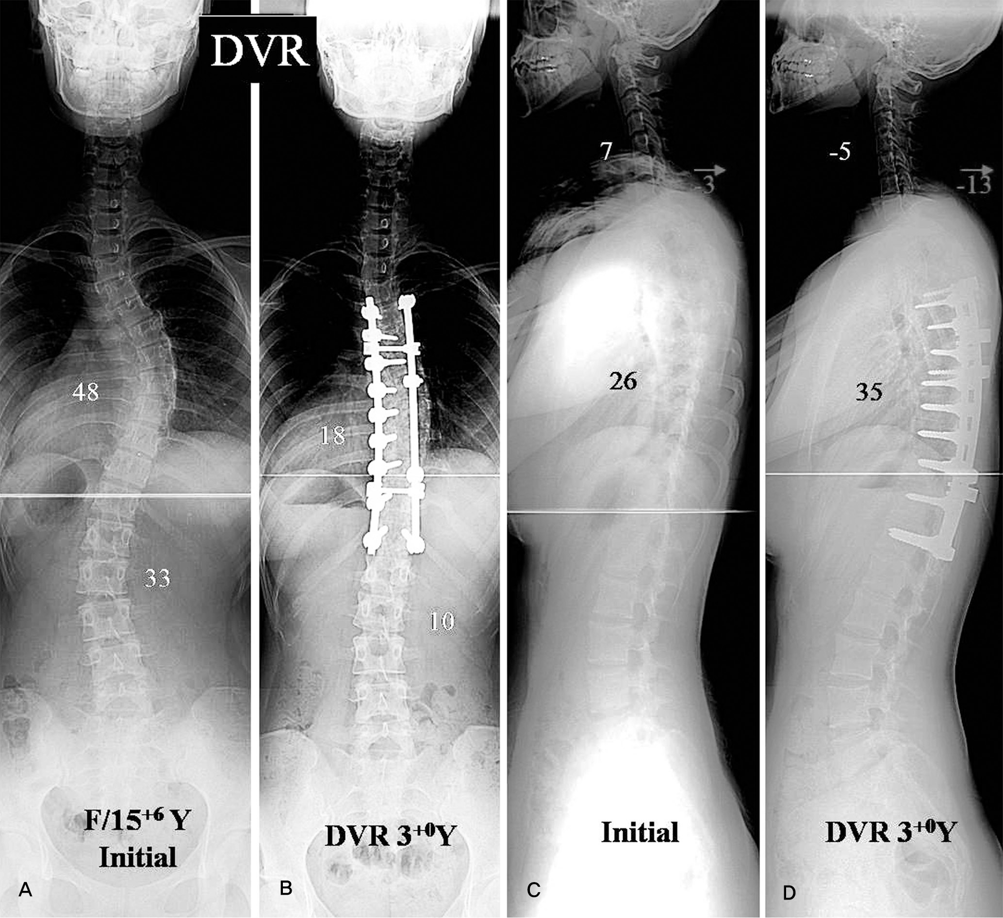
 | Fig. 4.(A, B) A fifteen-year-old AIS patient with King type II curve showed cervical kyphosis of 7 degrees and thoracic hypokyphosis of 26 degrees at the initial radiographs. (C, D) She was operated by direct vertebral rotation (DVR). Postoperative cervical curve in the sagittal plane showed a noticeable change. Preoperative cervical kyphosis of 7 degrees was changed to cervical lordosis of 5 degrees after the operation with a statistically significant difference. |
Table 1.
Characteristics in Cervical Curves Groups
Table 2.
Changes of Curves Angles and Types according to the Treatment Methods


 PDF
PDF ePub
ePub Citation
 Print
Print


 XML Download