Abstract
Objectives
The purpose of this study is to confirm the clinical usefulness of utilizing ProSet imaging for checking the nerve root compression and swelling in extraforaminal disc herniation.
Summury of Literature Review
Diagnosing extraforaminal disc herniations can be neglected with using a conventional MRI.
Materials and Methods
A retrospective analysis was performed on 25 patients, who underwent both conventional & Principles of the selective excitation technique (ProSet) MR imaging for the evaluation of extraforaminal disc herniation, from April 2008 to October 2010. Radiographic analysis was based on the notion that the degree of nerve root compression and swelling was decided by Pfirrmann's classification.
Results
Severe compression in the ProSet 3D rendering image was observed in 21 subjects, as compared with 8 subjects in the conventional axial image. Especially, nothing was ever detected in the conventional sagittal image. Severe compression in the ProSet 3D rendering image was observed in 4 subjects, while their nerve root compression was not clear in the conventional axial image. Severe compression and severe swelling in the ProSet 3D & coronal image was observed in 15 subjects, while their nerve root compression was none or not clear in the conventional sagittal image. The swelling degree of the ProSet coronal image turned out bigger than the swelling degree of conventional axial image, and the signal intensity change was also obvious.
Go to : 
REFERENCES
1.Benini A. Der Zugang zu den lateralen lumbalen diskush-ernien ambeispiel einer hernie L4/L5. Operat Orthop Trau-matol. 1988. 10:103–16.
2.Ohmori K., Kanamori M., Kawaguchi Y., Ishihara H., Kimura T. Clinical features of extraforaminal lumbar disc herniation based on the radiographic location of the dorsal root ganglion. Spine (Phila Pa 1976). 2001. 26:662–6.

3.Jackson RP., Clah JJ. Foraminal and extraforaminal lumbar disc herniation: Diagnosis and treatment. Spine (Phila Pa 1976). 1987. 12:577–85.

4.Junichi K., Mitsuo H. Diagnosis and operative treatment of intraforaminal & extraforaminal nerve root compression. Spine (Phila Pa 1976). 1991. 16:1312–20.
5.Macnab I. Negative disc exploration: An analysis of the causes of nerve-root involvement in sixty-eight patients. J Bone Joint Surg. 1971. 53:891–903.
6.Abdullah AF., Ditton EW III., Byrd EB. Extreme-lateral lumbar disc herniations: Clinical syndrome and special problems of diagnosis. J Neusurg. 1974. 41:229–34.
7.Kim MH., Suh KJ., Lee JY., Min SH., Yoo HY. Usefulness of Coronal MR Image in Diagnosis of Foraminal and Extraforaminal Disc Herniation. J Korean Soc Spine Surg. 2008. 15:165–73.

8.Pfirrmann CW., Dora C., Schmid MR., Zanetti M., Holdler J., Boos N. MR image-based grading of lumbar nerve root compromise due to disk herniation: Reliability study with surgical correlation. Radiology. 2004. 230:583–8.

9.Lindblom K. Protrusions of disks and nerve compression in the lumbar region. Acta Radiol. 1944. 25:195–212.

10.Bronx NY. Foraminal and far lateral lumbar disc herniations: Surgical alternatives outcome measures. Spinal Cord. 2002. 40:491–500.
11.Faust SE., Ducker TB., VanHassent JA. Lateral lumbar disc herniations. J Spinal Disord. 1992. 5:97–103.

12.Lee CK., Rauschning W., Glenn W. Lateral lumbar canal stenosis: Classification, pathologic anatomy and surgical decompression. Spine (Phila Pa 1976). 1988. 13:313–20.
13.Forristal RM., Marsh HO., Pay NT. MRI & CT of the lumbar spine: Comparison of diagnotic methods and correlation with surgical findings. Spine (Phila Pa 1976). 1988. 13:1049–54.
14.Byun WM., Kin JW., Lee JK. Differentiation between symptomatic and asymptomatic extraforaminal stenosis in lum-bosacral transitional vertebra: role of three-dimensional magnetic resonance lumbosacral radiculography. Korean J Radiol. 2012. 13:403–11.

15.Byun WM., Ahn SH., Ahn MW. Value of 3D MR lum-bosacral radiculopathy in the diagnosis of symptomatic chemical radiculitis. Am J Neuroradiol. 2012. 33:529–34.
Go to : 
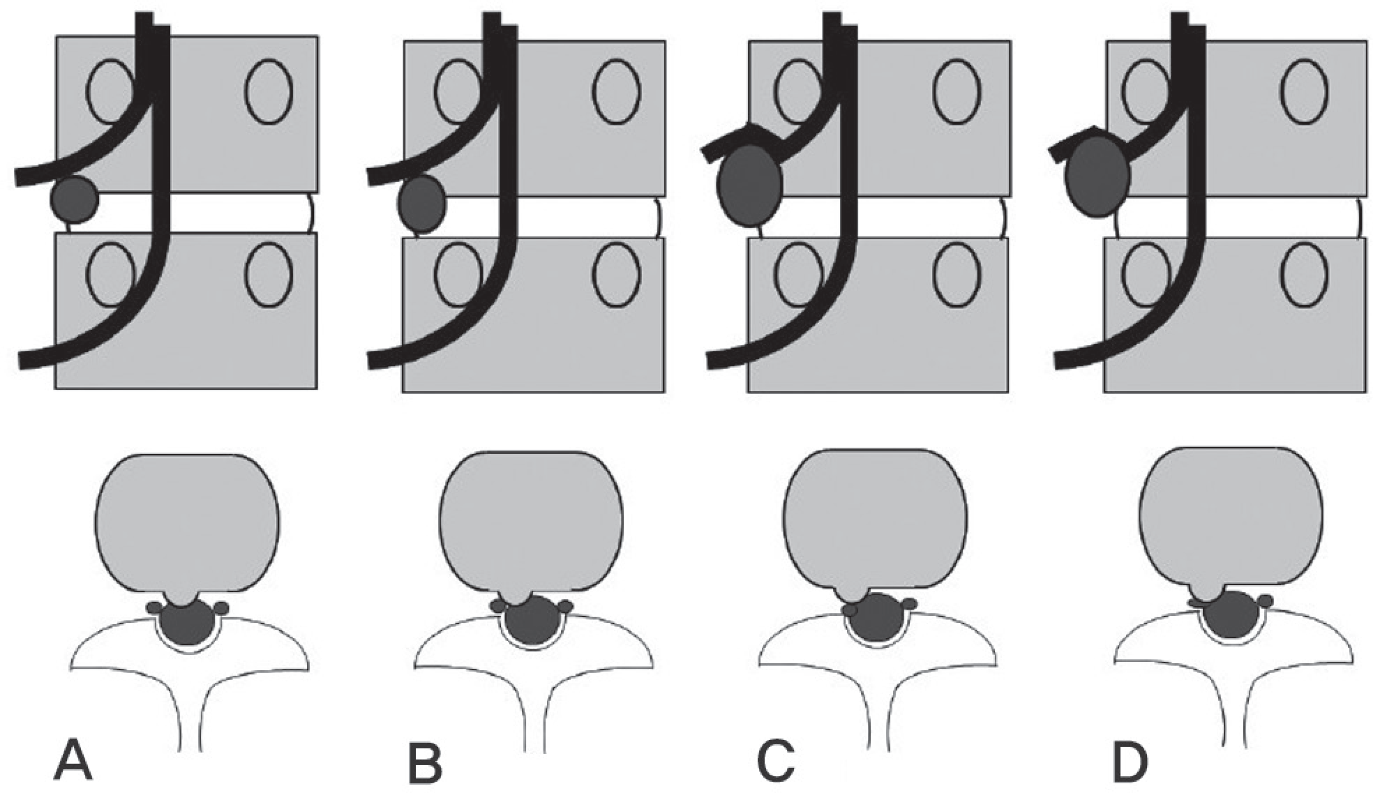
 | Fig. 1.Schematic diagram of a system for grading lumbar nerve root compromise in coronal plane and axial plane. (A) No compromise of the nerve root. (B) Contact of disc material with the right exiting nerve root. The nerve root is in the normal position and is not dorsally deviated. (C) Dorsal deviation of the right exiting nerve root caused by contact with disc material. (D) Compression of the right exiting nerve root between disc material and the surrounding structure (e.g. pedicle). It may be ap-pear flattened or be indistinguished from disc material. |
 | Fig. 2.Comparison of conventional MRI axial view and Proset coronal view, extraforaminal herniation extended to the foramen at L5-S1. (A) Axial T2-weighted image shows left extraforaminal disc herniation with extension to foramen at L5-S1. (B,C) Swelling of the L5 nerve root(arrow) on Proset coronal view and 3D MR radiography is seen. |
 | Fig. 3.Comparison of conventional MRI axial view and Proset coronal view of right extraforaminal herniation at L4-5. (A) Axial T2-weighted image shows contact of disc material with the right L4 exiting nerve root. The nerve root is in the normal position and is not dorsally deviated. (Pfirmann stage 1). (B,C) Swelling and compression of the right L4 exiting nerve root (arrow) between disc material and surrounding structure on Proset coronal view and 3D MR radiography is seen (Pfirmann stage 3). |
Table 1.
Compression grades according to classification with relation to the types of MR image in extraforaminal disc herniation of the lumbar spine
| Pfirrmann | Image type | Total | ||
|---|---|---|---|---|
| Conventional MRI | ProSet image | |||
| Axial | Sagittal | Coronal | ||
| 0 | 0 | 12 | 0 | 12 |
| 1 | 7 | 7 | 3 | 17 |
| 2 | 10 | 1 | 1 | 12 |
| 3 | 8 | 5 | 21 | 34 |
| Total | 25 | 25 | 25 | 75 |
Table 2.
Nerve root compression grade in the conventional axial image with relation to the ProSet 3D rendering image grade according to Pfirrmann's classification.
| Pfirrmann's grade | ProSet 3D rendering image | Total | |||
|---|---|---|---|---|---|
| 1 | 2 | 3 | |||
| Conventional axial image | 1 | 2 | 1 | 4 | 7 |
| 2 | 1 | 0 | 9 | 10 | |
| 3 | 0 | 0 | 8 | 8 | |
| Total | 3 | 1 | 21 | 25 | |
Table 3.
Nerve root compression grade in the conventional sagittal image with relation to the ProSet 3D rendering image grade according to Pfirrmann's classification.
| Pfirrmann's grade | ProSet 3D rendering image | Total | |||
|---|---|---|---|---|---|
| 1 | 2 | 3 | |||
| Conventional sagittal image | 0 | 2 | 0 | 10 | 12 |
| 1 | 1 | 1 | 5 | 7 | |
| 2 | 0 | 0 | 1 | 1 | |
| 3 | 0 | 0 | 5 | 5 | |
| Total | 3 | 1 | 21 | 25 | |


 PDF
PDF ePub
ePub Citation
 Print
Print


 XML Download