Abstract
Objectives
To look into the anatomical and pathophysiological features of cauda equina and support their basic knowledge of treating cauda equina syndrome.
Summary of Literature Review
Cauda equina has different anatomical and pathophysiological features to peripheral nerve.
Go to : 
REFERENCES
1. Konno S, Olmarker K, Byrod G, Rydevik B, Kikuchi S. Intermittent cauda equina compression: an experimental study of the porcine cauda equina with analyses of nerve impulse conduction properties. Spine (Phila Pa 1976). 1995; 20:1223–6.

2. Konno S, Yabuki S, Sato K, Olmarker K, Kikuchi S. A model for acute, chronic and delayed graded compression of the dog cauda equina. Presentation of the gross, microscopic and vascular anatomy of the dog cauda equina and accuracy in pressure transmission of the compression model. Spine (Phila Pa 1976). 1996; 20:2758–64.
3. Olmarker K, Rydevik B. Single versus double level nerve root compression: an experimental study on the porcine cauda equina with analyses of nerve impulse conduction properties. Clin Orthop. 1992; 279:35–9.
4. Olmarker K, Rydevik B, Hansson T, Holm S. Compression-induced changes of the nutritional supply to the porcine cauda equina. J Spinal Disord. 1990; 3:25–9.

5. Olmarker K, Rydevik B, Holm S. Edema formation in spinal nerve roots induced by experimental, graded compression. An experimental study on the pig cauda equina with special reference to differences in effects between rapid and slow onset of compression. Spine (Phila Pa 1976). 1989; 14:569–73.
6. Olmarker K, Rydevik B, Holm S, Bagge U. Effects of experimental graded compression on blood flow in spinal nerve roots. A vital microscopic study on the porcine cauda equina. J Orthop Res. 1989; 7:817–23.

7. Olmarker K, Takahashi K, Rydevik B. Anatomy and compression-pathophysiology of the nerve roots of the lumbar spine. Anderson G.B.J., MacNeill T., editors(Eds.),. Spinal Stenosis. St. Louis: Mosby Year Book;1992. p. 77–90.
8. Orendacova J, Cızkova D, Kafka J, et al. Cauda equina syndrome. Progress in Neurobiology. 2011. 613–37.

9. Yonetake T, Sekiguchi M, Konno S, Kikuchi S, Kanaya F. Compensatory Neovascularization After Cauda Equina Compression in Rats. Spine (Phila Pa 1976). 2008; 33:140–5.

10. Pedowitz RA, Garfin SR, Massie JB, et al. Effects of magnitude and duration of compression on spinal nerve root conduction. Spine (Phila Pa 1976). 1992; 17:194–9.

11. Takahashi K, Olmarker K, Holm S, Porter RW, Rydevik B. Double-level cauda equina compression: an experimental study with continuous monitoring of intraneural blood flow in the porcine cauda equina. J. Orthop. Res. 1993; 11:104–9.
12. Parke WW, Gammel K, Rothman RH. Arterial vascularization of the cauda equine. J Bone Joint Surg Am. 1981; 63:53–62.
13. Parke WW, Watanabe R. The intrinsic vasculature of the lumbosacral spinal nerve roots. Spine (Phila Pa 1976). 1985; 10:508–15.
Go to : 
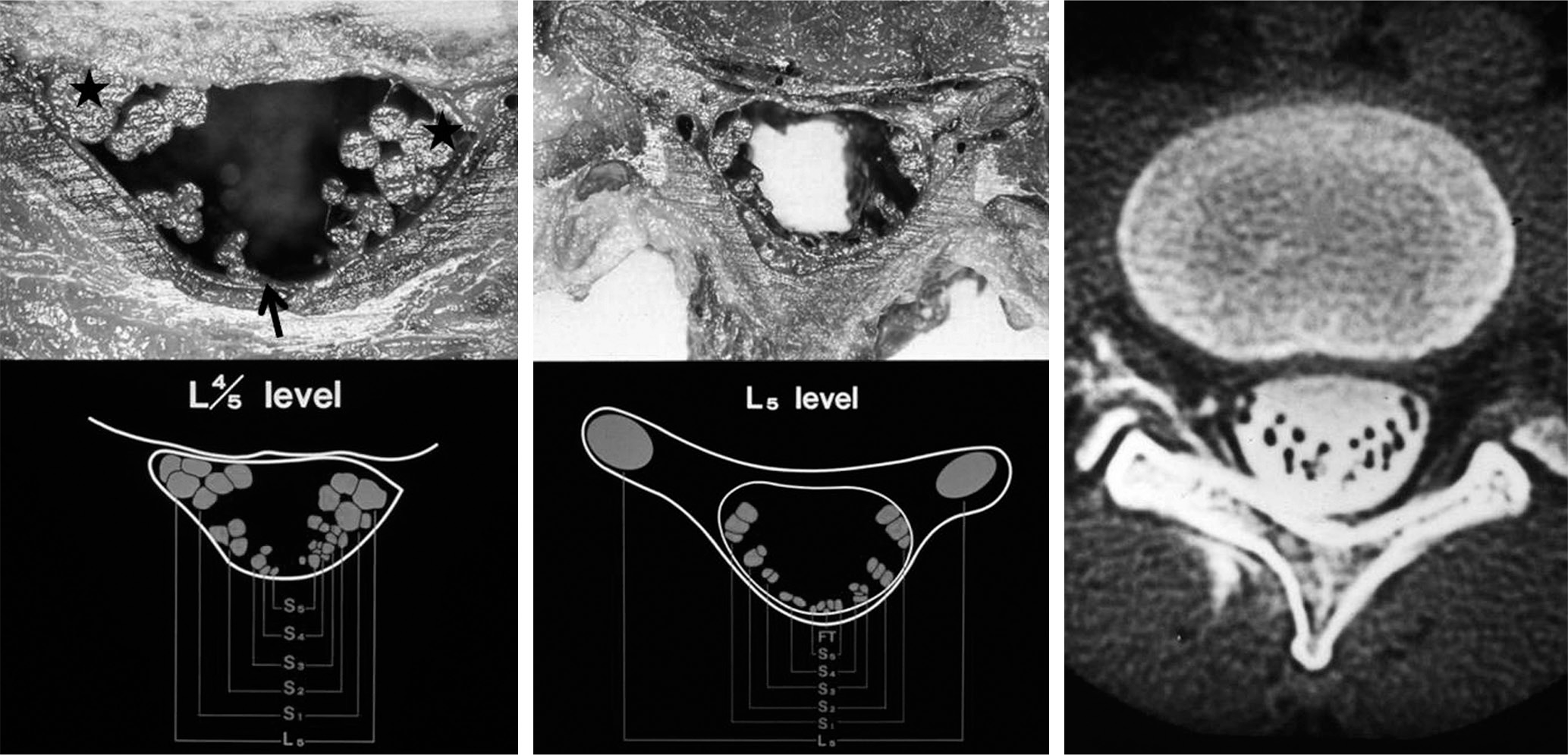
 | Fig. 1.Intradural arrangement of cauda equina. The most caudal roots (black arrow) is located in central and posterior position, and the most cephalad roots (asterix) is located in lateral and anterior position. In addition, motor fiber is located in medial and anterior to sensory fiber (Photography by Shinich Kikuchi, MD, PhD). |
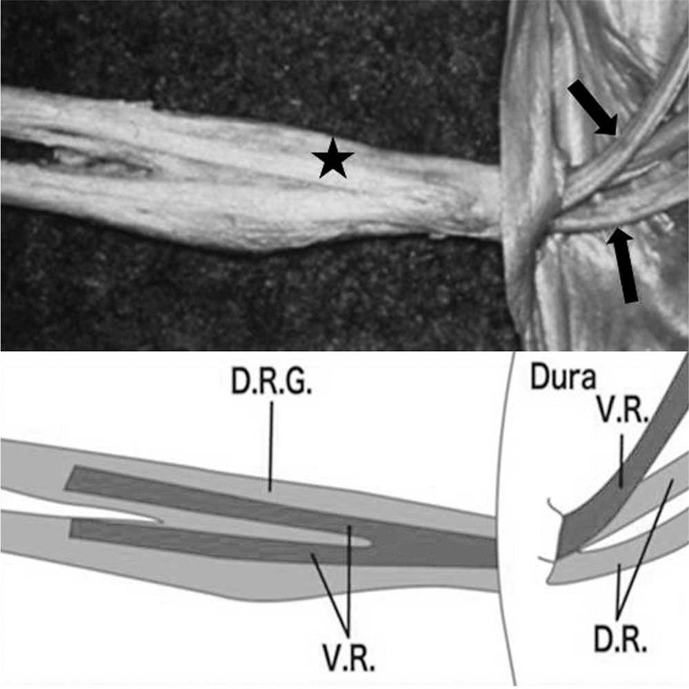
 | Fig. 2.Photo showing the differences between spinal nerve root (asterix) and cauda equine (black arrows). Cauda equina has no connective tissue, adipose tissue, perineurium, epineurium, and endonurium (VR: ventral root, DR: dorsal root, DRG: dorsal root ganglion, Photography by Shinich Kikuchi, MD, PhD). |
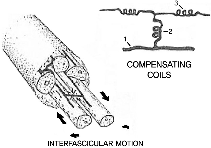
 | Fig. 3.Inter- and intrafascicular arteries showing compensating coils to allow interfascicular movement (1:Radicular artery, 2: interfascicular artery, 3: intrafascicular artery). Cited from Parke WW, Watanabe R, Spine (Phila Pa 1976). 1985;10:508-15. |
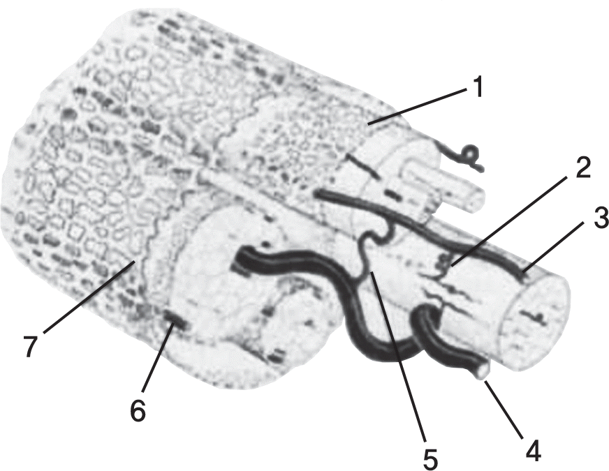
 | Fig. 4.Graphic complilation showing the structure of a typical lumbosacral nerve root derived from data obtained by injection studies. 1. Fas-cicular pia; 2. Inter- and intrafascicular arteries 3. Longitudinal radicular artery; 4. Large radicular vein; 5. Arteriovenous anastomosis; 6. Collateral radicular artery; 7. Gauzelike pia-arachnoid that permits percolation of CSF to assist in metabolic support. Cited from The Spine Fifth Ed. Page 49 Figure 2-42. |


 PDF
PDF ePub
ePub Citation
 Print
Print


 XML Download