

 PDF
PDF ePub
ePub Citation
 Print
Print


Abstract
Objectives
To evaluate the effectiveness of ultrasound guidance in caudal epidural block and sonographic feature of sacral hiatus.
Summary of Literature Review
High success rate of ultrasound-guided regional nerve block has been reported and recently, ultrasound-guided nerve block in spinal field has been introduced.
Materials and Methods
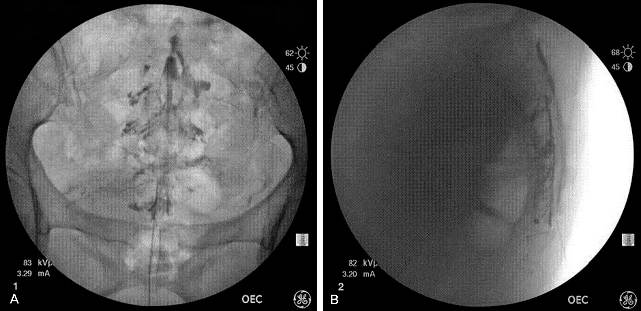
Ultrasound-guided caudal epidural block was performed in 48 patients with radiating pain to leg. Patient was placed in the prone position and sonographic image of sacral hiatus was obtained using linear probe. After measuring the intercornual distance, thickness of sacrococcygeal membrane and depth of sacral canal in transverse view, then the probe was rotated 90° to obtain the longitudinal view of the sacral hiatus. Under ultrasound guidance, a 21-gauge needle was inserted into the sacral hiatus in parallel with sacrum base. After contrast dye injection, needle placement was checked by the fluoroscopy and then medication was injected into the caudal epidural space. We investigated the change of radiating pain after caudal epidural block using visual analogue scale(VAS).
Results
The intercornual distance was mean 16.4±2.3mm, thickness of sacrococcygeal membrane was mean 2.8±0.9mm and depth of sacral hiatus was mean 2.6±0.9mm. There was 97.9% success rate of the caudal epidural block under ultrasound guidance. The mean VAS for radiating pain was improved from 7.5±0.7 before the block to 2.8±1.5 after the block.
Go to : 
REFERENCES
1. White AH, Derby R, Wynne G. Epidural injections for the diagnosis and treatment of low-back pain. Spine (Phila Pa 1976). 1980; 5:78–86.

2. Weinstein SM, Herring SA, Derby R. Contemporary con-cepts in spine care. Epidural steroid injections. Spine (Phila Pa 1976). 1995; 20:1842–6.
3. Standring S. Gray's anatomy. 39th ed.Spain: Churchill Livingstone:;2005. p. 750.
4. Stitz MY, Sommer HM. Accuracy of blind versus fluoroscopically guided caudal epidural injection. Spine (Phila Pa 1976). 1999; 24:1371–6.
5. Lewis MP, Thomas P, Wilson LF, Mulholland RC. The ‘whoosh’ test. A clinical test to confirm correct needle placement in caudal epidural injections. Anaesthesia. 1992; 47:57–8.
6. Tsui BC, Tarkkila P, Gupta S, Kearney R. Confirmation of caudal needle placement using nerve stimulation. Anesthesiology. 1999; 91:374–8.
7. Robecjji A, Capra R. L’ idrocortisone (composto F): prime esperienze clinche in campo rheumatlogico. Min Med. 1952; 98:1259–63.
8. Kim DH, Kim KH, Kim YC. Minimally invasive percutaneous spinal techniques. 1. Korea: Elsevier Inc;2011. p. 111–23.
9. Sekiguchi N, Yabuki S, Satoh K, Kikuchi S. An anatomic study of the sacral hiatus: a basis for successful caudal epidural block. Clin J Pain. 2004; 20:51–4.
10. Guinard JP, Borboen M. Probable venous air embolism during caudal anesthesia in a child. Anesth Analg. 1993; 76:1134–5.
11. Klocke R, Jenkinson T, Glew D. Sonographically guided caudal epidural steroid injections. J Ultrasound Med. 2003; 22:1229–32.
12. Chen CP, Tang SF, Hsu TC, et al. Ultrasound guidance in caudal epidural needle placement. Anesthesiology. 2004; 101:181–4.
13. Roh JH, Kim WO, Yoon KB, Yoon DM. The success rate of caudal block under ultrasound guidance and the direction of the needle in the sacral canal. Korean J Pain. 2007; 20:40–5.
14. Roberts SA, Guruswamy V, Galvez I. Caudal injectate can be reliably imaged using portable ultrasound – a preliminary study. Paediatr Anaesth. 2005; 15:948–52.
15. Rhee HD, Yoon DM, Park EY, et al. The optimal angle of needle insertion for caudal block in adults. Korean J Anesthesiol. 2008; 54:295–9.
16. Dadure C, Raux O, Rochette A, et al. Interest of ultrasono-graphic guidance in paediatric regional anaesthesia. Ann Fr Anesth Reanim. 2009; 28:878–84.
17. de Josemaria B, Galvez I, Reinoso-Barbero F. Ultrasound guidance in pediatric regional anesthesia. Rev Esp Anestesiol Reanim. 2009; 56:170–9.
18. Schwartz D, Raghunathan K, Dunn S, Connelly NR. Ultrasonography and pediatric caudals. Anesth Analg. 2008; 106:97–9.
Go to :
Figures and Tables%
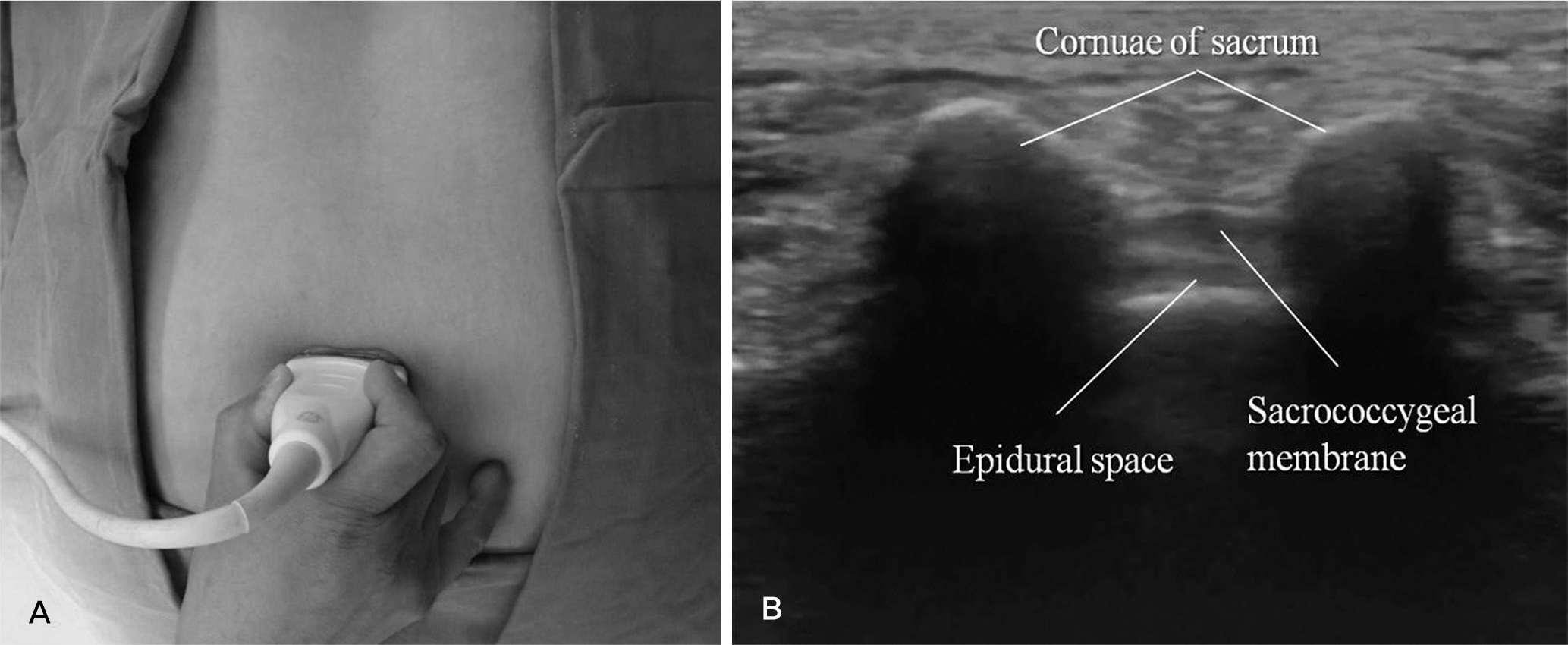 | Fig. 1.The transducer was placed transversely on the sacral hiatus and checked intercornual distance, thickness of sacrococcygeal membrane, depth of caudal space. (A) Photo, (B) Ultrasound finding. |
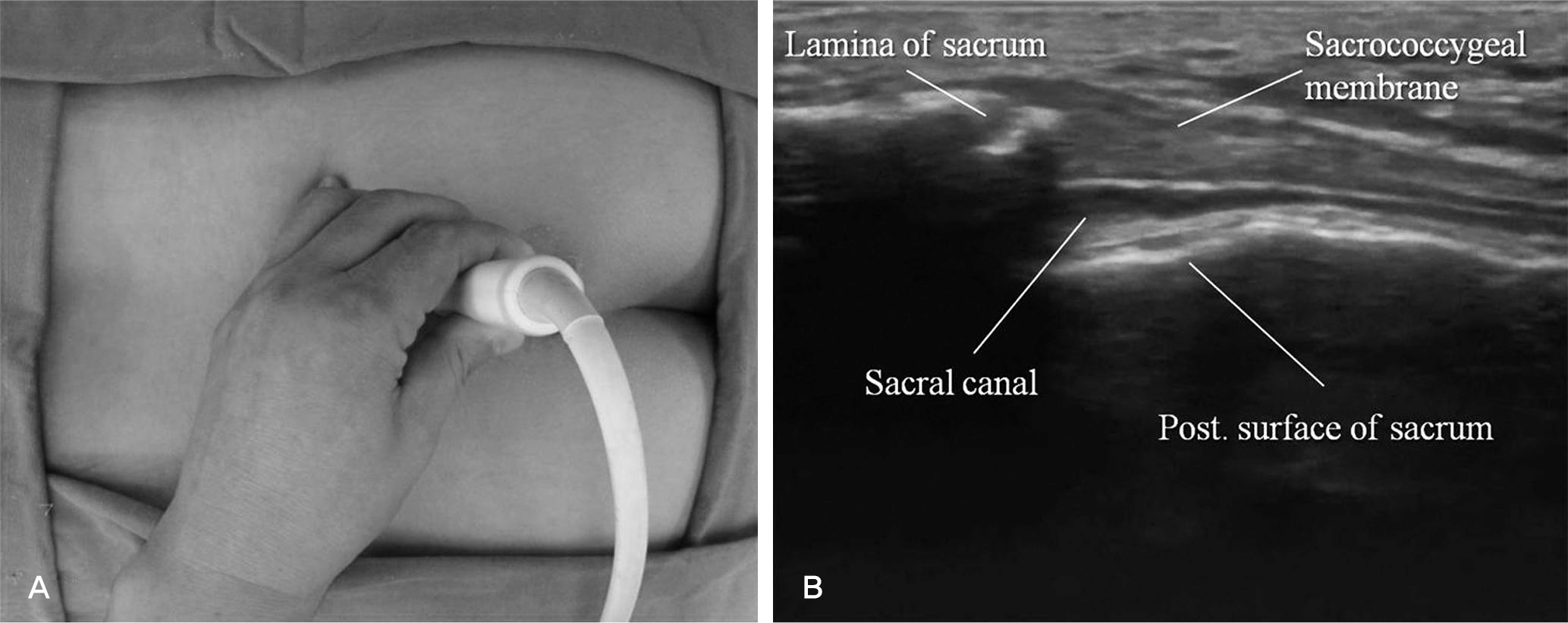 | Fig. 2.The transducer was rotated 90 degrees to obtain the longitudinal view of sacral hiatus. (A) Photo, (B) Ultrasound finding. |
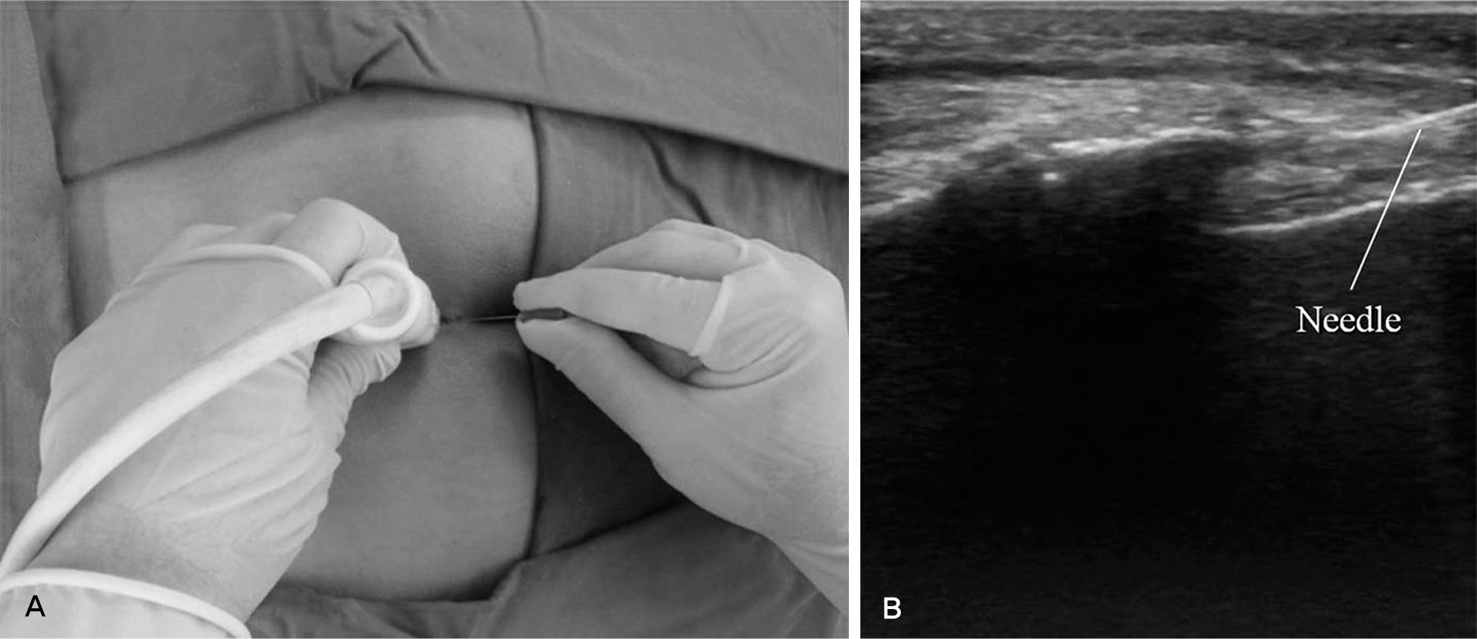 | Fig. 3.Needle was inserted to caudal epidural space under ultrasound guidance. (A) Photo, (B) Ultrasound finding. |
 XML Download