Abstract
Study Design
To analyze the relationship between the adjacent superior segment disease and facet joint violations after lumbar fusion.
Objectives
We retrospectively analyzed the relationship between the adjacent superior segment disease and facet joint violations after lumbar fusion.
Summary of Literature Review
Among numerous literatures regarding adjacent superior segment disease, there is no analysis concerning the relationship between adjacent superior segment disease and facet joint violations after lumbar fusion.
Materials and Methods
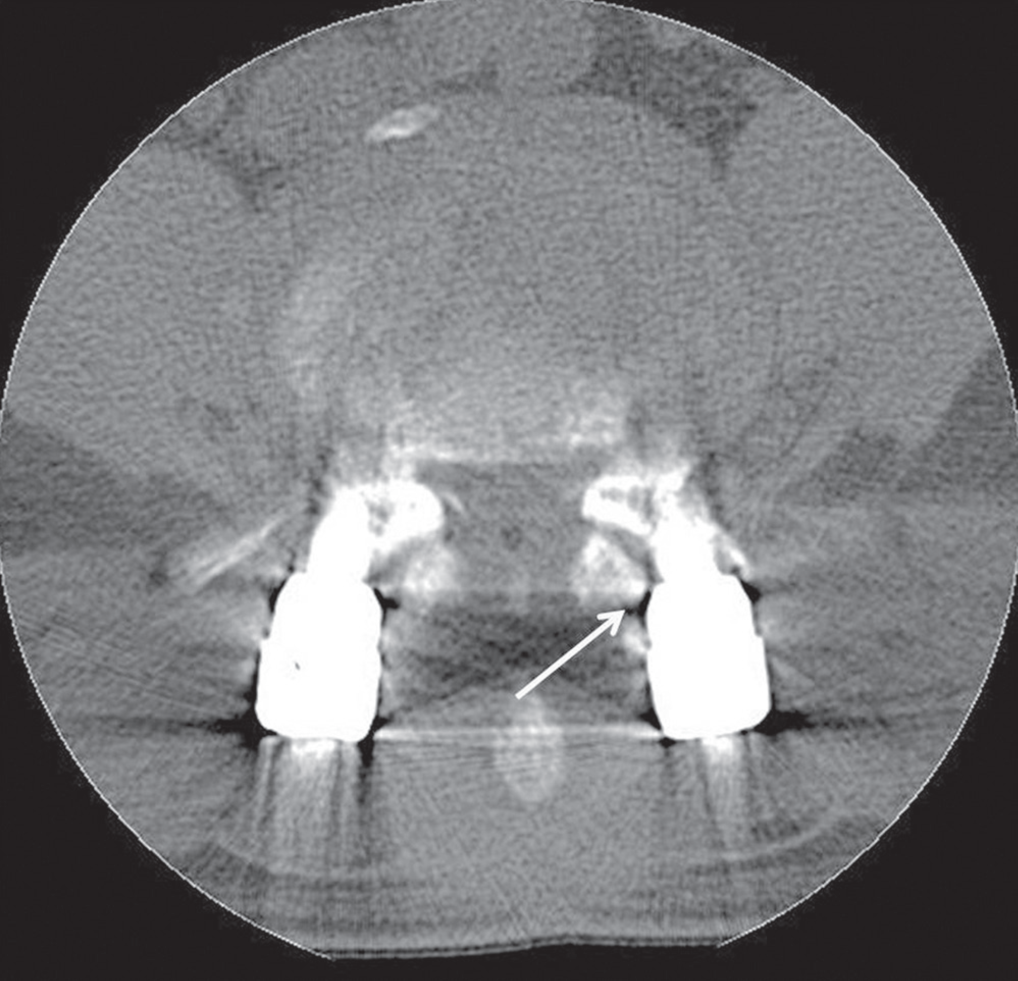
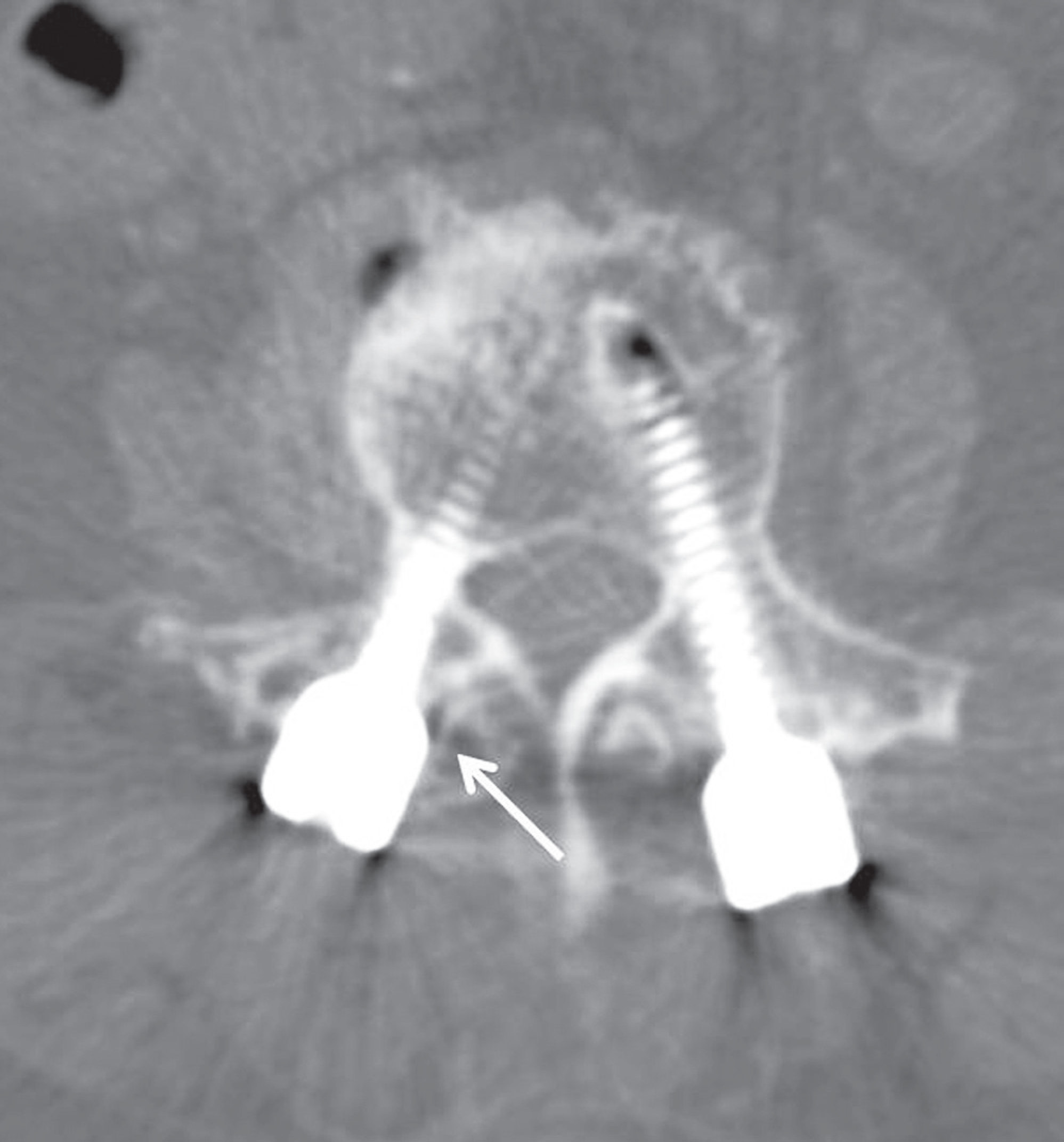
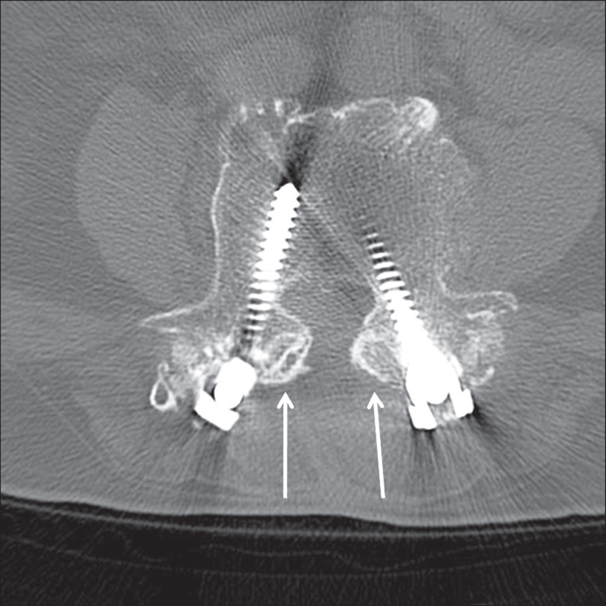
We reviewed 2056 patients who underwent lumbar fusion, between March 2004 and April 2009. Analysis was performed for 79 (3.8%) of the 2056 patients with adjacent superior segment disease and needed a second operation. A facet joint was considered as 3 types of violations with computed tomography scans if any of the following situations were encountered: pedicle screw clearly within the facet joint; pedicle screw head clearly within the facet joint; and pedicle screw and/or screw head within 1mm from or abutting the facet joint, without clear joint involvement.
Results
The incidence of the violations was 45% (36/79) of all patients and 28% (44/158) of all screws. The incidence of L4-5 facet joint violations was 35% (28/79) of patients with adjacent superior segment disease, statistically.
Conclusions
Facet joint violations were observed in patients with the adjacent superior segment disease after posterolateral lumbar fusion. Because L3-4 facet joint violations increased when L4-5 fusion was done, more care should be taken to avoid facet joint violations when the surgeon is considered for insertion of the pedicle screws at L4-5.
Go to : 
REFERENCES
1.Aota Y., Kumano K., Hirabayashi S. Postfusion instability at the adjacent segments after rigid pedicle screw fixation for degenerative lumbar spinal disorders. J Spinal Disord. 1995. 8:464–73.

2.Fritsch EW., Heisel J., Rupp S. The failed back surgery syndrome: reasons, intraoperative findings, and long-term results: a report of 182 operative treatments. Spine (Phila Pa 1976). 1996. 21:626–33.
3.Frymoyer JW., Hanley EN Jr., Howe J., Kuhlmann D., Matteri RE. A comparison of radiolographic findings in fusion and nonfusion patients ten or more years following lumbar disc surgery. Spine (Phila Pa 1976). 1979. 4:435–40.
4.Guigui P., Lambert P., Lassale B., Deburge A. [Long-term outcome at adjacent levels of lumbar arthrodesis]. Rev Chir Orthop Reparatrice Appar Mot. 1997. 83:685–96.
5.Ha KY., Sung TP. Changes of the Adjacent Mobile Seg-ment After Cat Spine Fixation. J Korean Orthop Assoc. 1997. 32:1808–16.

6.Ha KY., Moon MS., Paek SY. Effect of Instumental stabili-zation and fusion of degenerative lumbar scoliosis on? un-fused adjacent segment. J Korean Spine Surg. 1995. 2:270–8.
7.Etebar S., Cahill DW. Risk factors for adjacent-segment failure following lumbar fixation with rigid instrumentation for degenerative instability. J Neurosurg. 1999. 90(2 Suppl):163–9.

8.Kim SS., Michelsen CB. Revision surgery for failed back surgery syndrome. Spine (Phila Pa 1976). 1992. 17:957–60.

9.Bastian L., Lange U., Knop C., Tusch G., Blauth M. Evalu-ation of the mobility of adjacent segments after posterior thoracolumbar fixation: A biomechanical study. Eur Spine J. 2001. 10:295–300.

10.Frymoyer JW., Hanley E., Howe J., Kuhlmann D., Matteri R. Disc excision and spine fusion in the management of lum-bar disc disease. A minimum 10 year follow up. Spine (Phila Pa 1976). 1978. 13:1–6.
11.Hambly MF., Wiltse LL., Raghavan N., Schneiderman G., Koenig C. The transition zone above a lumbosacral fusion. Spine (Phila Pa 1976). 1998. 23:1785–92.

12.Whitecloud TS 3rd., Davis JM., Olive PM. Operative treat-ment of the degenerated segment adjacent to a lumbar fusion. Spine (Phila Pa 1976). 1994. 19:531–6.

13.Morgan FP., King T. Primary instability of lumbar vertebrae vertebrae as a common cause of low back pain. J Bone Joint Surg Br. 1957. 39:6–22.
14.Dupuis PR., Yong-Hing K., Cassidy JD., Kirkaldy-Willis WH. Radiologic diagnosis of degenerative lumbar spinal instability. Spine (Phila Pa 1976). 1985. 10:262–76.

15.Lee CK., Langrana NA. Lumbosacral spinal fusion. A bio-mechanical study. Spine (Phila Pa 1976). 1984. 9:574–81.

16.Yang SW., Langrana NA., Lee CK. Biomechanics of lumbo-sacral spinal fusion in combined compression-torsion loads. Spine (Phila Pa 1976). 1986. 11:937–41.
17.Frymoyer JW., Hanley EN Jr., Howe J., Kuhlmann D., Mat-teri RE. A comparison of radiographic findings in fusion and nonfusion patients ten or more years following lumbar disc surgery. Spine (Phila Pa 1976). 1979. 4:435–40.

18.Moshirfar A., Jenis LG., Spector LR, et al. Computed to-mography evaluation of superior-segment facet-joint violation after pedicle instrumentation of the lumbar spine with a midline surgical approach. Spine (Phila Pa 1976). 2006. 31:2624–9.

19.Shah RR., Muhammed S., Saifuddin A., Taylor BA. Radio-logic evaluation of adjacent superior segment facet joint violation following transpedicular instrumentation of the lumbar spine. Spine (Phila Pa 1976). 2003. 28:272–5.

20.Park P., Garton HJ., Gala VC., Hoff JT., McGillicuddy JE. Adjacent segment disease after lumbar or lumbosacral fusion: review of the literature. Spine (Phila Pa 1976). 2004. 29:1938–44.

21.Jeffrey B. Knox, Joseph M. Dai III. Joseph R. Orchowski. Superior segment facet joint violation and cortical violation after minimally invasive pedicle screw placement. Spine (Phila Pa 1976). 2011. 11:213–7.
22.Kim WJ., Kang JW., Kam BS, et al. Analysis of risk factors and surgical results of lumbar adjacent segment disease. J Korean Spine Surg. 2010. 17:74–81.

23.Kettler A., Wilke HJ., Haid C., Claes L. Effects of specimen length on the monosegmental motion behaviour of the lumbar spine. Spine (Phila Pa 1976). 2000. 25:543–50.
Go to : 
 | Fig. 1.Type 1 violations. The white arrow indicates that the pedicle screw is clearly within the facet joint. |
 | Fig. 2.Type 2 violations. The white arrow indicates that the pedicle screw head is apparently within the facet joint. |
 | Fig. 3.Type 3 violations. The white arrow indicates that the pedicle screw and/or screw head are within 1 mm from or abutting the facet joint without clear joint involvement. |
Table 1.
Distribution of superior facet joint violations by patient clinical characteristics


 PDF
PDF ePub
ePub Citation
 Print
Print


 XML Download