Abstract
Objectives
To evaluate the correlation of adjacent segmental disease with tilt angles of the upper and lower instrumented vertebra after instrumented posterolateral fusion for degenerative lumbar scoliosis.
Summary of Literature Review
There has been no study of radiologic measurement and decision of fusion level using the angle of pedicle screws inserted for treatment of degenerative lumbar scoliosis.
Materials and Methods
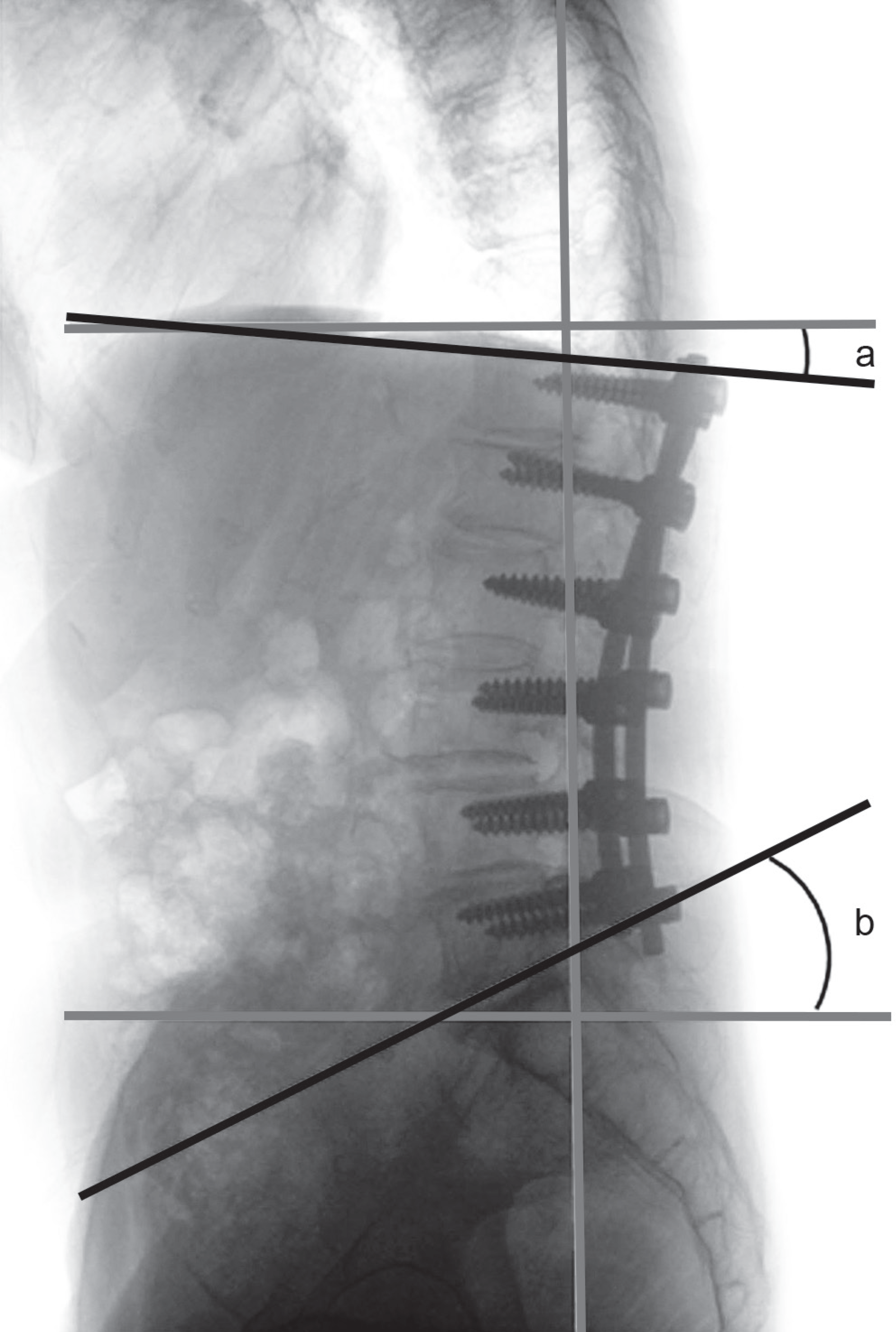
From 2004 to 2008, 74 patients that underwent decompression and posterolateral fusion for degenerative lumbar scoliosis were included in this study. In all cases, instrumentation and posterolateral fusion were both performed. The sex ratio was 31:43, the mean age was 68.7 years and the mean follow up duration was 37.4 months. The angle between each upper end plate of the upper vertebral body and lower end plate of the lower vertebral body of the fusion, and the line parallel to the axis of the sagittal line of vertebrae was each defined as UIV-a and LIV-b. The correlation of development of adjacent segment disease and UIV-a, and LIV-b angle was investigated.
Go to : 
REFERENCES
1.Epstein JA., Epstein BS., Jones MD. Symptomatic lumbar scoliosis with degenerative changes in the elderly. Spine (Phila Pa 1976). 1979. 4:542–7.

2.Song KJ., Choi BW., Song JH., Kim GH. The Causes of Re-vision Arthrodesis for the Degenerative Changes at the Adjacent Segment after Lumbosacral Fusion for Degenerative Lumbar Diseases. J Korean Soc Spine Surg. 2008. 15:230–5.

3.Grubb SA., Lipscomb HJ. Diagnostic findings in painful adult scoliosis. Spine (Phila Pa 1976). 1992. 17:518–27.

6.Aebi M. Correction of degenerative scoliosis of the lumbar spine. A preliminary report. Clin Orthop Relat Res. 1988. 232:80–6.
8.Hilibrand AS., Robbins M. Adjacent segment degeneration and adjacent segment disease: the consequence of spinal fusion? Spine J. 2004. 4(6 Suppl):190S–4S.
9.Booth KC., Bridwell KH., Eisenberg BA., Baldus CR., Lenke LG. Minimum 5-year results of degenerative spondylolis-thesis treated with decompression and instrumented posterior fusion. Spine (Phila Pa 1976). 1999. 24:1721–7.

10.Lenke LG., Bridwell KH., Bullis D., Betz RR., Baldus C., Schoenecker PL. Results of in situ fusion for isthmic spon-dylolisthesis. J Spinal Disord. 1992. 5:433–42.

11.Kumar MN., Jacquot F., Hall H. Long-term follow-up of functional outcomes and radiographic changes at adjacent levels following lumbar spine surgery for denerative disc disease. Eur Spine J. 2001. 10:309–13.
12.Ahn DK., Lee S., Jeong KW., Park JS., Cha SK., Park HS. Adjacent Segment failure after Lumbar Spine Fusion: Con-trolled Study for Risk Factors. J Korean Orthop Assoc. 2005. 40:203–8.

13.Wu CH., Wong CB., Chen LH., Niu CC., Tsai TT., Chen WJ. Instrumented posterior lumbar interbody fusion for patients with degenerative lumbar scoliosis. J Spinal Disord Tech. 2008. 21:310–5.

14.Rohlman A., Neller S., Bergmann G., Graichen F., Claes L., Wilke HJ. Effect of an internal fixator and a bone graft on instersegmental spinal motion and intradiscal pressure in the adjacent region. Eur Spine J. 2001. 10:301–8.
15.Gillet P. The fate of the adjacent motion segment after lum-bar fusion. J Spinal Disord Tech. 2003. 16:338–45.
16.Lee CK. Lumbar spinal instability (olisthesis) after exten-sive posterior spinal decompression. Spine (Phila Pa 1976). 1983. 8:429–33.

17.Schlegel JD., Smith JA., Schleusener RL. Lumbar motion segment patholoty adjacent to thoracolumbar, lumbar, and lumbosacral fusions. Spine (Phila Pa 1976). 1996. 21:970–81.
18.Simmons ED., Simmons EH. Spinal stenosis with scoliosis. Spine (Phila Pa 1976). 1992. 17(6 Suppl):S117–20.

19.Herkowitz HN., Kurz LT. Degenerative lumbar spondylo-listhesis with spinal stenosis. A prospective study comparing decompression with decompression and intertransverse process arthrosis. J Bone Joint Surg Am. 1991. 73:802–8.
20.Cho KJ., Park SL., Kim MG., Yoon YH., Lee JS., Suk SI. Proximal Adjacent Segment Disease following Posterior Instrumentation and Fusion for Degenerative Lumbar Scoliosis. J Korean Orthop Assoc. 2009. 44:109–17.

21.Shufflebarger H., Suk SI., Mardjetko S. Debate: determining the upper instrumented vertebra in the management of adult degenerative scoliosis: stopping at T10 versus L1. Spine (Phila Pa 1976). 2006. 31(19 Suppl):S185–94.
22.Tsuchiya K., Bridwell KH., Kuklo TR., Lenke LG., Baldus C. Minimum 5-year analysis L5-S1 fusion using sacropelvic fixation (bilateral S1 and iliac screws) for spinal deformity. Spine (Phila Pa 1976). 2006. 31:303–8.
23.Edwards CC 2nd., Bridwell KH., Patel A, et al. Thoraco-lumbar deformity arthrodesis to L5 in adults: the fate of the L5-S1 disc. Spine (Phila Pa 1976). 2003. 28:2122–31.

24.Dekutoski MB., Schendel MJ., Ogilvie JW., Olsewski JM., Wallace LJ., Lewis JL. Compression of in vivo and in vitro adjacent segment motion after lumbar fusion. Spine (Phila Pa 1976). 1994. 19:1745–51.
25.Grouw AV., Nadel CI., Weieman RJ., Lowell HA. Long term follow-up of patients with idiopathic scoliosis treated surgi-cally: a preliminary subjective study. Clin Orthop Relat Res. 2001. 117:197–201.

26.Cho JL., Choi SW., Lee JM., Park YS. The Changes of Ad-jacent Segments after Long Segment Posterolateral Fusion: Comparative Study of 3 year versus over the 7 year Follow-up Patients. J Korean Orthop Assoc. 2005. 40:38–43.

27.Lee CK., Langrana NA. Lumbosacral spinal fusion. A bio-mechanical study. Spine (Phila Pa 1976). 1984. 9:574–81.

28.Weinhoffer SL., Guyer RD., Herbert M., Griffith SL. Intra-discal pressure measurements above an instrumented fusion. A cadaveric study. Spine (Phila Pa 1976). 1995. 20:526–31.
29.Cunningham BW., Kotani Y., McNulty PS., Cappuccino A., McAfee PC. The effect of spinal destabilization and instrumentation on lumbar intradiscal pressure: an in vitro bio-mechanical analysis. Spine (Phila Pa 1976). 1997. 22:2655–63.
Go to : 
 | Fig. 1.UIV-a and UIV-b are defined angles formed by 2 lines of the 1st line along the upper end plate of the most upper instrumented vertebra or the lower end plate of the most lower instrumented vertebra and the 2nd sagittal vertical axis of the vertebrae respectively. |
 | Fig. 2.67-year-old patient with degenerative lumbar scoliosis and stenosis (A) Preoperative radiographs (B) Postoperative radiographs after decompression and posterolateral fusion on L3-S1. UIV-a was 15.2 degrees and LIV-b was 17.9 degrees in this radiographs (C) Last Follow-up radiographs 27 months after the surgery show degenerative changes and development of proximal adjacent segment disease (disc space narrow and sclerosis) at the L2-3 level. |
Table 1.
Baseline Demographic findings of patients Baseline patient de-mographics in this study.
Table 2.
Tilt angles of the upper and lower instrumented end vertebra with relation to adjacent segment disease
| ASD‡ (n=11) | ASD (-)(n=63) | p-value | |
|---|---|---|---|
| UIV-a∗ | 14.5±5 | 6.7±4 | |
| LIV-b† | 16.9±8 | 12.5±9 | |
| | UIV-a |+| LIV-b | | 31.4±11 | 19.2±7 | 0.047 |
Table 3.
Tilt angles of the upper and lower instrumented end vertebra with relation to proximal or distal adjacent segment disease respectively
| Proximal ASD‡ | Proximal ASD (-) | p-value | |
|---|---|---|---|
| No. of patients | 7 | 67 | |
| UIV-a∗ | 15.3±7 | 7.9±6 | 0.042 |
| Distal ASD | Distal ASD (-) | p-value | |
| No. of patients | 4 | 70 | |
| LIV-b† | 17.4±7 | 19.6±8 | 0.274 |


 PDF
PDF ePub
ePub Citation
 Print
Print


 XML Download