Abstract
Objectives
To evaluate the prevalence and associated factors of the concurrent lower thoracic lesions in patients who have a lumbar spine disease, using the extended lumbar MRI.
Summary of Literature Review
There are no studies regarding the concurrent thoracic lesions with lumbar disease.
Materials and Methods
All the patients, who had visited the out-patient department (OPD) of orthopaedic surgery in our hospital and underwent lumbar spine MRI, were studied during 1 year. Totally, 750 patients were included. The extended lumbar spine MRI contained additional extended T2-weighted sagittal images that cover the lower thoracic vertebrae with 35 centimeters long. We analyzed the highest observable level, characteristics of detected thoracic lesions. Those lesions were classified according to the severity of compression of the spinal cord and investigation for associated factors of patients. Also, the times for additional tests were measured.
Results
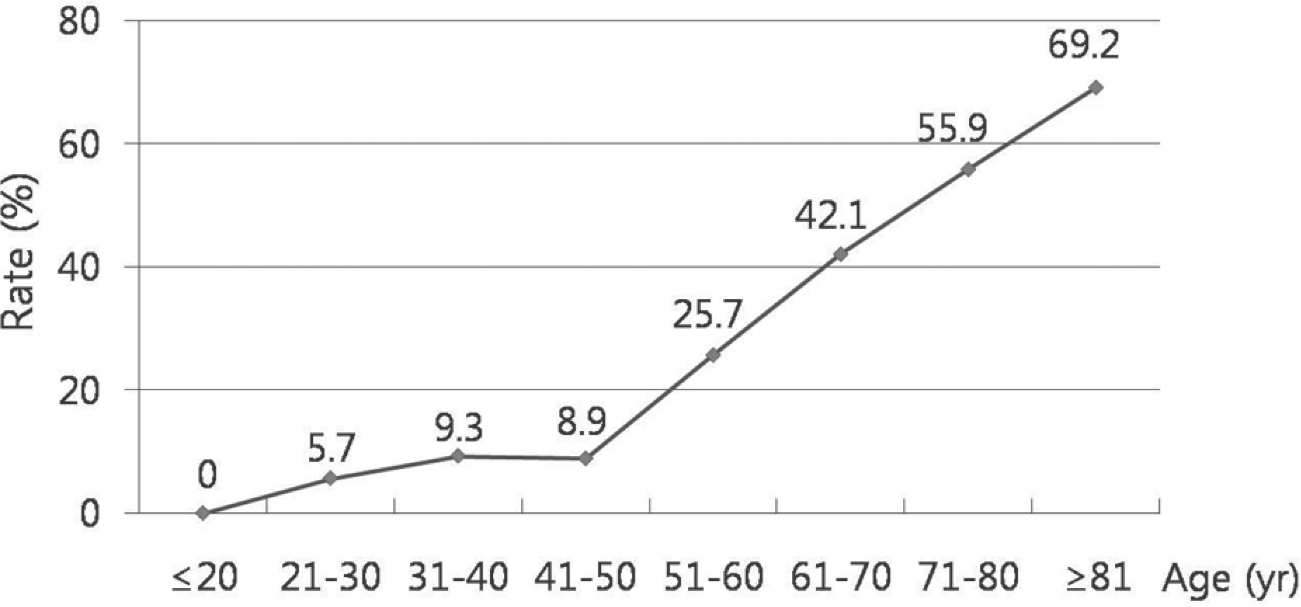
Additional tests were able to observe up to the 7th thoracic vertebrae. In 257 cases (34.3%), the lower thoracic lesions were detected and increased with aging (p<0.001). A total of 48 patients (6%) had the lesion compressing the spinal cord and 28 patients needed further evaluation for the lower thoracic lesion. Further, 2 cases were treated surgically for lower thoracic lesions. Scanning extra time for additional test were 3 minutes.
REFERENCES
1.Epstein NE., Epstein JA., Carras R., Murthy VS., Hyman RA. Coexisting cervical and lumbar spinal stenosis: diagnosis and management. Neurosurgery. 1984. 15:489–96.

2.Smith DE., Godersky JC. Thoracic spondylosis: an unusual cause of myelopathy. Neurosurgery. 1987. 20:589–93.

3.Moon ES., Kim HS., Park JO, et al. The incidence of new vertebral compression fractures in women after kyphoplasty and factors involved. Yonsei Med J. 2007. 48:645–52.

5.Barnett GH., Hardy RW Jr., Little JR., Bay JW., Sypert GW. Thoracic spinal canal stenosis. J Neurosurg. 1987. 66:338–44.

6.Niemelainen R., Battie MC., Gill K., Videman T. The prevalence and characteristics of thoracic magnetic reso-nance imaging findings in men. Spine (Phila Pa 1976). 2008. 33:2552–9.
7.Abhaykumar S., Tyagi A., Towns GM. Thoracic vertebral osteophyte-causing myelopathy: early diagnosis and treatment. Spine (Phila Pa 1976). 2002. 27:E334–6.

8.Bosacco SJ., Berman AT., Raisis LW., Zamarin RI. High lumbar disk herniations. Case reports. Orthopedics. 1989. 12:275–8.

9.Fukuta S., Miyamoto K., Iwata A, et al. Unusual back pain caused by intervertebral disc degeneration associated with schmorl node at Th11/12 in a young athlete, successfully treated by anterior interbody fusion: a case report. Spine (Phila Pa 1976). 2009. 34:E195–8.
10.Okada Y., Shimizu K., Ido K., Kotani S. Multiple thoracic disc herniations: case report and review of the literature. Spinal Cord. 1997. 35:183–6.

12.Chen CF., Chang MC., Liu CL., Chen TH. Acute noncontig-uous multiple-level thoracic disc herniations with myelopathy: a case report. Spine (Phila Pa 1976). 2004. 29:E157–60.
13.Otani K., Yoshida M., Fujii E., Nakai S., Shibasaki K. Thoracic disc herniation. Surgical treatment in 23 patients. Spine (Phila Pa 1976). 1988. 13:1262–7.
14.Fink HA., Milavetz DL., Palermo L, et al. What proportion of incident radiographic vertebral deformities is clinically di-agnosed and vice versa? J Bone Miner Res. 2005. 20:1216–22.

15.Lindsay R., Silverman SL., Cooper C, et al. Risk of new vertebral fracture in the year following a fracture. JAMA. 2001. 285:320–23.

16.Adachi JD., Adami S., Gehlbach S, et al. Impact of prevalent fractures on quality of life: baseline results from the global longitudinal study of osteoporosis in women. Mayo Clin Proc. 2010. 85:806–13.

17.Delmas PD., van de Langerijt L., Watts NB, et al. Underdi-agnosis of vertebral fractures is a worldwide problem: the IMPACT study. J Bone Miner Res. 2005. 20:557–63.

18.Bates D., Ruggieri P. Imaging modalities for evaluation of the spine. Radiol Clin North Am. 1991. 29:675–90.
Fig. 1.
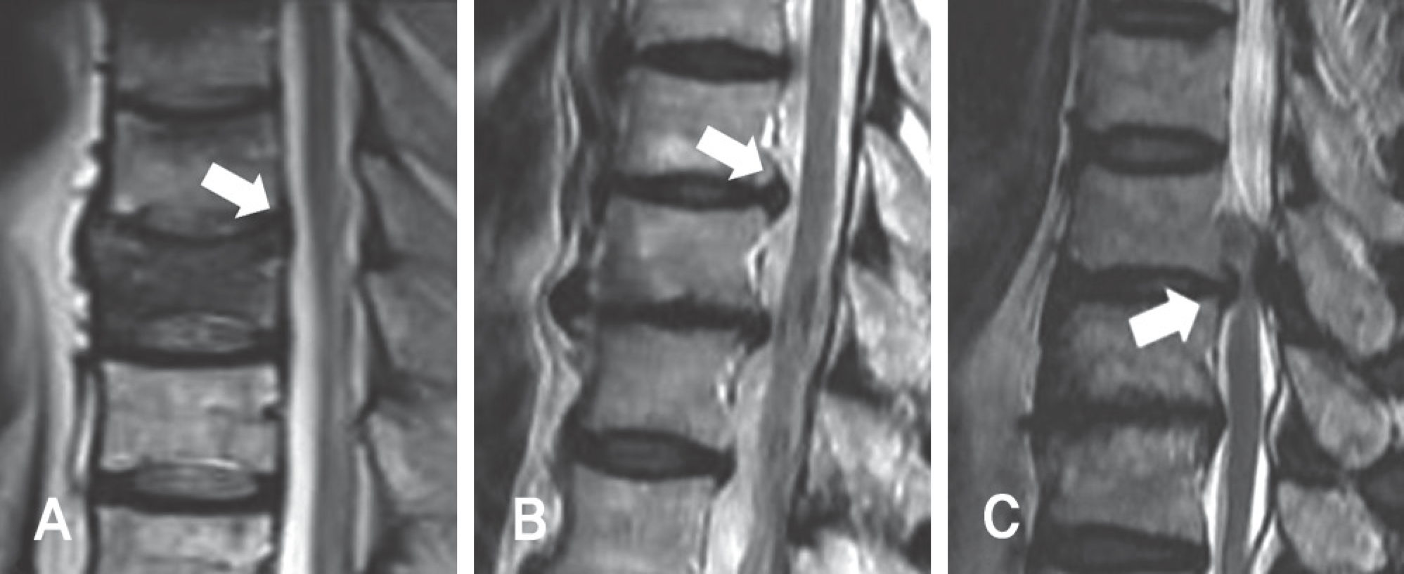
Grades of the severity of lower thoracic lesion(white arrow). (A) Grade 0: No compressive lesion. (B) Grade 1: The herniated thoracic intervertebral disc compressed the thecal sac but there is no cord contact. (C) Grade 3: The herniated thoracic intervertebral disc and hypertrophic ligamentum flavum compress the spinal cord.

Fig. 3.
The lower thoracic spine lesion(white arrow) detected in extended lumbar spine MRI, T2 sagittal images. (A) Hypertrophic ligamentum flavum involving T11/12 in a 72-year-old man. (B) Osteoporotic fracture at T7 and T12 in a 68-year-old woman. (C) Disc herniation at T10/11 in a 81 year-old woman. (D) Metastatic tumor lesion at T9 in a 56-year old man.

Table 1.
Grade for severity of thoracic lesion.
| Grade | Criteria |
|---|---|
| 0 | No compress the thecal sac |
| 1 | Lesion compress the only thecal sac not spinal cord |
| 2 | Lesion compress the spinal cord |
Table 2.
Data according to the age groups of the concurrent thoracic lesion.
| Age (yr) | No. of total cases | No. of concurrent lower thoracic lesion |
|---|---|---|
| ≤20 | 29 | 0 |
| 21-30 | 35 | 2 |
| 31-40 | 43 | 4 |
| 41-50 | 78 | 7 |
| 51-60 | 148 | 38 |
| 61-70 | 221 | 93 |
| 71-80 | 170 | 95 |
| ≥81 | 26 | 18 |
Table 3.
Classification and severity of the concurrent thoracic lesion.
Table 4.
Details of the concurrent thoracic lesion.


 PDF
PDF ePub
ePub Citation
 Print
Print


 XML Download