

 PDF
PDF ePub
ePub Citation
 Print
Print


Abstract
Objectives
We report a case of a female patient who had only upper back pain without neurological symptoms and was later diagnosed with spine tuberculosis in combination with a compression fracture.
Summary of Literature Review
Spine tuberculosis is the most common type of musculoskeletal tuberculosis. However, the indolent nature of tuberculous bone and joint disease often leads to delayed diagnosis and severe neurologic complications.
Material and Methods
A 37-year-old female with only upper back pain for five months was admitted. She had no signs, symptoms or past histories related to tuberculosis. She had taken conservative management, but symptoms persisted.
REFERENCES
1.WHO Library Cataloguing-in-Publication Data. World health statistics. 2010.
2.Friedman B. Chemotherapy of tuberculosis of the spine. J Bone Joint Surg Am. 1966. 48:451–74.

3.Steven K., Denise IC., Joel AD. Spinal cord medicine. Philadelphia: Lippincott Williams & Wilkins;2002. p. 500–02.
4.Du Plessis J., Andronikou S., Theron S., Wieselthaler N., Hayes M. Unusual forms of spinal tuberculosis. Childs Nerv Syst. 2008. 24:453–7.
5.Garg RK. Tuberculosis of the central nervous system. Postgrad Med J. 1999. 75:133–40.
6.Teo EL., Peh WC. Imaging of tuberculosis of the spine. Singapore Med J. 2004. 45:43–4.
7.Skendros P., Kamaria F., Kontopoulos V., Tsitouridis I., Sidiropoulos L. Intradural, extramedullary tuberculoma of the spinal cord as a complication of tuberculous meningitis. Infection. 2003. 31:115–7.
8.Cruickshank GS., Johnston RA. Intradural, extramedullary spinal cord compression from tuberculous granuloma. Br J Neurosurg. 1996. 10:93–5.
9.Chang DJ., Yoon DM., Kang YS., Yoon KB. Chronic Back Pain Proven to Be Spinal Tuberculosis: A report of 2 cases. Korean J Pain. 2008. 21(74):9.
10.Ahn JS., Lee JK., Jeon TS., Kwon YS., Kwak SK. Changes of Kyphotic Angle Following Operative Treatment of Tuberculous Spondylitis. J Korean Soc Spine Surg. 2001. 8:148–55.
11.Crenshaw AH. Campbell's Operative Orethopedics. 8th ed.Saint Louis:The CB Mosby company;1992. p. 3802–23.
12.Liebenson Craig. Rehabilitation of the spine. 2nd ed.Baltimore: Lippincott Williams & Wilkins;2007. p. 586–611.
Fig. 1.
Preoperative posteroanterior view of the chest radiography shows no active lesion in both lungs.
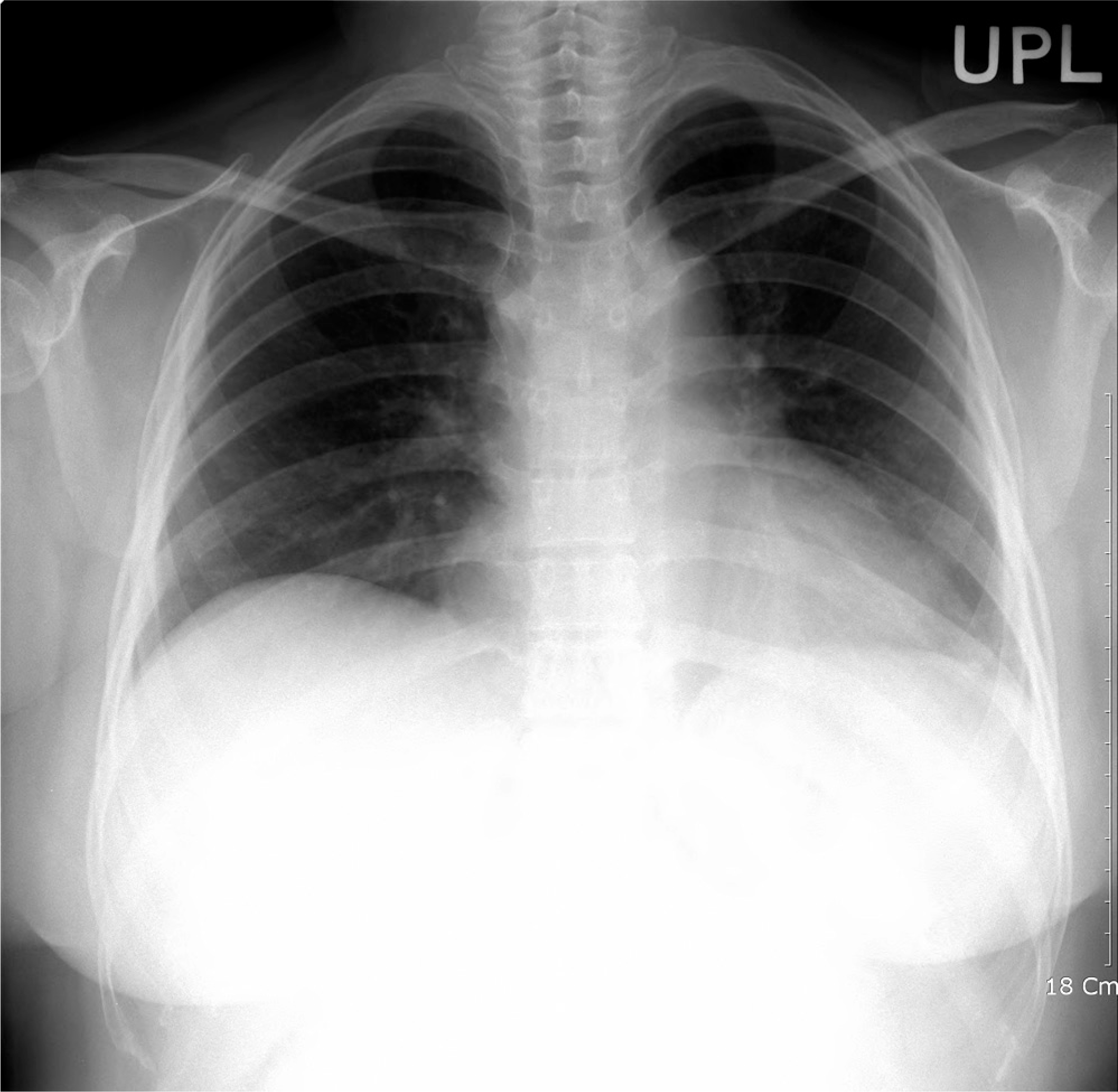
Fig. 2.
Preoperative posteroanterior and lateral view of the cervical X ray shows no remarkable finding.

Fig. 3.
Magnetic resonance Sagittal T2-weighted images shows high signal in T4-6, which is abscess formation and inflammation involvement of Rt. posterior epidural space and posterior elements and compression fracture T5.

Fig. 4.
Sensory evoked potentials. (A) Right median nerve SEP (B) Left median nerve SEP (C) Right tibial nerve SEP (D) Left tibial nerve SEP
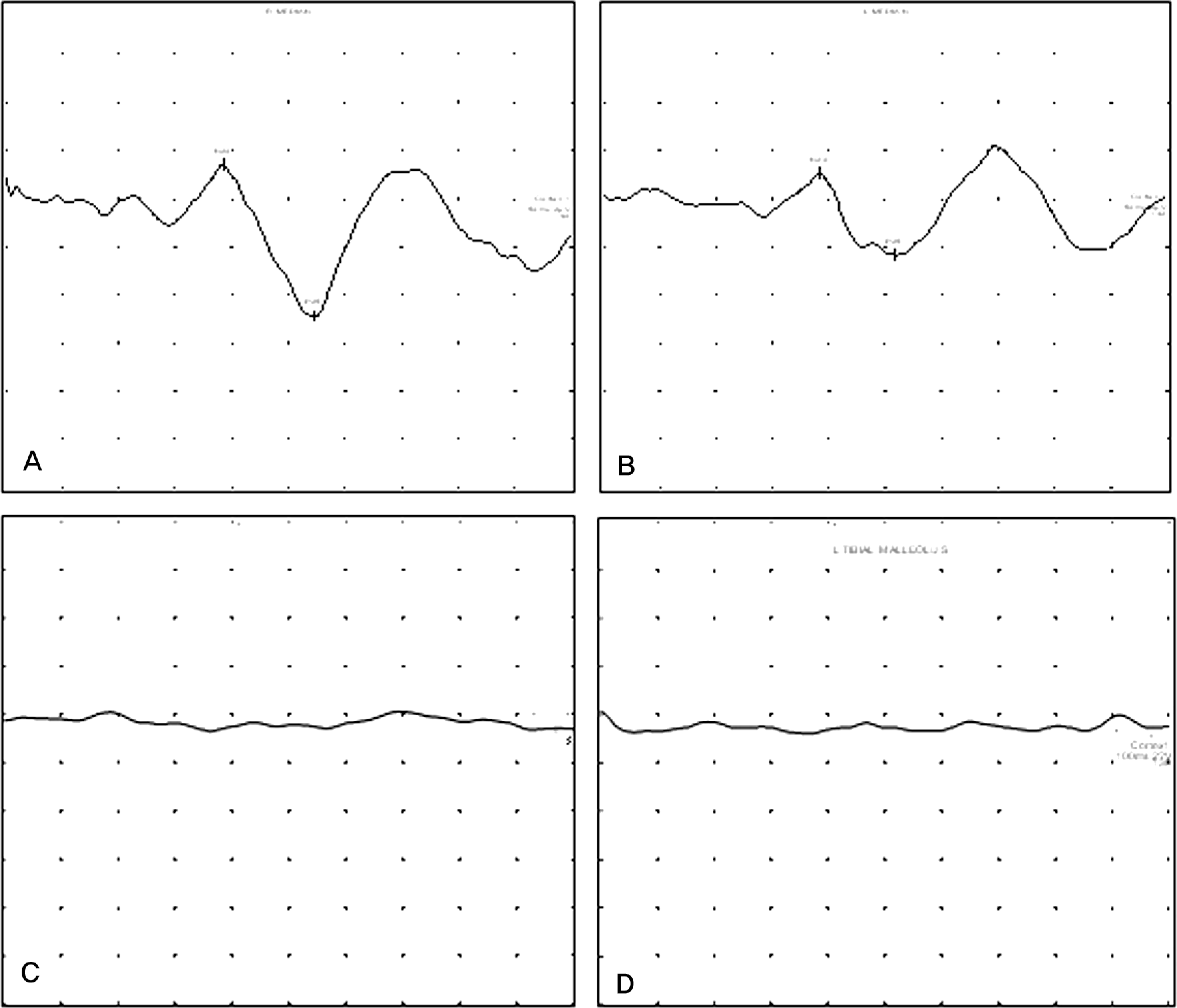
Table 1.
Results of sensory evoked potentials (SEPs)
| Location | Median SEP | Tibial SEP | ||||
|---|---|---|---|---|---|---|
| Latency (ms) | Amplitude (μV) | Latency (ms) | Amplitude (μV) | |||
| CZ-N20 | CZ-P25 | N20 | CZ-N20 | CZ-P25 | N20 | |
| Right | 19.20 | 27.20 | 27.20 | No response | ||
| Left | 19.20 | 25.75 | 25.75 | No response | ||
Table 2.
Results of motor evoked potentials (MEPs)
 XML Download