Abstract
Objectives
We report a case of thoracic intradural extramedullary tumor that has been misdiagnosed as the cerebral infarction.
Summary of Literature Review
Spinal meningioma is one of the common spinal tumors. Clinical symptoms were characteristically progressive myelopathy, rather than radiculopathy.
Materials and Methods
A 66-year-old female patient who had a history of cerebral infarction admitted as suffering from progressive lower extremities weakness for 6 months. The patient was diagnosed and has been treated as the cerebral infarction at another hospital. However, the patient showed worsening symptoms.
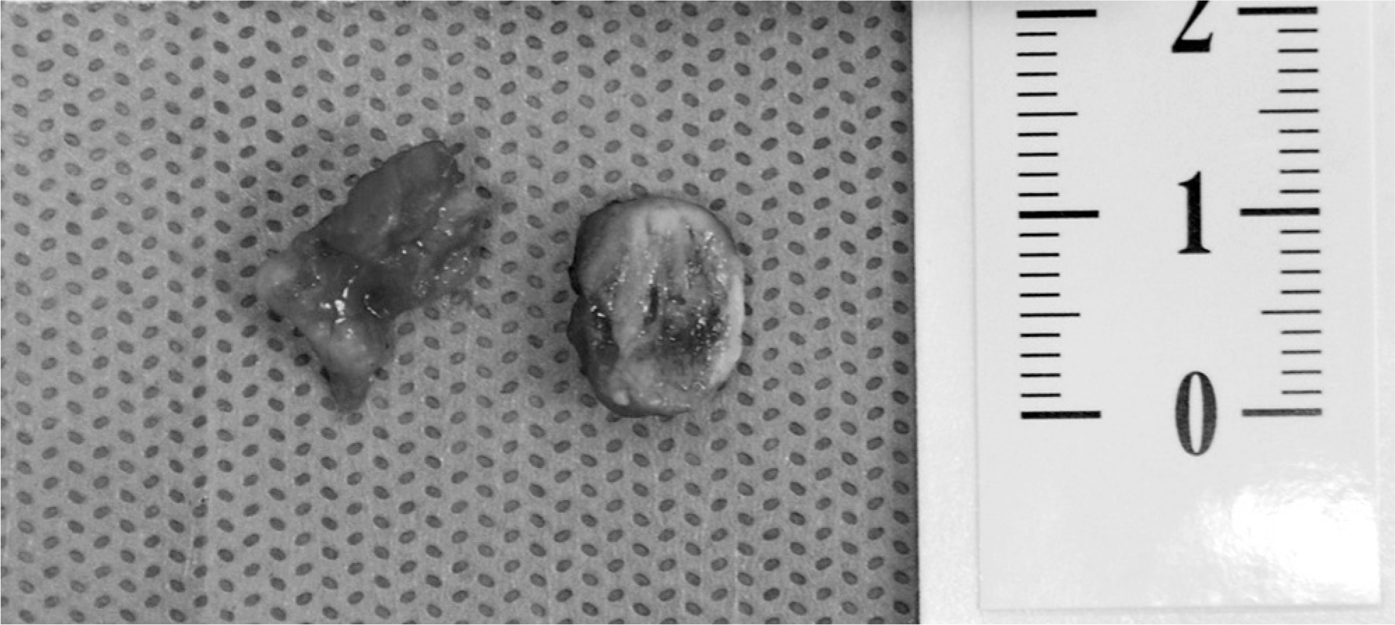
In magnetic resonance imaging, an intradural extramedullary space occupying mass compressing the spinal cord, between T8 and T9 level, was shown. By undergoing an operation, resected the mass. In a pathologic report, mass was confirmed to be meningioma.
REFERENCES
2.Souweidance MM., Benjamin V. Spinal cord meningiomas. Neurosurg Clin N Am. 1994. 5:283–91.
3.Solero CL., Fornari M., Giombini S, et al. Spinal meningiomas: review of 174 operated cases. Neurosurgery. 1989. 25:153–60.

4.Levy WJ., Lanchaw J., Hahn JF., Sawhny B., Bay J., Dohn DF. Spinal Neurofibromas: a report of 66 cases and a comparison with meningiomas. Neurosurgery. 1986. 18:331–4.

5.Shin BJ., Lee JC., Yoon TK, et al. Surgical treatment of intradural extramedullary tumor. J Korean Soc Spine Surg. 2002. 9:230–7.
6.Stechison MT., Tasker RR., Wortzman G. Spinal meningioma en plaque. Report of two cases. J Neurosurg. 1987. 67:452–5.
7.Park SH., Hwang SK., Park YM. Intramedullary clear cell meningioma. Acta Neurochir(Wien). 2006. 148:463–6.

8.Chung JY. Spinal Tumor. J Korean Soc Spine Surg. 1999. 6:316–25.
Fig. 1.
The tumor is located at the T8-9 level. (A) T1-weighted sagittal MR image shows peripheral low signal intensity and central iso-signal intensity mass. (B) T2-weighted sagittal MR image shows iso-signal intensity mass. (C) Gadolinium enhanced T1-weighted sagittal MR image shows high signal intensity mass. (D, E, F) T1, T2-weighted, Gadolinium enhanced T1-weighted axial MR image shows oval shape intradural extramedullary mass compressing spinal cord.

Fig. 2. (A)
Intraoperative finding shows well exposed dural sac. (B) Intraoperative finding shows oval shape mass compressed spinal cord in dura matter.


 PDF
PDF ePub
ePub Citation
 Print
Print


 XML Download