

 PDF
PDF ePub
ePub Citation
 Print
Print


Abstract
Objectives
We report a very rare case of the inferior accessory ossicle of the anterior arch of the atlas misdiagnosed as anterior arch fracture.
Summary of Literature Review
It is necessary to know the existence of inferior accessory ossicle of the anterior arch of the atlas, even though it is extremely rare.
Materials and Methods
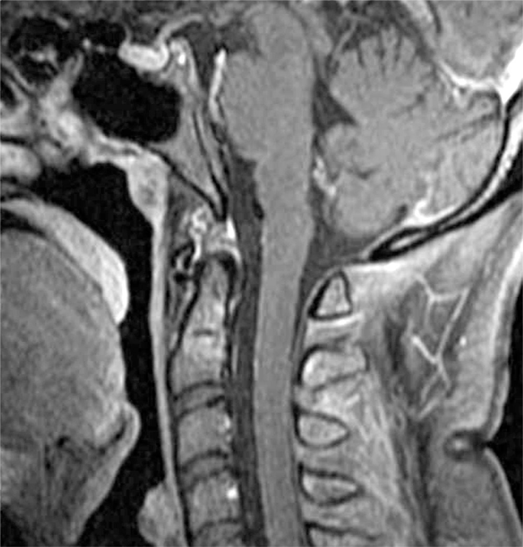
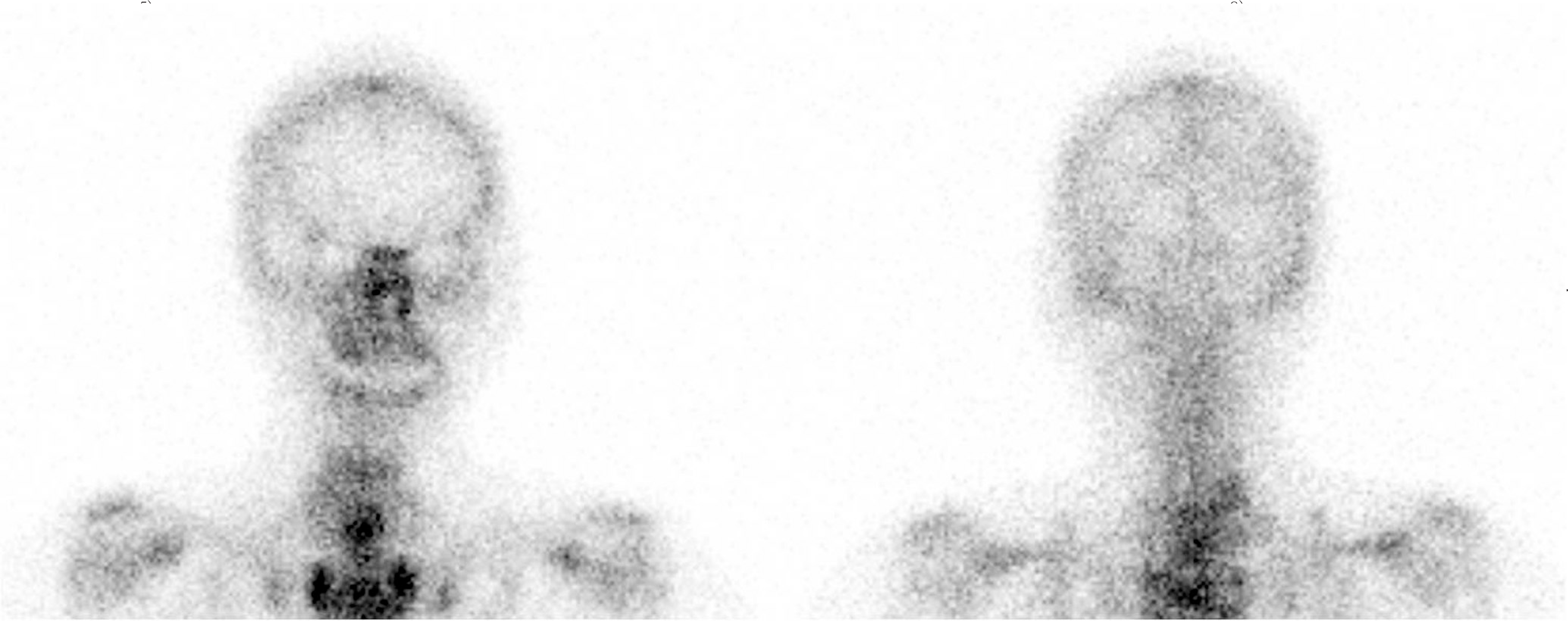
A 29-year-old woman was referred to our emergency service unit with symptoms of neck pain and scalp laceration, after being involved in a car accident. She was diagnosed as the inferior accessory ossicle of the anterior arch of the atlas, by multiple diagnostic mordalities.
REFERENCES
1.Naji MF., Bhat R. The typical appearance of the inferior accessory ossicle of the anterior arch of the atlas. Surg Radiol Anst. 2009. 31:69–71.

2.Kohler A., Zimmer EA. Borderlands of the normal and early pathologic in skeletal roentgenology. Tenth edition. Grune & Stratton Inc., New York,. 1956. 85:206.
3.Keats TE. The inferior accessory ossicle of the anterior arch of the atlas. Am J Roentgenol Radium Ther Nucl Med. 1967. 101:834–6.
4.Chow K., Motamedi K., Seeger LL., Kalantari BN. Accessory ossicles and sesamoid bones: spectrum of pathology and imaging evaluation. Appl Radiol. 2007. 36:28–37.
5.Kim NH., Choi CH., Koh GH. Acute Fractures and Dislocations of the Cervical Spine in Children and Adolescents. J Korean Soc Spine Surg. 1994. 1:19–27.
6.Lustrin ES., Karakas SP., Ortiz AO, et al. Pediatric cervical spine: normal anatomy, variants, and trauma. Radiographics. 2003. 23:539–60.
7.Von Ludinghausen M., Fahr M., Prescher A, et al. Accessory joints between basiocciput and atlas/axis in the median plane. Clin Anat. 2005. 18:558–71.
8.Omezzine SJ., Hafsa C., Lahmar I, et al. Calcific tendinitis of the longus colli: diagnosis by CT. Joint Bone Spine. 2008. 75:90–1.
9.Feldman VB. Eagle's syndrome: a case of symptomatic calcification of the styloid ligaments. J Can Chiropr Assoc. 2003. 47:21–7.
10.Jevtich V. Horizontal fracture of the anterior arch of the atlas. Case report. J Bone Joint Surg Am. 1986. 68:1094–5.
Fig.1.
Lateral radiograph of cervical spine shows a well corticated bone fragment inferior to the anterior arch of atlas (black arrow)
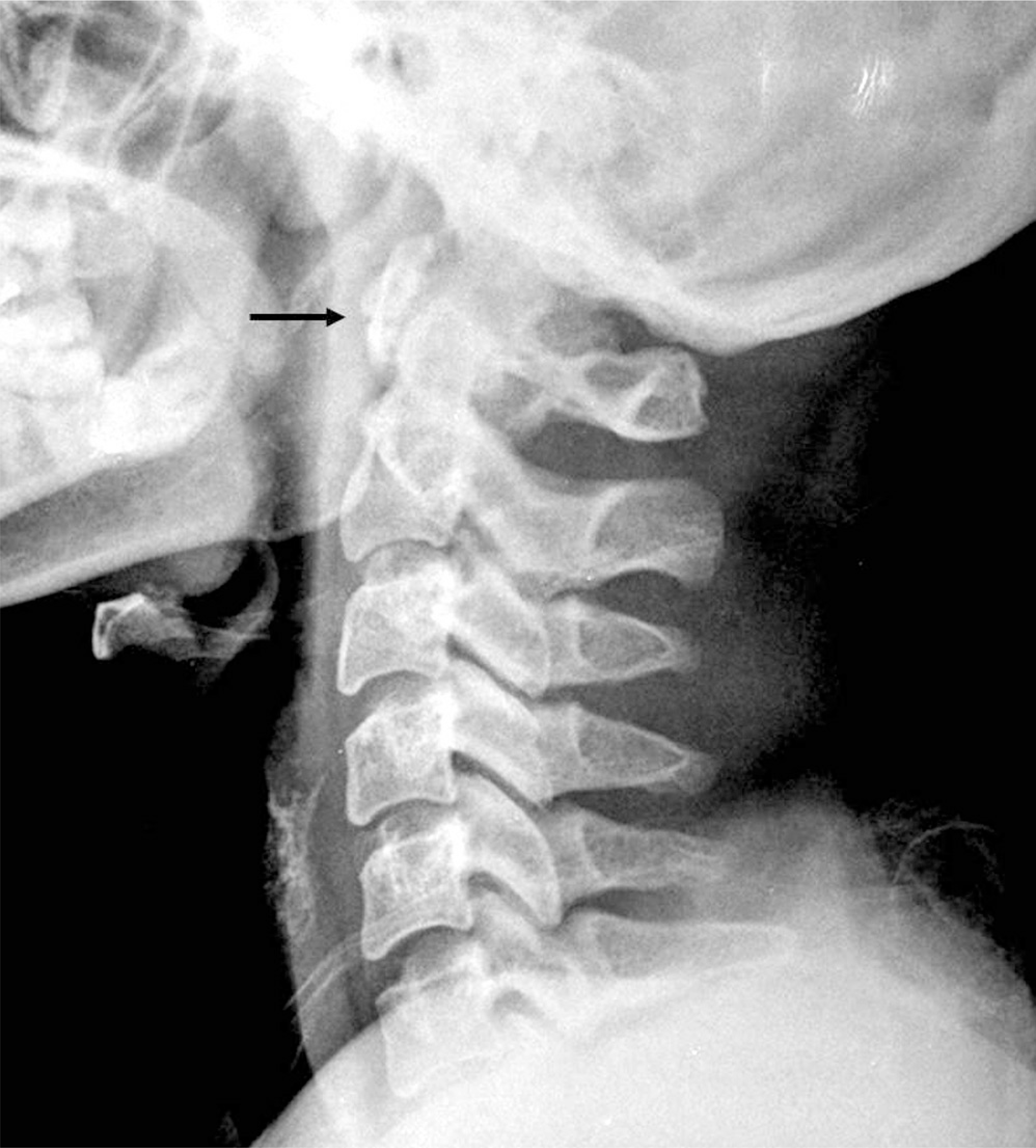
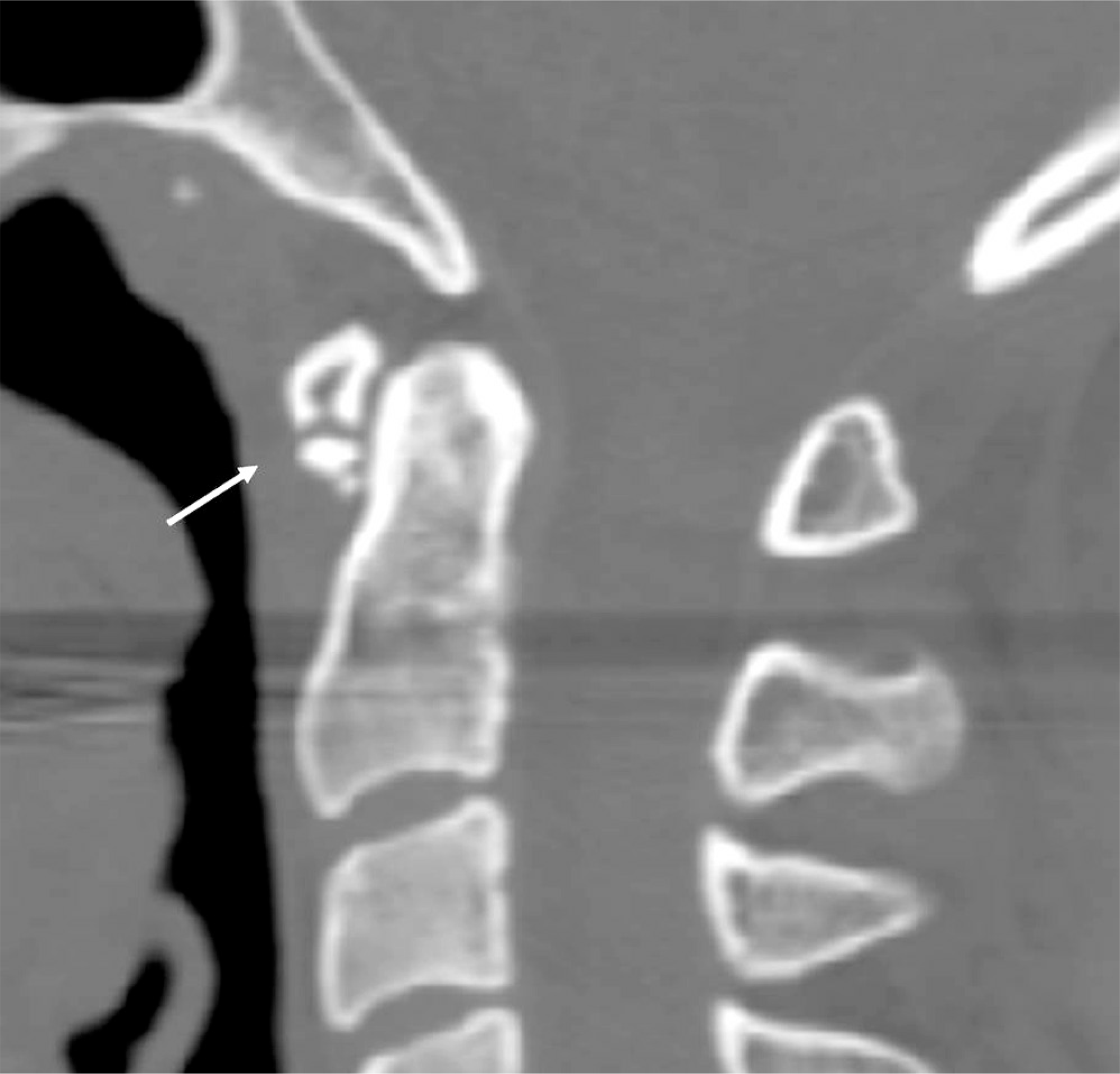
Fig.2.
Lateral CT at the level of C1 showing inferior accessory ossicle of the anterior arch of atlas (white arrow).
 XML Download