

 PDF
PDF ePub
ePub Citation
 Print
Print


Abstract
Objectives
To demonstrate the fusion rate, degree of subsidence and donor site morbidity of anterior cervical interbody fusion with autogenous bicortical iliac bone graft and anterior cervical locking plate.
Summary of Literature Review
In anterior cervical discectomy and fusion with autogenous tricortical iliac bone graft, a large percentage of patients report chronic donor site pain.
Materials and Methods
Retrospective research was done for 39 patients who underwent anterior cervical interbody fusion with autogenous bicortical iliac bone graft, from January 2006 to July 2011, with a follow up period of longer than 1 year. Fusion rates and subsidece of the graft is estimated with radiographs. Neck pain and donor site pain was estimated with visual analogue scale (VAS) and dysfunction was estimated with the neck disability index (NDI).
Results
A 95% of patients who underwent anterior cervical interbody fusion with autogenous bicortical iliac bone graft revealed definitive fusion with little amount of subsidence. The mean VAS score was 0.7 on the donor site and the mean NDI score was 3.8 at the final visit. There was excellent clinical outcome without complication at the donor site or the recipient site.
Go to : 
REFERENCES
1.Wang JM., Kim DJ. Clinical Course of Iliac Bone Graft Donor Site Morbidity. J Korean Soc Spine Surg. 1996. 3:154–60.
2.Kim DJ., Shin MC. Long-Term Follow-up of Posterior Pedicle Instrumentation and Anterior Lumbar Fusion using a Bicortical Iliac Allograft. J Korean Orthop Assoc. 2003. 38:594–600.

3.Song KJ., Choi BW., Kim SJ., Yoon SJ. Cross-Cultural Ad-aptation and Validation of the Korean Version of the Neck Disability Index. J Korean Orthop Assoc. 2009. 44:350–9.
4.Brantigan JW. Pseudoarthrosis rate after allograft posterior lumbar interbody fusion with pedicle screw and plate fixation. Spine (Phila Pa 1976). 1994. 19:1271–9.
5.Sasso RC., LeHuec JC., Shaffrey C. Iliac Crest Bone Graft Donor Site Pain After Anterior Lumbar Interbody Fusion: A Prospective Patient Satisfaction Outcome Assessment. J Spinal Disord Tech. 2005. 18:77–81.
6.Myeroff C., Archdeacon M. Autogenous Bone Graft: Donor Sites and Techniques. J Bone Joint Surg Am. 2011. 93:2227–36.
7.Fasolis M., Boffano P., Ramieri G. Morbidity associated with anterior iliac crest bone graft. Oral Surg Oral Med Oral Pathol Oral Radiol. 2012. 114:586–91.
8.Lee JK., Ahn JS., Kim SB., Hong CH., Lee JB. Influence of Plate Position on Fusion Time and Clinical Outcomes after Anterior Cervical Interbody Fusion. J Korean Soc Spine Surg. 2005. 12:22–7.
9.Wang JC., McDonough PW., Endow K., Kanim LE., Dela-marter RB. The effect of cervical planting on single-level anterior cervical discectomy and fusion. J Spinal Disord. 1999. 12:467–71.
10.Fernandes HM., Mendelow AD., Choksey MS. Anterior cervical discectomy: an improvement in donor site operative technique. Br J Neurosurg. 1994. 8:201–3.
Go to :
 | Fig. 2. (A)Preoperative lateral radiograph of a 51–year-old woman with cervical herniated nucleus pulposus of C 5-6. (B) Lateral radiograph of the same patient after an one level anterior fusion operation. (C) Lateral radiograph of the same patient at the last follow-up 1 year after operation. The films show definitive fusion at C5-6 and without grafted bicortical iliac bone collapse. |
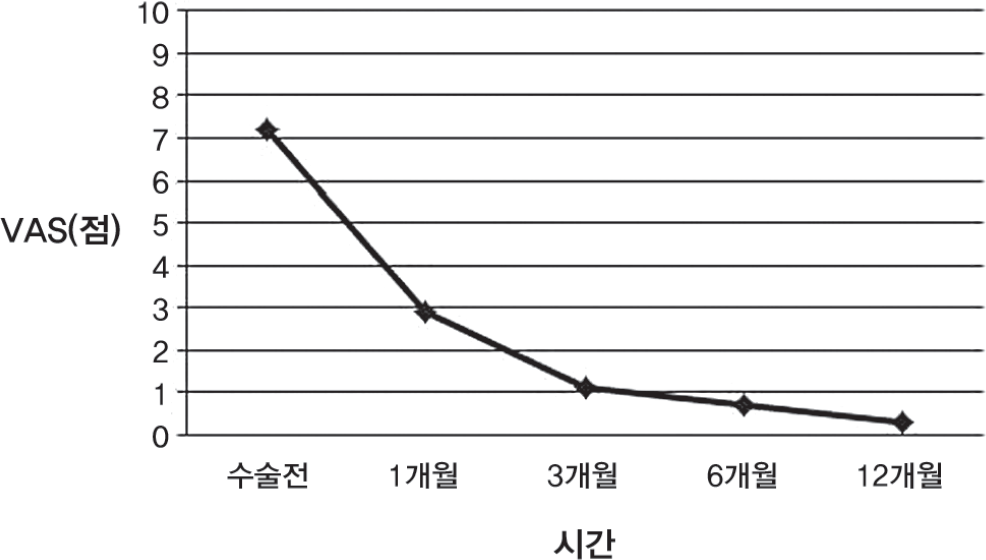
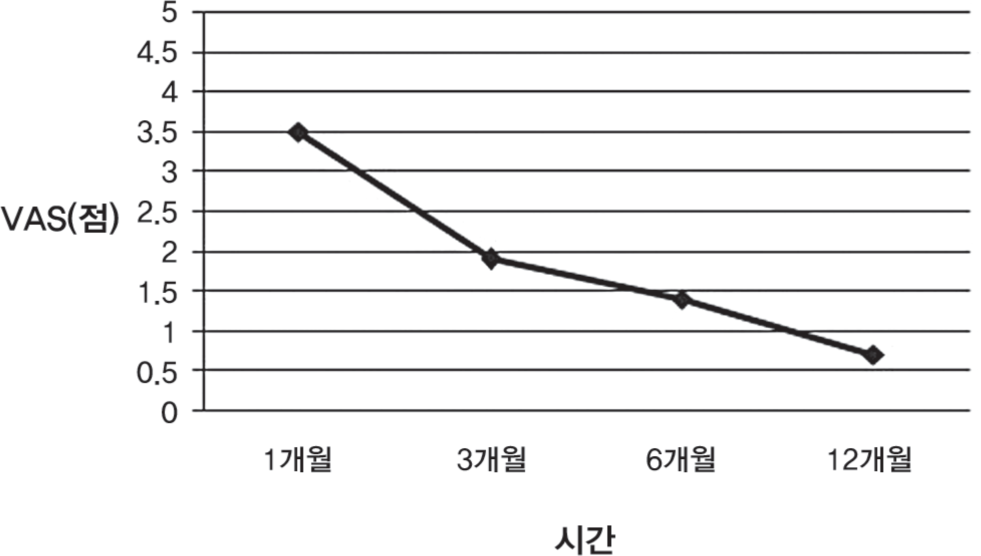
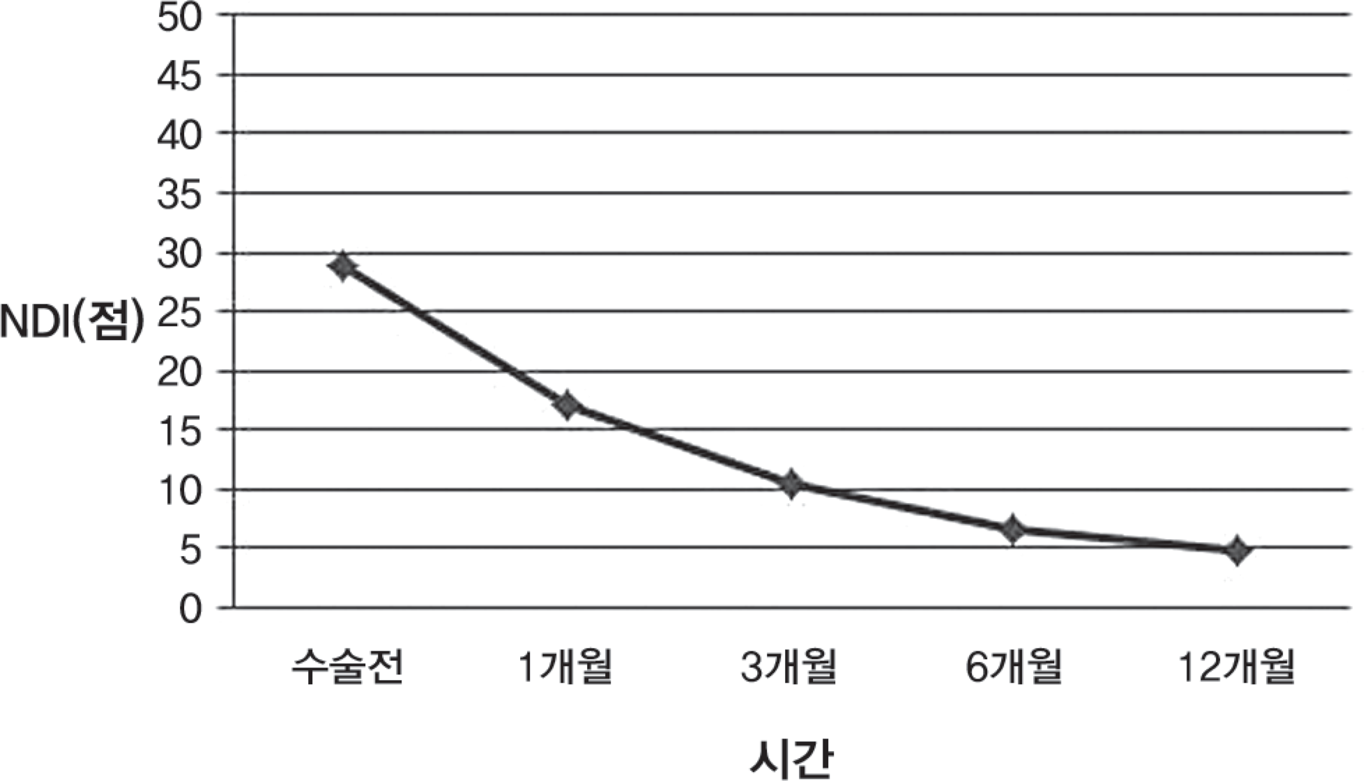
Table 1.
Description of Radiographic Fusion Result (by Brantigan,1994)
 XML Download