Abstract
Objectives
We investigated whether the lumbosacral sagittal curvature have any relation to the patterns of lumbar disc degeneration.
Summary of Literature Review
Recently, there have been many studies on the correlations between the changes of lumbar disc degeneration and associated factors, such as age, gender, weight, occupation, cigarette smoking, and genetics; but, it is hard to find research into lumbosacral sagittal alignments.
Materials and Methods
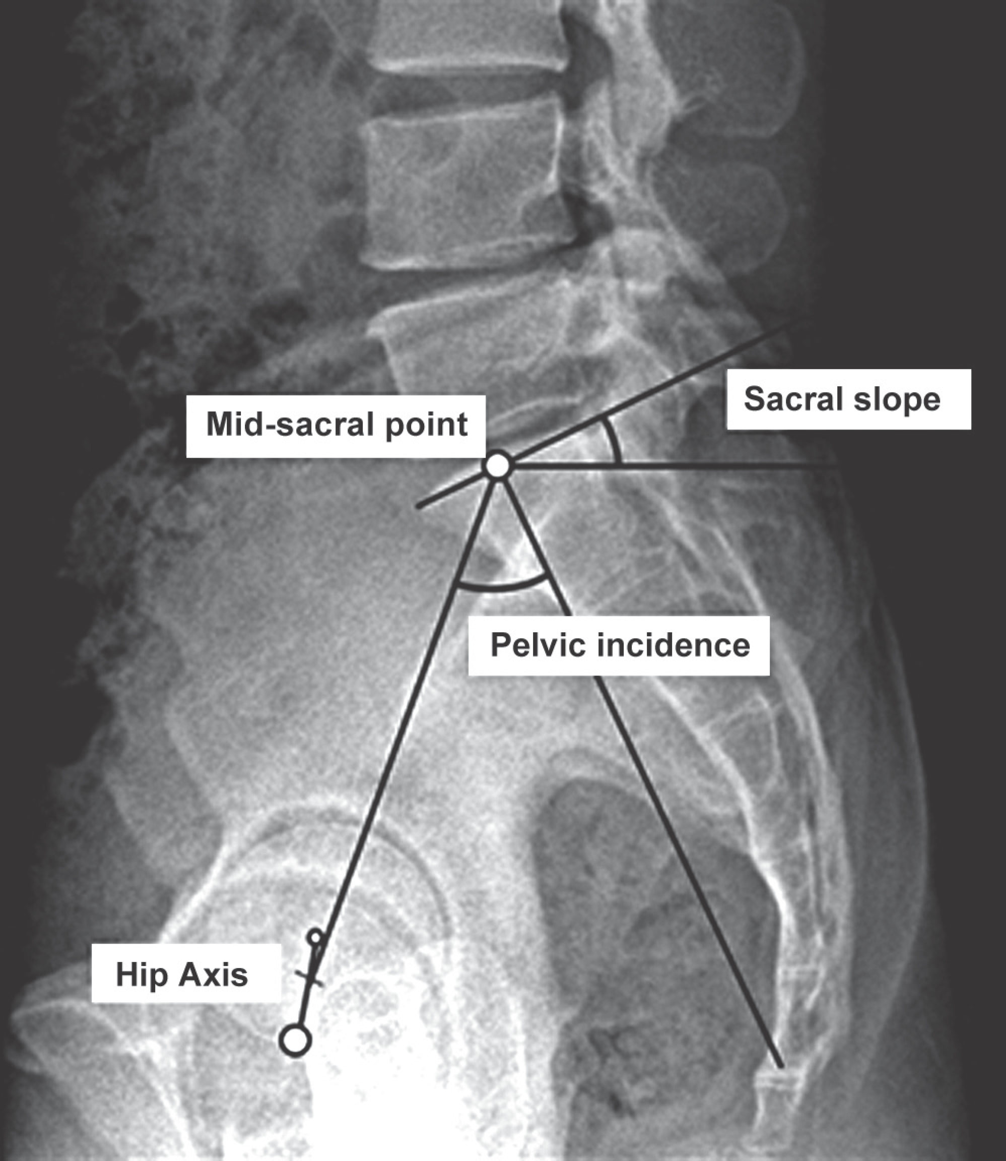
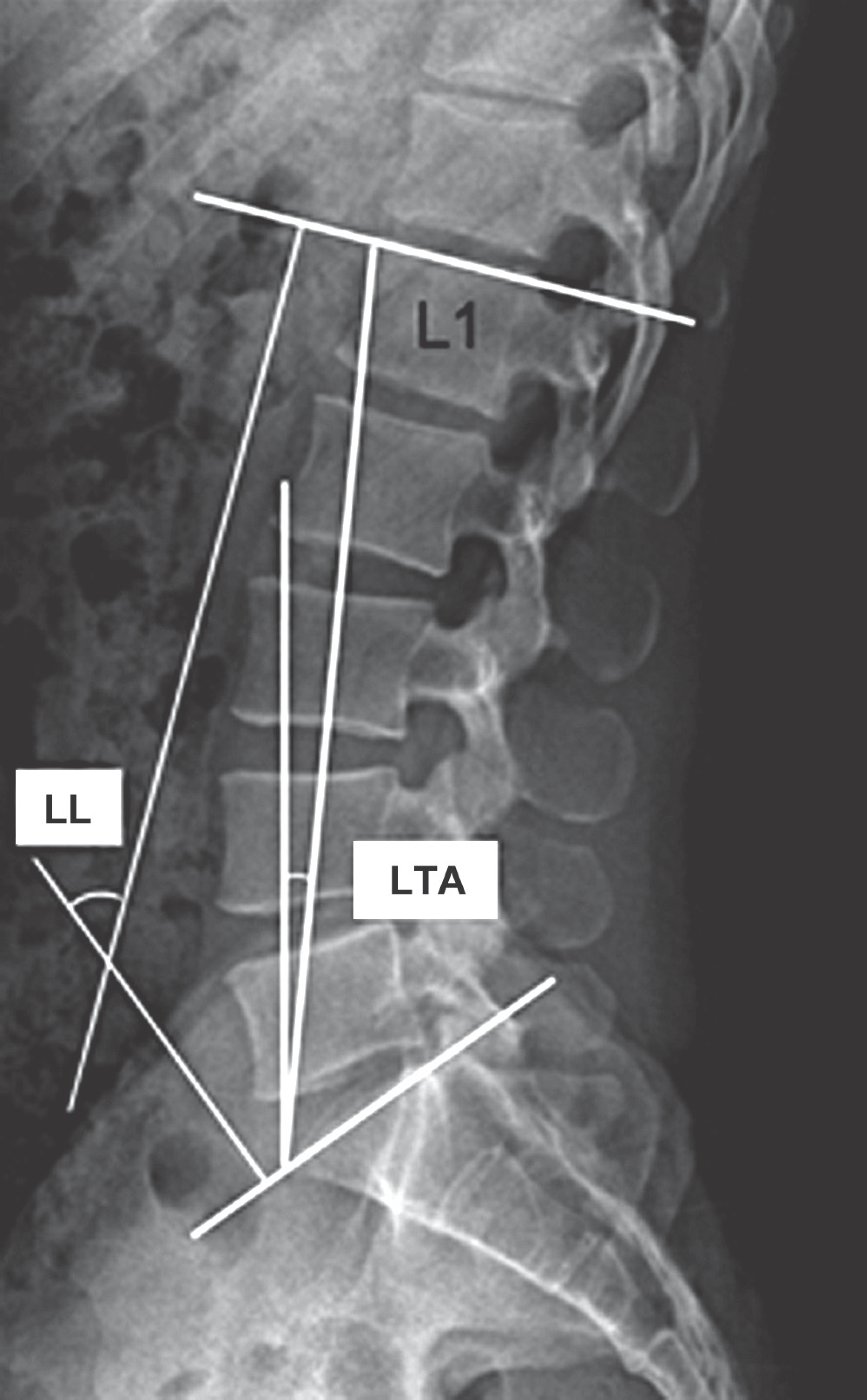
This study enrolled 117 young adult patients limited by age (18-35 years), BMD (<30kg/m2), no smoking, occupation except heavy worker, no prior lumbar surgery and no combined spinal deformity. By measuring the pelvic incidence, sacral slope, lumbar tilt angle, lumbar lordosis and lumbar axis indicating the parameters of sagittal alignments, we investigated the correlation between the number and severity of lumbar disc degeneration and the number of herniated intervertebral discs.
Results
This study found a moderate correlation between pelvic incidence, sacral slope, lumbar lordosis, and the number of lumbar degenerative disc (r=-0.451, p<0.001; r=-0.433, p<0.001; r=-0.425, p<0.001). We calculated the most proper cut-off value of pelvic incidence associated with more than three segments of multiple lumbar disc degeneration, using a minimum p-value approach.
Conclusions
As pelvic incidence, sacral slope, and lumbar lordosis indicating the parameters of lumbosacral sagittal alignments get smaller, the numbers of lumbar disc degenerations and herniated intervertebral discs increase. When pelvic incidence is below 45.6 degrees, it is more likely for degenerative changes of lumbar disc to affect more than three segments.
Go to : 
REFERENCES
1.Salminen JJ., Erkintalo MO., Pentti J., Oksanen A., Kormano MJ. Recurrent low back pain and early disc degeneration in the young. Spine (Phila Pa 1976). 1999. 24:1316–21.

2.Bezer M., Erol B., Kocaoglu B, et al. [Low back pain among children and adolescents]. Acta Orthop Traumatol Turc. 2004. 38:136–44.
3.Videman T., Battie MC. The influence of occupation on lumbar degeneration. Spine (Phila Pa 1976). 1999. 24:1164–8.
5.Elfering A., Semmer N., Birkhofer D., Zanetti M., Hodler J., Boos N. Risk factors for lumbar disc degeneration: a 5-year prospective MRI study in asymptomatic individuals. Spine (Phila Pa 1976). 2002. 27:125–34.
6.Battie MC., Videman T. Lumbar disc degeneration: epidemiology and genetics. J Bone Joint Surg Am. 2006. 88(Suppl 2):3–9.
7.Djurasovic MO., Carreon LY., Glassman SD., Dimar JR 2nd., Puno RM., Johnson JR. Sagittal alignment as a risk factor for adjacent level degeneration: a case-control study. Or-thopedics. 2008. 31:546.

8.Battie MC., Videman T., Gill K, et al. 1991 Volvo Award in clinical sciences. Smoking and lumbar intervertebral disc degeneration: an MRI study of identical twins. Spine (Phila Pa 1976). 1991. 16:1015–21.
9.Sward L., Hellstrom M., Jacobsson B., Nyman R., Peterson L. Disc degeneration and associated abnormalities of the spine in elite gymnasts. A magnetic resonance imaging study. Spine (Phila Pa 1976). 1991. 16:437–43.
10.Battié MC., Videman T., Gibbons LE., Fisher LD., Manninen H., Gill K. 1995 Volvo Award in clinical sciences. Determi-nants of lumbar disc degeneration. A study relating lifetime exposures and magnetic resonance imaging findings in identical twins. Spine (Phila Pa 1976). 2006. 88(Suppl):3–9.
11.Ong A., Anderson J., Roche J. A pilot study of the prevalence of lumbar disc degeneration in elite athletes with lower back pain at the Sydney 2000 Olympic Games. Br J Sports Med. 2003. 37:263–6.

12.Liuke M., Solovieva S., Lamminen A, et al. Disc degeneration of the lumbar spine in relation to overweight. Int J Obes (Lond). 2005. 29:903–8.

13.Rajnics P., Templier A., Skalli W., Lavaste F., Illes T. The im-portance of spinopelvic parameters in patients with lumbar disc lesions. Int Orthop. 2002. 26:104–8.
14.Barrey C., Jund J., Noseda O., Roussouly P. Sagittal bal-ance of the pelvis-spine complex and lumbar degenerative diseases. A comparative study about 85 cases. Eur Spine J. 2007. 16:1459–67.

15.Ergun T., Lakadamyali H., Sahin MS. The relation between sagittal morphology of the lumbosacral spine and the degree of lumbar intervertebral disc degeneration. Acta Orthop Traumatol Turc. 2010. 44:293–9.

16.Schneiderman G., Flannigan B., Kingston S., Thomas J., Dillin WH., Watkins RG. Magnetic resonance imaging in the di-agnosis of disc degeneration: correlation with discography. Spine (Phila Pa 1976). 1987. 12:276–81.
17.Kawaguchi Y., Kanamori M., Ishihara H., Ohmori K., Matsui H., Kimura T. The association of lumbar disc disease with vitamin-D receptor gene polymorphism. J Bone Joint Surg Am. 2002. 84-A:2022–8.

18.Cheung KM., Chan D., Karppinen J, et al. Association of the Taq I allele in vitamin D receptor with degenerative disc disease and disc bulge in a Chinese population. Spine (Phila Pa 1976). 2006. 31:1143–8.

19.Sambrook PN., MacGregor AJ., Spector TD. Genetic influences on cervical and lumbar disc degeneration: a magnetic resonance imaging study in twins. Arthritis Rheum. 1999. 42:366–72.

20.Solovieva S., Lohiniva J., Leino-Arjas P, et al. COL9A3 gene polymorphism and obesity in intervertebral disc degeneration of the lumbar spine: evidence of gene-environment interaction. Spine (Phila Pa 1976). 2002. 27:2691–6.

21.Battie MC., Videman T., Parent E. Lumbar disc degeneration: epidemiology and genetic influences. Spine (Phila Pa 1976). 2004. 29:2679–90.
22.Seki S., Kawaguchi Y., Chiba K, et al. A functional SNP in CILP, encoding cartilage intermediate layer protein, is associated with susceptibility to lumbar disc disease. Nat Genet. 2005. 37:607–12.

23.Videman T., Sarna S., Battie MC, et al. The long-term effects of physical loading and exercise lifestyles on back-related symptoms, disability, and spinal pathology among men. Spine (Phila Pa 1976). 1995. 20:699–709.

24.Miyakoshi N., Itoi E., Murai H., Wakabayashi I., Ito H., Mi-nato T. Inverse relation between osteoporosis and spon-dylosis in postmenopausal women as evaluated by bone mineral density and semiquantitative scoring of spinal degeneration. Spine (Phila Pa 1976). 2003. 28:492–5.

25.Jhawar BS., Fuchs CS., Colditz GA., Stampfer MJ. Cardio-vascular risk factors for physician-diagnosed lumbar disc herniation. Spine J. 2006. 6:684–91.

26.Lee CS., Lee CK., Kim YT., Hong YM., Yoo JH. Dynamic sagittal imbalance of the spine in degenerative flat back: significance of pelvic tilt in surgical treatment. Spine (Phila Pa 1976). 2001. 26:2029–35.
27.Roussouly P., Gollogly S., Berthonnaud E., Dimnet J. Clas-sification of the normal variation in the sagittal alignment of the human lumbar spine and pelvis in the standing position. Spine (Phila Pa 1976). 2005. 30:346–53.

28.Lee CS., Chung SS., Kang KC., Park SJ., Shin SK. Normal patterns of Sagittal Alignment of the Spine in Young Adults Radiological analysis in a Korean population. Spine (Phila Pa 1976). 2011. 36:1648–54.

29.Duval-Beaupere G., Robain G. Visualization on full spine radiographs of the anatomical connections of the centres of the segmental body mass supported by each vertebra and measured in vivo. Int Orthop. 1987. 11:261–9.
30.Lazennec JY., Ramare S., Arafati N, et al. Sagittal alignment in lumbosacral fusion: relations between radiological parameters and pain. Eur Spine J. 2000. 9:47–55.

31.Gelb DE., Lenke LG., Bridwell KH., Blanke K., McEnery KW. An analysis of sagittal spinal alignment in 100 asymptomatic middle and older aged volunteers. Spine (Phila Pa 1976). 1995. 20:1351–8.

Go to : 
 | Fig. 1.Methods for measuring the pelvic parameters. Pelvic incidence: angle between the perpendicular to the sacral plate at its midpoint and the line connecting this point to the middle axis of the femoral heads. Sacral slope: angle between the superior plate of S1 and a horizontal line. |
 | Fig. 2.Methods for measuring the spinal parameters. Lumbar lordosis (LL): angle between the upper end plates of the first lumbar vertebra (L1) and first sacrum vertebra (S1). Lumbar tilt angle (LTA): angle between the line connecting the anterosuperior edge of the L1 to the anterosuperior edge of the S1 and the vertical line. |
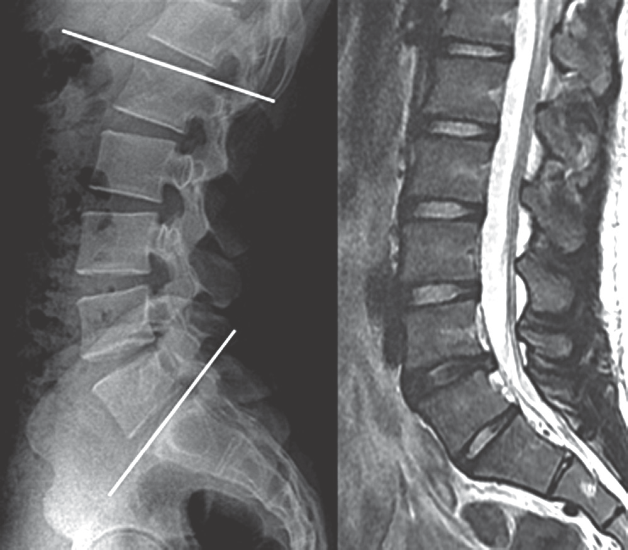
 | Fig. 3.25-year-old female patient. In standing lateral X-ray L1-S1 lordosis was checked by 68.1° and pelvic incidence by 61.7°. MRI scan showed that the lumbar spine had the one degenerated level. |
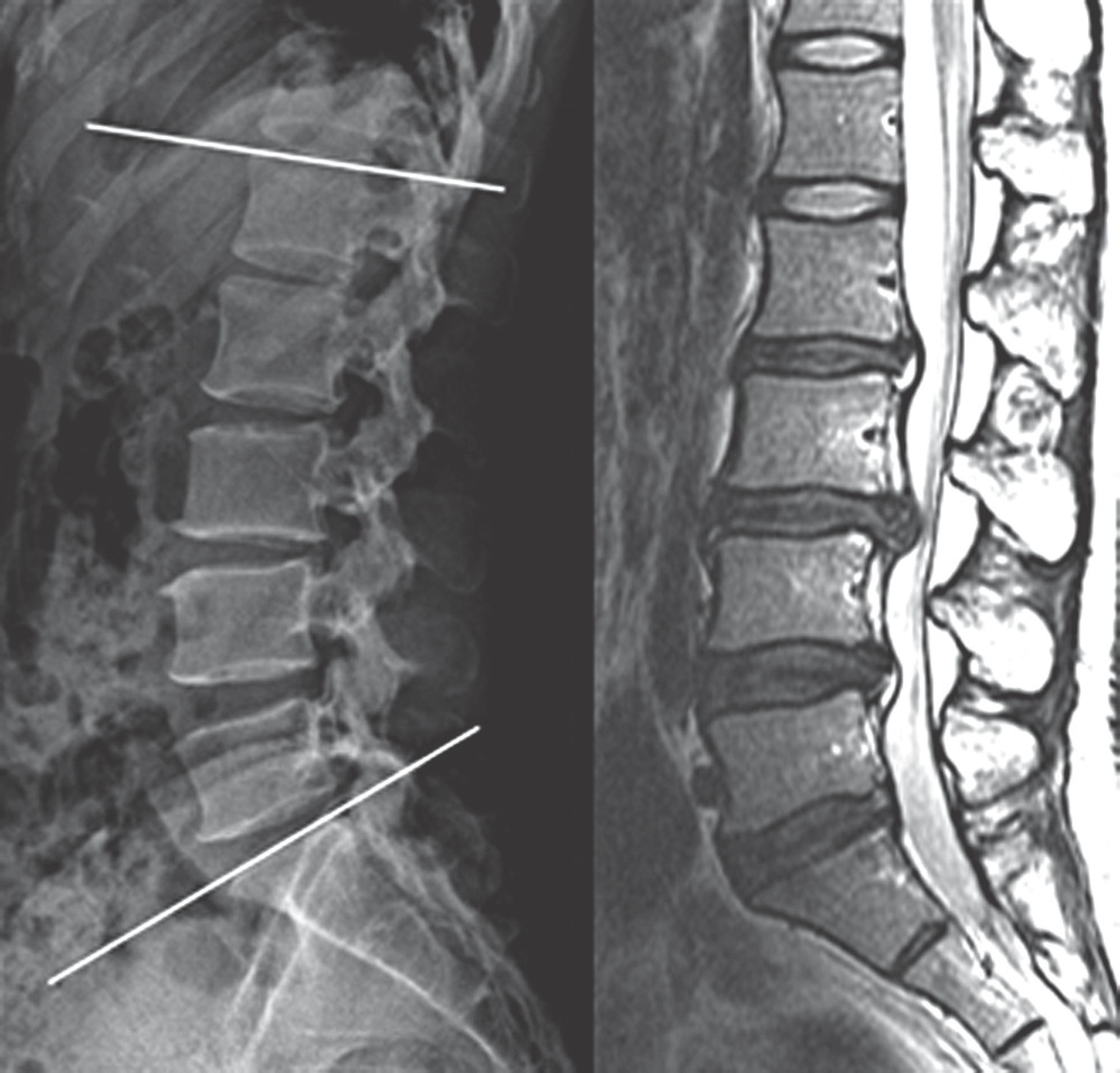
 | Fig. 4.Figure 4. 32-year-old female patient. In standing lateral X-ray L1-S1 lordosis was checked by 38.3° and pelvic incidence by 39.5°. MRI scan showed that the lumbar spine had the four degenerated levels. |
Table 1.
Mean, Minimum, Maximum and Standard Deviations of the Parameters
Table 2.
The Mean of the Parameters in the 3 groups
| Parameters | Group I (N=43) | Group II (N=47) | Group III (N=25) | P-value (ANOVA) |
|---|---|---|---|---|
| Pelvic incidence (°) | 51.9 (±8.0) | 47.6(±7.2) | 42.1 (±8.2) | <0.001∗ |
| Sacral slope (°) | 36.0 (±6.9) | 32.7(±7.3) | 27.4 (±4.9) | <0.001∗ |
| Lumbar lordosis (°) | 50.0 (±9.3) | 44.9(±11.2) | 37.6 (±8.1) | <0.001∗ |
| Lumbar tilt angle (°) | 3.1 (±5.8) | 2.9(±4.7) | 4.6 (±5.3) | 0.431 |
| Lumbar apex | 3.6 (4.0,3.0) | 3.6(4.0,3.0) | 3.6 (4.0,3.0) | 0.946† |
| LDD score | 2.1 (3,2) | 4.1(5,4) | 6.1 (7,5) | <0.001∗† |
| BMI (kg/m2) | 23.7 (±3.1) | 23.2(±2.8) | 23.6 (±2.9) | 0.718 |
| Age | 25.5 (±4.2) | 27.6(±5.3) | 30.9 (±5.2) | <0.001∗ |
Table 3.
The HIVD(N) in the 3 groups
| HIVD(N) | Group I (N=43) | Group II (N=47) | Group III (N=25) | Total |
|---|---|---|---|---|
| 0 | 1 | 0 | 0 | 1 |
| 1 | 42 | 23 | 11 | 76 |
| 2 | 0 | 24 | 12 | 36 |
| 3 | 0 | 0 | 2 | 2 |
| Total | 43 | 47 | 25 | 115 |
Table 4.
Correlation between the sagittal parameters and lumbar disc degeneration
| Parameters | LDD(N) | LDD score | LDD score /LDD(N) | HIVD(N) | ||||
|---|---|---|---|---|---|---|---|---|
| R∗ | P† | R∗ | P† | R∗ | P† | R∗ | P | |
| Pelvic incidence | -0.451 | <0.001 | -0.396 | <0.001 | 0.058 | 0.536 | -0.210 | 0.024 |
| Sacral slope | -0.433 | <0.001 | -0.439 | <0.001 | -0.046 | 0.626 | -0.252 | 0.006 |
| Lumbar lordosis | -0.425 | <0.001 | -0.397 | <0.001 | 0.044 | 0.640 | -0.263 | 0.004 |
| Lumbar tilt angle | 0.098 | 0.297 | 0.094 | 0.316 | 0.007 | 0.942 | -0.010 | 0.918 |
| Lumbar apex | 0.009 | 0.921 | -0.037 | 0.697 | -0.092 | 0.326 | -0.070 | 0.457 |


 PDF
PDF ePub
ePub Citation
 Print
Print


 XML Download